

Transthoracic Echocardiogram
Learn how a transthoracic echocardiogram works, what to expect during this heart ultrasound in Guadalajara, and how to prepare.
What is a transthoracic echocardiogram?
A transthoracic echocardiogram is a non-invasive heart test that uses high-frequency sound waves to create detailed, real-time pictures of your heart. "Transthoracic" simply means the test is performed across the chest wall, or thorax. It is the most common type of cardiac imaging test ordered by cardiologists to evaluate heart structure, function, and overall cardiovascular health. For patients facing heart surgery, this test provides the surgical team with an essential visual map of the heart before any procedure begins.
Why does my heart need to be looked at? A transthoracic echocardiogram helps your doctor see the size of your heart chambers, the thickness of the heart muscle, and how well the valves are opening and closing. It allows the medical team to assess the pumping strength of the heart, ensuring that the heart muscle is contracting normally to push blood to the rest of your body.
Is the procedure safe and comfortable? Unlike X-rays or CT scans, an echocardiogram test does not use radiation. It relies entirely on harmless ultrasound waves, making it exceptionally safe for patients of all ages. The procedure is painless, requires no needles, and typically takes less than an hour to complete, allowing you to return to your normal activities immediately afterward.
How does a heart ultrasound work?
The principles behind a heart ultrasound are similar to the sonar used by ships to map the ocean floor. A small, handheld device called a transducer sends sound waves through the chest wall. These sound waves bounce off the solid structures of your heart and return to the transducer, which is connected to a computer. The computer then translates these returning echoes into moving images on a screen.
How does the machine create moving pictures? The transducer captures thousands of snapshots per second, piecing them together to show the heart in motion. This allows the physician to see the heart valves opening and closing, the muscle contracting and relaxing, and the blood flowing through the chambers. Seeing the heart in real-time is crucial for identifying mechanical problems or abnormal blood flow patterns.
What does the gel do during the test? The technician applies a clear, water-soluble gel to the skin before placing the transducer on your chest. This gel eliminates the tiny pocket of air that would otherwise exist between the transducer and your skin. Because air blocks sound waves, the gel acts as a bridge, ensuring the ultrasound waves travel smoothly from the device into your chest and back again to produce a clear image.
Why is a cardiac imaging test ordered?
Your doctor might order a cardiac imaging test for a variety of reasons, ranging from routine heart screening test protocols to investigating specific physical symptoms. If you experience shortness of breath, chest pain, irregular heartbeats, or unexplained fatigue, this test helps identify the underlying cause. It is also frequently used to monitor existing heart conditions over time to ensure they are not worsening.
What specific conditions can this test find? A transthoracic echocardiogram is highly effective at identifying structural issues like heart valve echocardiogram abnormalities, such as stenosis (stiff valves) or regurgitation (leaky valves). It can also detect fluid buildup around the heart, congenital heart defects, or signs of previous heart attacks that may have gone unnoticed by the patient.
How does this test help surgeons plan? For a cardiac surgeon, this imaging test provides a preoperative blueprint of your heart. By understanding the exact size of the chambers, the extent of valve damage, and the overall pumping capacity, the surgical team can plan the most effective approach for your operation.
What to expect during a TTE echocardiogram?
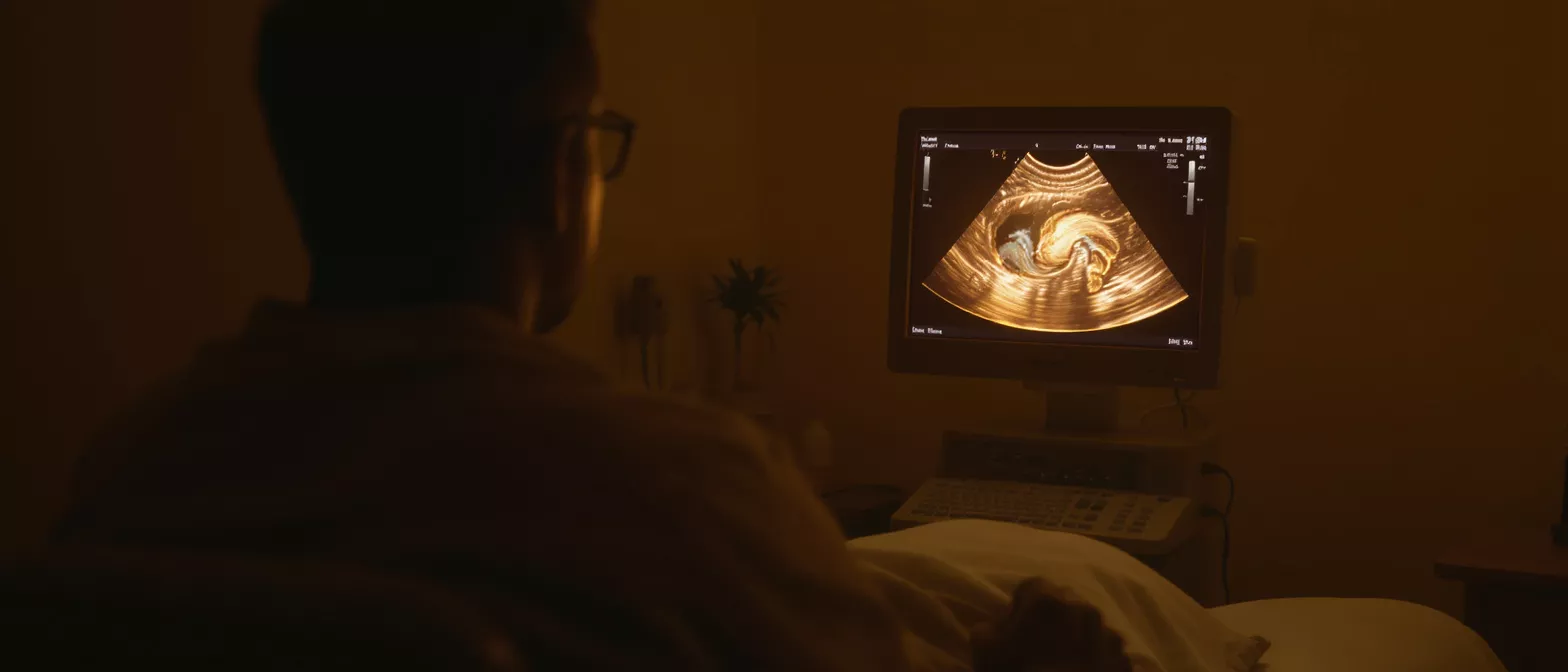
Knowing what will happen during a TTE echocardiogram can greatly reduce any anxiety you might feel before the appointment. When you arrive, you will be asked to change into a hospital gown to allow easy access to your chest. The echocardiography technician will guide you to a dimly lit room, which helps them see the ultrasound monitor more clearly. You will lie on an examination table, usually on your left side, which brings your heart closer to the chest wall for better imaging.
What will the technician do during the procedure? The technician will place a few sticky patches called electrodes on your chest to record an electrocardiogram (ECG) at the same time. This tracks your heart's electrical activity and helps time the ultrasound images with your heartbeat. The technician will then apply the ultrasound gel and press the transducer firmly against different areas of your chest to get multiple viewing angles.
Will the procedure hurt? The test is entirely painless, though you may feel a slight pressure as the technician presses the transducer against your ribs to get a clear view of your heart. You might be asked to hold your breath for a few seconds at various points during the scan to improve image quality. Once the technician has collected all the necessary images, the gel is wiped off, and the test is complete.
How to prepare for your echo test for heart?
Preparing for an echo test for heart evaluation is straightforward and requires minimal adjustment to your daily routine. Unlike some other medical tests, a standard transthoracic echocardiogram usually requires no fasting, no dietary restrictions, and no special bowel preparations. You can eat, drink, and take your regular medications exactly as you normally would unless your doctor specifically advises otherwise.
What should I wear to the appointment? It is best to wear comfortable, loose-fitting clothing that can be easily removed from the waist up. A two-piece outfit, such as a shirt and pants, is highly recommended because you will need to undress from the waist up to expose your chest. Avoid one-piece garments like dresses or overalls to make the changing process quicker and more comfortable.
- Check medications: Confirm with your cardiologist whether to take morning doses.
- Avoid heavy lotions: Skip thick creams or powders on your chest, as they can interfere with the ultrasound gel.
- Arrive early: Give yourself time to check in, change into a gown, and relax before the scan.
Understanding echocardiogram results
Getting echocardiogram results is the most important part of the process, as it translates the visual data into actionable medical insights. After the test, a cardiologist specialized in imaging will review the recorded images and measurements. They will compile a detailed report that assesses the size and shape of your heart, the function of your heart valves, and the strength of your heart muscle contractions.
How soon will I get my results? The preliminary images are available immediately to the technician, but a full interpretation takes time. The reviewing cardiologist must carefully analyze the data, measure specific heart chambers, and calculate important metrics like the ejection fraction, which measures how much blood your heart pumps out with each beat. You will usually discuss these findings with your referring physician within a few days of the test.
What do the numbers on the report mean? A normal result shows that your heart chambers are standard sizes, your valves are opening and closing properly, and your heart is pumping blood effectively. Abnormal results might show thickened muscle, enlarged chambers, or leaky valves, which require careful monitoring or surgical intervention.
Transthoracic echocardiogram vs transesophageal
When comparing a transthoracic echocardiogram vs transesophageal approach, the key difference is the vantage point of the ultrasound probe. A transthoracic echocardiogram is performed externally, moving a probe across the chest. A transesophageal echocardiogram, or TEE, requires a specialized probe to be gently guided down the esophagus, placing the ultrasound sensor directly behind the heart for a closer look.
Why choose one type of echo over the other? The standard transthoracic approach is non-invasive, fast, and requires no sedation, making it the perfect first-line screening tool. However, the lungs and ribcage can sometimes obstruct the sound waves, producing blurry images. If your doctor needs an extremely detailed view of specific structures—like the presence of blood clots in the left atrium or tiny infections on a heart valve—they may recommend a TEE instead.
Does the TEE require different preparation? Yes, a transesophageal echo requires fasting and usually involves mild sedation to keep the patient comfortable during the procedure. Because the probe is much closer to the heart, the sound waves do not have to pass through bone or lung tissue, resulting in highly detailed images. Your surgeon will decide which imaging test is necessary based on your surgical needs.
Is a heart screening test safe?
Patients often ask, is a heart screening test safe? The short answer is yes, a transthoracic echocardiogram is exceptionally safe. Because the test relies on sound waves rather than ionizing radiation, there is no risk of cellular damage or radiation exposure. It can be safely repeated multiple times over a patient's life without any cumulative negative effects, which is why it is heavily relied upon for long-term cardiac monitoring.
Are there any risks or side effects to worry about? There are essentially no risks associated with a standard transthoracic echo. The ultrasound gel is hypoallergenic and water-soluble, and the pressure applied by the transducer does not harm the chest wall. Even patients with pacemakers or implantable defibrillators can safely undergo this ultrasound procedure without any interference to their devices.
Can pregnant women undergo this cardiac ultrasound procedure? Yes, an echocardiogram is the preferred cardiac imaging method for pregnant women, as it completely avoids exposing the developing fetus to radiation. If a pregnant patient experiences heart palpitations or chest pain, an echocardiogram is the safest way for doctors to evaluate her cardiovascular health. During recovery from any procedure, using a portable blood pressure monitor can help keep your entire cardiovascular profile on track safely.
Echocardiogram near me and next steps
If you are searching for an echocardiogram near me, it is important to look for a cardiology practice or hospital with specialized imaging technicians and board-certified cardiologists. The quality of the images relies heavily on the skill of the technician operating the ultrasound machine and the expertise of the physician interpreting the results.
How do I schedule an appointment? Scheduling your cardiac imaging test is a simple process that begins with a referral from your primary care physician or cardiologist.
What happens after my echocardiogram results are reviewed? Once the imaging report is finalized, your physician will discuss the findings with you in a follow-up consultation. If the results are normal, you may simply need routine monitoring. If the test reveals a structural issue requiring surgical repair, your medical team will outline the next steps, discussing potential surgical interventions and long-term treatment plans to restore your heart health.
Conclusion and key takeaways
In conclusion, a transthoracic echocardiogram is a vital, safe, and entirely painless diagnostic tool that gives your medical team a clear window into your heart's function and structure. By using harmless sound waves, this non-invasive heart test eliminates the risks associated with radiation while providing highly detailed images of your heart chambers, valves, and blood flow.
Understanding the process, from the application of the ultrasound gel to the measurement of your ejection fraction, helps alleviate anxiety before a heart surgery. Whether you are undergoing a routine heart screening test or evaluating a specific valve abnormality, this cardiac ultrasound procedure is the gold standard for assessing preoperative cardiovascular health and monitoring postoperative recovery.
By working closely with your cardiology team and following their preparation instructions, you ensure the most accurate results possible. This imaging supports safe, effective cardiac care before, during and after surgery.






