

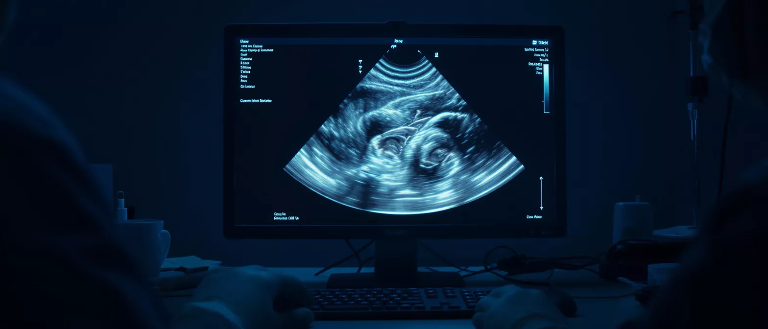
Cardiac Imaging Specialist
Learn how a cardiac imaging specialist supports heart surgery with advanced tests.
What is a Cardiac Imaging Specialist?
A cardiac imaging specialist is a medical doctor who focuses entirely on taking detailed, accurate pictures of the heart and blood vessels. They use advanced technology to look at how the heart muscle beats, how the valves open and close, and how blood flows through the chambers. For patients facing heart surgery, this doctor acts as the eyes of the surgical team, providing a clear roadmap before any operation begins.
Why this matters for cardiac surgery: Before a surgeon makes a single incision, they need to know exactly what they are dealing with inside the chest. The specialist provides the precise measurements of the heart valves, the exact location of blockages in the arteries, and the strength of the heart muscle. This information allows the surgical team to plan the safest, most effective approach for your specific condition.
These specialists do not just work in the background; they are an active part of your care team. They consult directly with your cardiovascular surgeon to interpret the scans, discuss any complexities, and ensure that every detail is understood before moving forward with treatment. Their goal is to remove uncertainty and replace it with clear, actionable facts about your heart.
How Does a Cardiac Imaging Specialist Support Heart Surgery?
When you are preparing for heart surgery, the cardiac imaging specialist plays a central role in both planning and follow-up care. They perform diagnostic cardiac imaging before the operation to give the surgeon a highly detailed, three-dimensional understanding of your heart. This pre-operative look ensures there are no surprises in the operating room and that the chosen surgical approach is tailored precisely to your anatomy.
Pre-surgery mapping: The heart imaging specialist uses scans to map out the exact size of your aorta, the degree of valve leakage, or the specific arteries affected by disease. This map is essentially a blueprint for the surgeon to study beforehand. By knowing the exact landscape of your chest and heart, the surgeon can anticipate challenges and operate with greater speed and safety.
Their job does not end when the surgery is complete. After the operation, the specialist performs follow-up scans to confirm that the repairs were successful and that the heart is functioning well in its new state. This careful monitoring helps the care team track your recovery, ensuring that your healing is progressing exactly as it should.
What Types of Cardiac Imaging Tests Are Used?
A heart imaging specialist has several different tools at their disposal, each designed to look at a specific part of the heart's anatomy or function. The choice of test depends entirely on what your doctor needs to see—whether it is the pumping strength of the muscle, the structure of the valves, or the presence of blocked arteries. These tests provide a complete picture of your cardiac health.
- Echocardiography: Uses sound waves to create moving images of the heart, ideal for checking valve function and blood flow.
- Cardiac MRI: Uses powerful magnets and radio waves to produce detailed pictures of the heart muscle, useful for spotting tissue damage.
- Cardiac CT Imaging: Uses X-rays to take cross-sectional pictures, often used to check for calcium buildup in the arteries.
- Nuclear Cardiology Imaging: Uses a small amount of radioactive tracer to show how well blood flows to the heart muscle.
Choosing the right heart imaging tests: Your doctor will select the test that gives the most useful information with the least amount of risk. Because these tests look at different things, you might need more than one to get a full evaluation. The specialist will use a cardiac imaging suite to perform these procedures, ensuring the highest quality images are captured for your surgical team.
Is Non-Invasive Cardiac Imaging Safe for Patients?
For most patients, non-invasive cardiac imaging is very safe and carries minimal risk. These procedures are designed to look inside the body without making any cuts or incisions. The technology allows doctors to gather critical information about your heart without putting you through the stress or recovery of an invasive procedure, which is especially important for patients who are already dealing with heart conditions.
Understanding the safety profile: Tests like echocardiograms use harmless sound waves, and MRIs use magnets instead of radiation. Even tests that do use radiation, like CT scans or nuclear imaging, use very small, carefully controlled doses. The benefits of getting an accurate diagnosis and a safe surgical plan far outweigh the tiny risks associated with the imaging tests themselves.
If you have concerns about the tests—such as claustrophobia in an MRI machine or a history of kidney issues that might affect contrast dye use—you should discuss them with your care team. The imaging specialist can often adjust the technique or provide medication to keep you comfortable. Your safety and peace of mind are always the top priority during the diagnostic process.
How Does an Echocardiography Specialist Evaluate Heart Function?
An echocardiography specialist focuses on one of the most common and useful tests in cardiology: the echo. This test uses high-frequency sound waves to create a live, moving picture of your heart. It allows the doctor to see the heart valves opening and closing in real time and to measure how strongly the heart muscle squeezes with each beat.
What the echo shows: This test is excellent for diagnosing valve disease, which is a common reason for heart surgery. It shows if a valve is narrowed, leaky, or structurally abnormal. It also measures the ejection fraction, which is a number that tells the doctor how well the main pumping chamber of the heart is working, giving a clear picture of overall heart health.
The procedure is painless and does not require any needles or special preparation in most cases. A technician places a small probe on your chest that sends sound waves into the heart, and the specialist interprets the results. Because it is so safe and easy to perform, it is often the first test used to get a baseline picture of your heart before deciding if more advanced imaging is needed.
When Is a Cardiac MRI Specialist Needed?
A cardiac MRI specialist is called in when the care team needs a highly detailed look at the heart muscle tissue itself. While an echo shows how the heart moves, an MRI shows the structure of the muscle in fine detail. It is often used to look for scarring from old heart attacks, inflammation of the heart muscle, or complex congenital heart defects that require a closer look.
Detailed tissue analysis: The MRI can tell the difference between healthy heart tissue and tissue that has been damaged by disease. This is crucial for surgical planning because it tells the surgeon whether a muscle can be repaired or if it has been too badly damaged to function well. It also provides very accurate measurements of the heart chambers, which helps in planning valve repairs or replacements.
During the test, you lie inside a large, tube-shaped machine for about an hour. It is noisy, but the technologist will give you earplugs and keep you informed throughout the process. The images produced give your surgical team a level of detail that is simply not possible with other tests, making the MRI a vital tool for advanced diagnostic equipment in complex heart cases.
What to Expect During Cardiac CT Imaging
Cardiac CT imaging is a test that uses X-rays to take very thin, cross-sectional pictures of the heart. These pictures are then combined by a computer to create a 3D model of the heart and its blood vessels. It is especially useful for looking at the coronary arteries—the vessels that supply blood to the heart muscle—to check for blockages or calcium deposits.
Checking for blockages: If your doctor suspects you have coronary artery disease, a CT scan can show exactly where the arteries are narrowed. This helps the surgical team decide if you need bypass surgery, a stent, or just medication. The test is fast, taking only a few minutes once the scan begins, and it is completely painless.
For some CT scans, a contrast dye is injected into a vein in your arm to make the blood vessels show up clearly on the images. You might feel a warm flush or a metallic taste in your mouth for a few seconds when the dye goes in. The specialist will monitor you closely during the scan to make sure everything goes smoothly and that the images are clear and useful for your surgical plan.
How Nuclear Cardiology Imaging Reveals Blood Flow
Nuclear cardiology imaging is a specialized test that shows how well blood is flowing to the heart muscle at rest and during stress. A tiny amount of a safe radioactive substance, called a tracer, is injected into your bloodstream. The tracer travels to your heart, and a special camera detects the radiation to create pictures of the blood flow.
Spotting poor circulation: This test is excellent for finding areas of the heart that are not getting enough blood due to blocked arteries. If an area of the heart shows up poorly on the scan, it tells the doctor that the artery supplying that muscle is narrowed or blocked. This information is very important for a surgeon who is planning a bypass operation to improve blood flow to the heart.
The test usually involves two parts: one set of pictures taken while you are resting, and another set taken after you have exercised on a treadmill or been given medicine to make your heart beat faster. By comparing the two sets of images, the specialist can tell if blood flow is normal or if there are areas that need surgical attention. The radiation you receive is very low and is gone from your body within a day.
How to Prepare for Your Heart Imaging Tests
Preparing for heart imaging tests is usually very straightforward, but the exact instructions depend on which test you are having. For most scans, you will be asked to avoid eating or drinking for a few hours before the appointment. This helps ensure that the images are clear and not affected by a full stomach or digestion.
Day-of-test instructions: You should wear comfortable, loose-fitting clothing that is easy to take off and put back on, as you may need to change into a hospital gown. Leave jewelry and metal objects at home, especially if you are having an MRI, because metal can interfere with the magnets. If you are having a stress test, wear shoes that are good for walking on a treadmill.
It is also important to bring a list of all the medications you take and to ask your doctor if you should take them on the day of the test. Some medications can affect your heart rate or interact with contrast dyes. Always follow the specific instructions given to you by the imaging center, and do not hesitate to call them if you have any questions about what to do before your appointment.
Working With Your Heart Scan Specialist
Your heart scan specialist is a key member of your care team, even if you only meet them briefly. They work behind the scenes to read your images, write detailed reports, and consult with your surgeon about the findings. Their expertise ensures that every decision made about your surgery is based on solid, objective evidence about the condition of your heart.
A team-based approach: The imaging specialist does not work alone. They discuss your scans with your cardiologist, your surgeon, and your anesthesiologist to make sure everyone is on the same page. This team approach means that your surgery is planned with input from experts in every area of heart care, giving you the best possible chance for a successful outcome.
In conclusion, the cardiac imaging specialist provides the vital information that makes modern heart surgery safe and effective. By using advanced technology to look inside your heart, they give your surgical team the knowledge they need to plan your operation and monitor your recovery. When you go in for your scans, you can feel confident knowing that these experts are giving your surgeon the clearest possible picture of your heart.






