

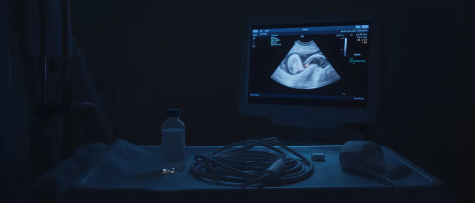
Transoesophageal Echocardiogram
Learn how a transoesophageal echocardiogram provides detailed heart images for diagnosis, plus what to expect during the procedure, risks, and recovery.
TEE vs Transthoracic Echo: Key Differences
Patients often wonder why they need a transoesophageal echocardiogram when they have already had a standard echo. The most common type of echocardiogram is the transthoracic echo (TTE), which involves moving a wand across the chest wall. While TTE is excellent for routine screening and general heart function checks, it has physical limitations. Sound waves must travel through the ribs, chest muscles, and lungs, which can scatter the waves and obscure fine details.
Image clarity and anatomical proximity: The primary difference lies in image quality and the physical barriers the sound waves must cross. Because the TEE probe sits directly behind the heart in the oesophagus, there is no interference from bone or air-filled lungs. This proximity provides a much higher resolution image, allowing doctors to spot small blood clots, subtle valve leaks, or tiny structural defects that a TTE might easily miss.
In summary, a TTE is the standard first step for general cardiac diagnosis, but a TEE is the definitive step when surgical precision is required. Your surgical team relies on the superior visualisation of a TEE to make final decisions in the operating room. It is a complementary, step-up test rather than a redundant one.
What to Expect During Transoesophageal Echo Prep
Proper preparation is crucial for a safe and successful transoesophageal echo. Because the probe passes through your mouth and into your oesophagus, your stomach must be completely empty to prevent any risk of aspiration, which is when stomach contents enter the lungs. You will typically be instructed to stop eating and drinking for at least six to eight hours before the test or before your scheduled surgery.
Medication and fasting guidelines: You should continue taking most of your regular medications with a tiny sip of water on the morning of the procedure, unless your surgeon specifically tells you otherwise. Medications like blood thinners may be managed separately, and your surgical team will give you a precise, personalised cardiac surgery recovery kit of instructions. Always follow the exact prep guidelines provided by Dr Alejandro Molina's clinical team.
When you arrive at the hospital, the nursing staff will review your medical history, check your vital signs, and place an intravenous line. This IV is used to administer the relaxing medications. If you are having the test as an outpatient rather than during surgery, you will need to arrange for a friend or family member to drive you home afterward, as you cannot drive for 24 hours after receiving sedation.
Is TEE Echocardiography Test Painful?
A very common concern among patients is whether the TEE echocardiography test is painful. The short answer is no, you should not feel pain. If you are having this test while awake, the medications you receive through your IV will make you very sleepy and relaxed. You will likely have little to no memory of the actual procedure. The throat is numbed with a spray to suppress the gag reflex, ensuring the tube passes smoothly.
Managing throat discomfort and sedation effects: While you will not feel pain during the test, you might experience a mild sore throat for a day or two afterward. This is caused by the physical presence of the tube and usually resolves quickly without any specific treatment. If the TEE is performed while you are already under general anaesthesia for heart surgery, you will be entirely unconscious and will not feel or remember the insertion at all.
It is completely normal to feel anxious about a tube passing down your throat. However, understanding that modern anaesthesia and numbing techniques make this a highly tolerable experience can help ease your mind. The clinical team monitors your comfort and vital signs continuously throughout the entire process.
Understanding Transoesophageal Echocardiogram Results
Once the imaging is complete, the cardiologist interprets the moving pictures to provide a comprehensive report. The transoesophageal echocardiogram results give your surgeon a highly detailed map of your heart's internal anatomy. The report will detail the exact size of your heart chambers, the thickness of the heart muscle, and the precise function of your heart valves.
Assessing valve repair and structural integrity: A key focus of the results is evaluating how well your valves are working. The report will grade any valve leakage (regurgitation) or narrowing (stenosis) on a scale, helping the surgeon decide if a valve can be repaired or must be replaced. The images also highlight structural issues like septal defects or the presence of a thrombus (blood clot) in the left atrium.
Because these images are so clear, the results are often available immediately. If the TEE is performed during surgery, the surgeon uses these real-time findings to guide the operation. If it is done pre-operatively, the results are discussed with you in detail to finalise your surgical strategy and ensure you understand the exact approach being taken to treat your heart condition.
Transesophageal Echocardiogram Risks and Safety
While any medical procedure carries some degree of risk, a transoesophageal echocardiogram is widely considered a very safe and routine diagnostic test, particularly in the hands of an experienced cardiac team. The complication rate is exceptionally low. The most common side effects are minor and temporary, such as a mild sore throat or a slight groggy feeling from the sedation medications.
Monitoring for esophageal irritation: Because a flexible tube is guided into the oesophagus, there is a small risk of minor irritation. However, serious complications like tearing or perforation of the oesophagus are extraordinarily rare. Your medical team takes great care to ensure the probe is inserted gently, and your vital signs, including heart rate and oxygen levels, are continuously monitored to ensure your absolute safety.
- Common, mild side effects: Sore throat, mild hoarseness, or drowsiness from sedation.
- Rare but serious risks: Esophageal perforation, dental injury, or adverse reaction to anaesthesia.
- Safety measures: Continuous monitoring of heart rhythm, blood pressure, and oxygen saturation throughout the test.
Before the procedure, your doctor will review your medical history to ensure there are no underlying issues, such as severe esophageal disease, that might change the approach. If you have a history of swallowing difficulties or esophageal conditions, it is vital to inform your surgical team so they can determine the safest imaging method for your specific case.
Transesophageal Echo Recovery and Aftercare
Recovery from a TEE is generally quick and straightforward, especially if it is performed while you are awake. Once the probe is removed, you will be observed in a recovery area until the effects of the sedation have worn off, which usually takes about an hour. During this time, the nursing staff will check your vital signs and ensure you are comfortable and alert.
Resuming diet and normal activities: Because your throat was numbed and your stomach was empty, you must wait for the gag reflex to fully return before eating or drinking anything. The nurses will offer you a sip of water to test your swallow reflex. Once you can swallow comfortably, you can resume your normal diet. If you were given an outpatient sedative, you must avoid driving or making important decisions for the rest of the day.
If the TEE was performed as part of your cardiac surgery, your recovery will naturally be integrated into your overall postoperative care. The mild throat irritation usually disappears within 24 to 48 hours. If you experience difficulty swallowing, chest pain, or fever after returning home, you should contact your medical team immediately.
TEE Cardiac Diagnosis: A Patient Summary
A transoesophageal echocardiogram is a powerful, safe, and essential tool in modern cardiac surgery. By placing an ultrasound probe close to the heart, it provides images of unparalleled clarity, allowing Dr Alejandro Molina and the surgical team to make critical decisions with absolute confidence. It bridges the gap between initial diagnosis and definitive surgical treatment.
Bringing clarity to complex heart conditions: Whether it is evaluating the subtle mechanics of a heart valve, ruling out blood clots before surgery, or confirming the success of a repair in the operating room, the TEE is indispensable. It transforms uncertainty into a precise surgical roadmap, directly contributing to better outcomes and safer procedures for the patient.
Understanding what to expect, how to prepare, and what the results mean can significantly reduce the anxiety surrounding your heart surgery. By following your medical team's instructions and asking questions, you play an active role in your care. This test is a vital step in ensuring your surgical journey is as safe and successful as possible.






