

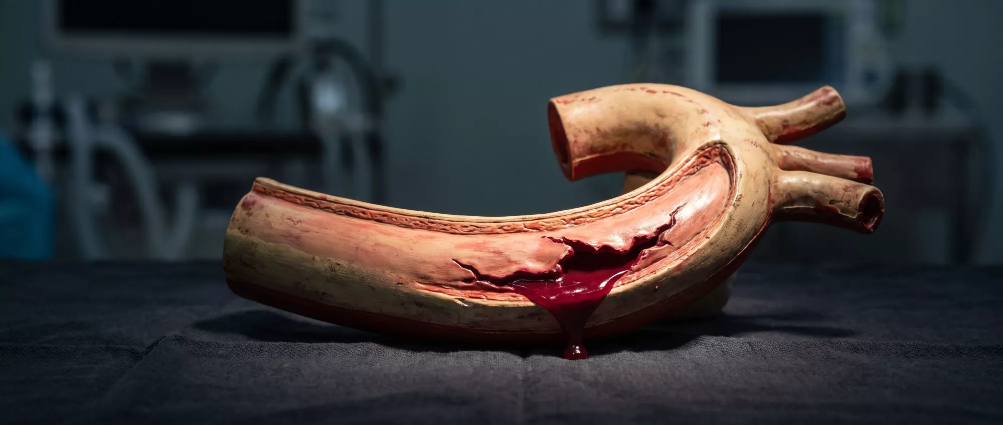
Aortic Rupture
Aortic rupture is a life-threatening cardiac emergency.
What is an aortic rupture?
An aortic rupture is a life-threatening condition that occurs when the wall of the aorta, the largest artery in the human body, tears or bursts completely. The aorta is responsible for carrying oxygen-rich blood directly from the heart to the rest of the body, meaning it handles high-pressure blood flow constantly. When this vital vessel ruptures, massive internal bleeding occurs, which can quickly become fatal if not treated immediately. For patients preparing for or recovering from cardiac surgery, understanding the severity of this condition helps clarify why vascular health is so closely monitored.
Unlike a simple tear in a minor blood vessel, a rupture of the aorta completely compromises the body's main blood supply highway. The rapid loss of blood means the heart and brain are starved of the oxygen they need to function. This is why immediate surgical intervention is always required. Understanding the mechanics of an aortic wall rupture can help patients recognize the importance of regular cardiac screenings and strict blood pressure management before any surgery takes place.
Patients often ask how this differs from an aneurysm. An aneurysm is a weakened, bulging area in the aortic wall, and it is usually a precursor to a rupture. Think of the aneurysm as a weak spot in a tire; the rupture is the actual blowout. Not all aneurysms rupture, but when they do, the situation immediately escalates to a critical cardiac emergency.
What causes an aortic rupture?
Understanding aortic rupture causes is essential for identifying individual risk levels. The most frequent cause is a severely weakened aortic wall, often due to an expanding aneurysm that eventually gives way under continuous blood pressure. Other common causes include aortic dissection, where the inner layer of the artery tears, forcing blood between the layers of the wall and causing it to split open. Chronic high blood pressure, genetic conditions affecting connective tissue like Marfan syndrome, and severe trauma can all trigger this catastrophic event.
Atherosclerosis and hypertension: Years of uncontrolled hypertension force the heart to pump harder, heavily stressing the aortic walls. Plaque buildup from atherosclerosis further degrades the tissue integrity, making the vessel stiff and brittle.
Genetic and inflammatory conditions: Connective tissue disorders directly compromise the structural strength of the blood vessels. Inflammatory diseases like giant cell arteritis can also inflame the blood vessels, creating dangerous weak points.
- Chronic high blood pressure weakening the arterial walls over many years.
- Inherited connective tissue disorders such as Marfan or Ehlers-Danlos syndrome.
- Traumatic injuries, often from severe falls or rapid-deceleration motor vehicle accidents.
- Advanced atherosclerosis causing plaque buildup and arterial stiffness.
What are common aortic rupture symptoms?
Recognizing aortic rupture symptoms can be the difference between life and death. The hallmark symptom is sudden, severe, and tearing pain in the chest, abdomen, or back. This pain is often described as ripping or shearing, and it typically radiates down the back or into the abdomen. Patients may also experience extreme shortness of breath, a sudden drop in blood pressure, dizziness, or a complete loss of consciousness as the body goes into shock from internal blood loss.
Thoracic presentation: If the rupture occurs in the chest cavity, patients typically feel intense chest pain radiating to the back. This is often accompanied by a noticeable difference in blood pressure readings between the right and left arms.
Abdominal presentation: When the rupture happens lower down in the abdominal aorta, the tearing pain is centered in the stomach or lower back. The abdomen may become rigid and tender to the touch, and the patient might feel a pulsating mass before collapse.
How do doctors diagnose an aortic rupture?
Diagnosing an aortic rupture requires rapid, highly specialized medical imaging because the patient is usually in critical condition upon arrival. Emergency room physicians act immediately, looking for signs of internal bleeding and cardiovascular instability. The diagnostic process for aortic rupture diagnosis prioritizes speed and accuracy to determine if emergency surgery is required to save the patient's life.
CT scans and ultrasounds: A computed tomography (CT) angiogram is the gold standard for quickly visualizing the aorta and identifying the exact location of the tear. In unstable patients, a bedside echocardiogram or abdominal ultrasound might be used immediately to assess fluid buildup around the heart or blood vessels.
Blood tests are often drawn simultaneously to check for rapid hemoglobin drops and assess organ function, though imaging remains the primary diagnostic tool. For patients interested in proactive monitoring, non-invasive cardiac imaging services are crucial for catching aneurysms long before a rupture can ever occur.
Why is aortic rupture a medical emergency?
Aortic rupture is inherently a medical emergency because the aorta is the main conduit for the body's blood supply. When it bursts, the massive internal bleeding prevents oxygen from reaching the brain, heart, and other vital organs. Without immediate surgical repair to stop the hemorrhage and restore vascular continuity, the body quickly enters hypovolemic shock. Death can occur within minutes to hours, making rapid transport to an experienced cardiac surgical center the highest priority.
Circulatory collapse: The rapid loss of blood volume causes blood pressure to plummet. The heart struggles to pump the remaining blood, leading to widespread tissue death.
Cardiac tamponade risk: If the rupture occurs near the heart, blood can leak into the pericardium, the sac surrounding the heart. This creates a life-threatening condition where the heart cannot expand properly to fill with blood.
What surgery treats an aortic rupture?
When a rupture occurs, aortic rupture surgery is the only viable treatment option. The specific surgical approach depends on the location and extent of the tear. In open surgery, the surgeon makes an incision to directly access the aorta, removes the damaged section, and replaces it with a durable synthetic tube called a graft. This highly complex procedure requires a skilled cardiac surgical team, a heart-lung bypass machine, and specialized perfusion techniques to maintain life while the aorta is repaired.
Endovascular stent grafting: For some lower-risk or descending aortic ruptures, surgeons might use a minimally invasive endovascular approach. They guide a stent graft up through a small incision in the groin and deploy it inside the damaged artery to seal the tear and reinforce the vessel wall.
In cases of ascending aortic rupture, the surgical team must act fast to open the chest, connect the patient to a heart-lung bypass machine, and replace the torn segment. The expertise of the surgical team in managing these specific, high-stakes cardiac emergencies is what gives patients a fighting chance at survival and long-term recovery.
What are the risk factors for aortic rupture?
Knowing the risk factors for aortic rupture helps patients and their families take proactive steps to protect their cardiovascular health. While some risks are unavoidable, many can be managed through lifestyle changes and consistent medical care. The most significant controllable risk factor is uncontrolled high blood pressure, which continuously stresses the aortic walls. Other factors include a family history of aneurysms, heavy tobacco use, and advancing age.
Smoking and lifestyle factors: Smoking damages blood vessels and accelerates the hardening of arteries. This drastically increases the likelihood of an aneurysm forming and eventually rupturing under pressure.
Age and family history: The risk of an aortic aneurysm increases significantly in men over the age of 65. Patients with a family history of aortic conditions are often advised to undergo regular screening ultrasounds.
How can aortic rupture be prevented?
Effective aortic rupture prevention relies heavily on early detection and strict management of cardiovascular risk factors. Because most aneurysms grow slowly without causing symptoms, regular screenings are critical for individuals with a family history of the condition or those diagnosed with genetic connective tissue disorders. Once an aneurysm is identified, a cardiac surgeon can monitor its size and recommend preemptive surgical repair before the vessel wall ever begins to tear.
Blood pressure management: Keeping blood pressure within a healthy range is the most effective way to reduce stress on the aorta. Patients are often prescribed antihypertensive medications to ensure the vessel walls are not overworked.
Cholesterol and lifestyle control: Managing cholesterol through diet, exercise, and medication prevents the plaque buildup that degrades the aorta. Patients must completely avoid smoking to reduce the rapid acceleration of vascular disease.
- Routine ultrasounds for high-risk patients to monitor aneurysm growth.
- Engaging in moderate, heart-healthy exercise without extreme heavy lifting.
- Adopting a low-sodium, heart-healthy diet to keep blood vessels flexible.
What is recovery like after aortic rupture surgery?
The aortic rupture recovery process is a long journey that requires deep dedication from the patient, their family, and the medical team. Immediately following surgery, the patient typically spends several days in the intensive care unit (ICU) under close monitoring. Here, medical staff ensure the heart is functioning well, blood pressure is strictly controlled, and there are no signs of internal bleeding or infection. Once stabilized, the patient moves to a standard hospital room before eventually going home.
Cardiac rehabilitation: After discharge, patients are strongly encouraged to enroll in a supervised cardiac rehabilitation program. This structured, gradual approach to physical activity helps safely rebuild cardiovascular strength under professional supervision.
Lifelong medical management: Recovery does not end when the surgical incision heals. Patients will require lifelong monitoring through regular imaging to ensure the graft remains intact and that no new weak points develop elsewhere in the aorta. Strict adherence to blood pressure medications is absolutely essential to protect the repaired vessel.
Understanding aortic rupture survival rate
When families research the aortic rupture survival rate, they are often met with sobering realities. Because the condition is so immediately life-threatening, survival depends entirely on how quickly the patient reaches a hospital with a specialized cardiac surgical team. If the rupture occurs outside a medical setting, the chances of survival drop precipitously. However, if the patient reaches the operating room in time, skilled surgeons can successfully repair the damage and give the patient a chance at a full life.
The role of immediate response: The first few hours are the most critical. The body cannot sustain massive internal bleeding for long, making rapid emergency medical transport the single most important factor in patient survival.
For a frightened patient or family, understanding these realities is not about losing hope, but rather highlighting the critical importance of preparation, prevention, and seeking expert care. Recognizing the symptoms early, managing risk factors proactively, and knowing where to find a specialized cardiac surgeon are the best defenses against the devastating impact of an aortic rupture.






