

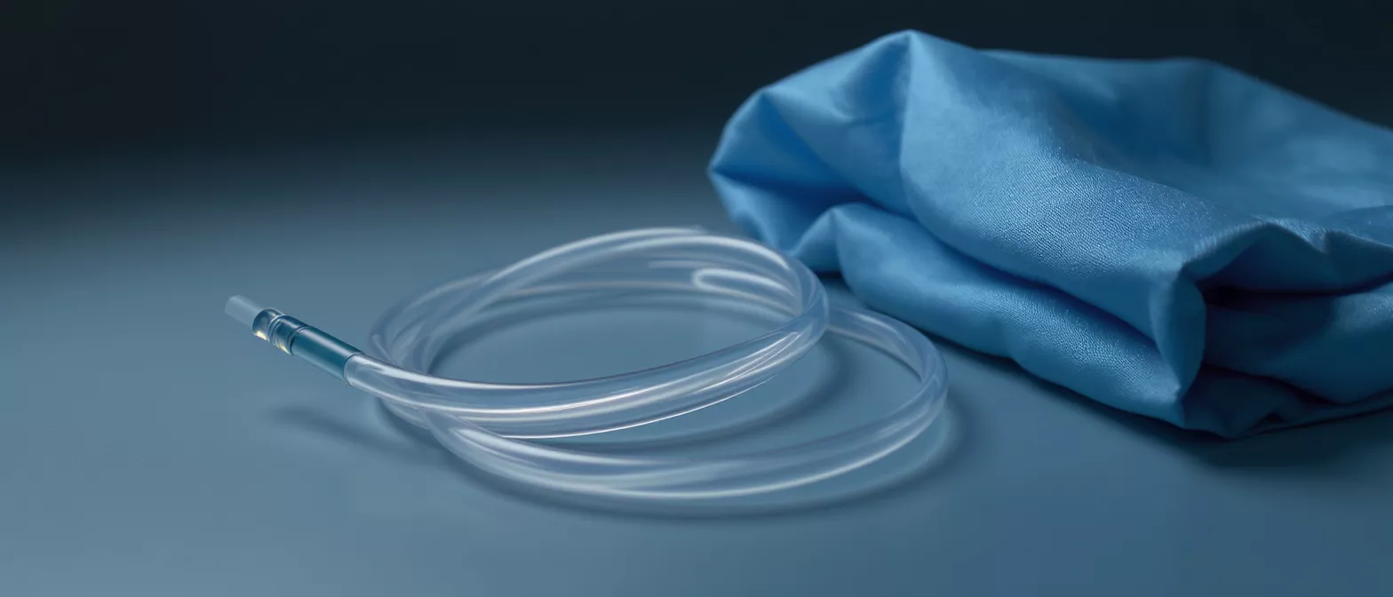
Endotracheal Tube
Learn how an endotracheal tube keeps patients breathing during heart surgery.
What Is an Endotracheal Tube?
An endotracheal tube is a flexible plastic medical device placed into the trachea, or windpipe, to help a patient breathe during and after surgery. For families awaiting a loved one’s cardiac procedure, seeing this tube for the first time can be unsettling, but it is one of the most important tools used in the operating room. It connects the patient to a mechanical ventilator, ensuring oxygen is delivered directly to the lungs while anesthesia keeps the body still.
When your loved one is under general anesthesia for heart surgery, the muscles that control breathing temporarily stop working. The endotracheal tube takes over the work of breathing, allowing the surgical team to focus entirely on repairing the heart without worrying about oxygen levels dropping.
It is helpful to think of the tube as a temporary bridge — one that carries air safely into the lungs when the body cannot do so on its own. Once the heart is repaired and anesthesia wears off, the tube is gently removed, and natural breathing returns.
Endotracheal Tube Purpose in Cardiac Surgery
Why is intubation necessary for heart surgery? Cardiac operations require the chest to be opened and the heart to be stopped or supported by a heart-lung machine. During this time, the patient cannot breathe independently. The endotracheal tube provides a secure, protected airway so the ventilator can control every breath, delivering precise amounts of oxygen and anesthetic gas.
The tube also protects the lungs from fluids. Because the patient is unconscious and lying flat, saliva or stomach contents could otherwise enter the airway. The tube’s inflatable cuff seals the trachea, keeping the lungs clean and reducing the risk of infection.
Without this secure airway, the complex work of coronary bypass or valve repair would not be possible. The breathing tube is not a sign of crisis — it is a planned and essential part of every cardiac surgery.
How Endotracheal Intubation Works
The process of placing the tube is called intubation. After the patient is given medication to fall asleep and relax, the anesthesiologist uses a lighted instrument called a laryngoscope to see the vocal cords. The tube is then guided gently between the cords and into the trachea.
Once in position, a small balloon at the tip of the tube is inflated. This balloon, called a cuff, creates a gentle seal against the walls of the windpipe. The seal ensures that the air pushed in by the ventilator goes fully into the lungs rather than leaking out through the mouth.
Throughout the surgery, the anesthesiologist listens to the lungs and watches monitors to confirm the tube remains perfectly positioned. This constant vigilance ensures that every breath the machine delivers reaches both lungs equally.
Types of Endotracheal Tubes Used in Heart Surgery
Are there different tubes for different patients? Yes. Tubes come in various sizes and materials to fit each patient safely. Adults, children, and infants each require different diameters, and the anesthesiologist selects the correct size based on the patient’s body and medical needs.
Most tubes used in cardiac surgery are made of soft, medical-grade plastic that flexes gently without harming the airway. Some have a spiral wire built into the wall to prevent kinking during long operations, which is especially useful when the patient’s head is repositioned.
Choosing the right tube is a careful decision made before the operation begins. The goal is always to provide the safest, most comfortable fit for the individual patient’s anatomy.
Endotracheal Tube Placement and Insertion Process
Placement happens immediately after anesthesia is induced, before the surgical incision is made. The patient is already asleep and unaware, so there is no discomfort or memory of the procedure. The anesthesiologist works quickly but carefully to secure the airway within seconds.
After insertion, the tube is taped or tied securely to prevent movement. The team confirms placement by listening to the chest with a stethoscope and watching carbon dioxide levels on the monitor — a reliable sign that the tube is in the windpipe and not the esophagus.
Only after the airway is secured and confirmed does the surgeon proceed. This step-by-step verification is a standard safety measure in every operating room.
Airway Management in Cardiac Surgery Explained
How does the breathing tube connect to the ventilator? The endotracheal tube extends from the patient’s mouth to a breathing circuit, which attaches to the ventilator. The machine pushes a measured mixture of oxygen and air into the lungs, then allows the lungs to empty naturally before delivering the next breath.
During heart surgery, the ventilator settings are adjusted continuously. The anesthesiologist changes the breath rate, oxygen concentration, and pressure based on what the surgeon is doing and how the patient’s body is responding. This precise control protects the heart and other organs.
- Secure airway: protects the lungs from fluid and ensures oxygen delivery.
- Ventilator control: delivers precise breaths tailored to the surgical stage.
- Continuous monitoring: confirms tube position and lung function throughout.
Endotracheal Tube Complications and Risks
While intubation is routine and generally very safe, the mouth and throat are sensitive areas. After the tube is removed, patients may experience a mild sore throat, a hoarse voice, or a dry cough. These symptoms usually clear within a day or two without treatment.
Rarely, the vocal cords can become irritated, or a tooth could be chipped during insertion. The anesthesia team takes great care to avoid these issues, using soft equipment and checking dental health beforehand. Serious complications are uncommon but monitored closely.
For cardiac patients, the benefits of a secure airway far outweigh the small risks. The breathing tube is what makes safe, still, and painless heart surgery possible.
Endotracheal Tube Care and Ventilator Support
What happens while the tube is in place after surgery? Some patients remain on the ventilator for a short time in the intensive care unit while their heart recovers. During this period, nurses and respiratory therapists check the tube, adjust ventilator settings, and suction secretions from the airway to keep it clear.
The patient cannot speak while the tube is in place because it passes through the vocal cords. However, they are often sedated or resting comfortably, and care teams use gestures, writing, or communication boards to interact once sedation is reduced.
This period of ventilator support allows the heart to rest and regain strength before it takes over full circulation again. It is a protective step, not a sign of setbacks.
Breathing Tube Removal After Surgery
When the heart is stable and the anesthesia has worn off, the team prepares to remove the tube. This process is called extubation. The patient is briefly sedated or fully awake, the cuff is deflated, and the tube is gently slid out.
Most patients do not remember the removal. They may cough briefly as the tube comes out, but this is a normal reflex. Within minutes, they begin breathing on their own, and oxygen is delivered through a mask or nasal prongs to ease the transition.
- Stable heart function: the primary condition for extubation.
- Reducing sedation: allows the patient to wake and breathe naturally.
- Supplemental oxygen: provided after removal to support recovery.
Endotracheal Tube Recovery and What to Expect
After the breathing tube is removed, the recovery phase begins. A mild sore throat or raspy voice is common for a day or two. Drinking warm liquids and resting the voice helps the throat heal quickly. If discomfort lasts longer, the medical team can recommend simple soothing remedies.
For families, it is reassuring to know the tube is a temporary support. It is placed with care, managed with constant attention, and removed as soon as the heart is ready. The endotracheal tube plays a vital but short-lived role in the larger journey of cardiac recovery, helping patients reach the moment they can breathe freely on their own once more.






