

Haller Index
Learn how the Haller Index measures pectus excavatum severity, its normal range, and surgery criteria.
What is the Haller Index?
A standard chest wall measurement: The Haller index is a numerical value doctors use to evaluate the shape of the chest wall and diagnose conditions like pectus excavatum. It gives surgeons a precise, objective metric to understand how deeply the sternum is sunken inward. By using this measurement, the medical team can move beyond a simple visual exam and accurately gauge the degree of chest deformity.
Why heart patients need it: For many patients with a sunken chest, the physical appearance is the most obvious concern, but the real clinical issue is what happens inside. A significantly depressed sternum can reduce the space available for the heart and lungs. The index helps a cardiac surgery team see whether the structural abnormality is compressing the heart and potentially interfering with cardiovascular function.
Understanding this measurement is often the first step in creating a clear, actionable treatment plan. It transforms a physical observation into a specific data point. From there, the surgical team can compare the patient's score against established diagnostic criteria to determine the best path forward.
How does the Haller Index calculation work?
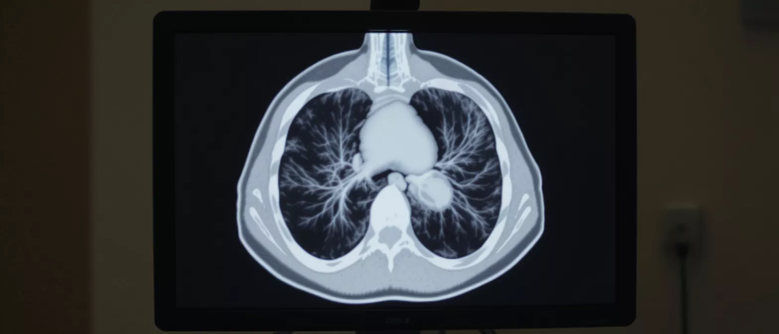
The basic formula: The Haller index formula is surprisingly straightforward. It divides the transverse diameter of the chest (the side-to-side width) by the anteroposterior diameter (the front-to-back depth). Both measurements are taken from the same axial slice of a CT scan, typically at the deepest point of the chest depression. The resulting ratio reveals the overall shape of the rib cage.
Reading the ratio: In a typical chest, the side-to-side width is larger than the front-to-back depth, meaning the resulting number is naturally greater than one. However, when the front of the chest is sunken, the front-to-back distance shrinks dramatically while the side-to-side distance often appears wider. This dynamic causes the overall ratio to climb higher, reflecting the severity of the inward depression.
- Transverse diameter: The horizontal width of the rib cage, measured from the inside of the ribs on one side to the inside of the ribs on the other.
- Anteroposterior diameter: The vertical depth of the chest, measured from the back of the vertebrae to the deepest point of the sternum.
- Final ratio: The width divided by the depth, producing a single number that grades the structural deformity.
Because the calculation relies entirely on precise imaging, a standard CT scan is essential for an accurate reading. A advanced chest CT imaging system provides the clear cross-sectional slices needed to measure these diameters perfectly. Without exact measurements, the final calculation could be skewed, leading to an inaccurate assessment of the patient's condition.
What is the Haller Index normal range?
Defining a normal chest shape: For individuals without a chest wall deformity, the Haller index normal range typically falls between 2.0 and 2.5. This means the width of the chest is roughly two to two and a half times greater than its depth. A score within this range indicates that the sternum sits at a typical distance from the spine and is not causing any structural crowding of the internal organs.
When the number climbs: Once the score exceeds 2.5, it begins to suggest a structural abnormality. A reading between 2.5 and 3.2 generally indicates a mild presentation of pectus excavatum. While these patients might notice a dip in their chest, the structural change is often not severe enough to cause significant compression of the heart or lungs, though monitoring is still recommended.
Cardiologists and surgeons pay close attention to this threshold because it helps differentiate between a purely cosmetic difference and a medically significant deformity. Understanding where a patient falls on this numerical scale provides immense peace of mind. It clearly outlines whether the heart has adequate room to function or if intervention is necessary.
Haller Index and pectus excavatum severity
Grading the depression: Pectus excavatum severity varies wildly from person to person. The Haller index pectus excavatum measurement serves as a universal grading tool, categorizing the condition into mild, moderate, and severe tiers. This stratification allows Dr Alejandro Molina and the surgical team to standardize their approach and ensure consistent, evidence-based decisions.
Connecting shape to physiology: As the severity increases, the likelihood of physiological symptoms rises as well. Patients with a moderate to severe score often experience shortness of breath, chest pain, or heart palpitations during exercise. The sunken sternum physically pushes the heart over to the left side of the chest and can compress the right ventricle, limiting the heart's ability to pump efficiently.
By measuring the severity of the chest deformity assessment accurately, doctors can correlate the physical structure with the patient's reported symptoms. This helps patients understand that their breathing and chest discomfort are real, physical consequences of the structural anomaly, not generalized anxiety or poor physical fitness.
What is a severe Haller Index score?
The critical threshold: A score of 3.25 or higher is widely recognized as the Haller index severe pectus threshold. At this level, the front-to-back depth of the chest has narrowed significantly, indicating a deep depression of the sternum. Reaching this score usually confirms that the structural deformity is substantial enough to warrant serious medical consideration.
Why this number matters for the heart: When a patient presents with a score this high, the risk of cardiac compression becomes very real. The right side of the heart, which pumps blood to the lungs, can be squeezed against the spine. This mechanical restriction can lead to decreased exercise tolerance, mitral valve prolapse, and a noticeable drop in cardiac output during physical exertion.
Hitting this severe threshold is often the turning point in a patient's medical journey. It transitions the conversation from simple observation to active treatment planning. The data provides the definitive proof required to justify surgical correction to insurance providers and medical review boards.
Haller Index, cardiac compression, and symptoms
Understanding the mechanical squeeze: The Haller index cardiac compression relationship is all about physical space. When the sternum pushes inward, it steals room from the chest cavity. The heart is a muscular pump that expands and fills with blood; if the chest cannot accommodate the heart's normal size and motion, the heart's pumping efficiency drops, leading to a cascade of cardiovascular symptoms.
What the patient experiences: Patients with a high index often complain of extreme fatigue, inability to keep up with peers in sports, and a racing heartbeat even with mild exertion. Some experience sharp chest pains or dizzy spells. Because the heart is displaced from its normal anatomical position, murmurs and irregular heartbeats are also commonly detected during a routine physical exam.
Recognizing these symptoms as structural rather than purely cardiac disease is vital. It is why a cardiac surgeon must evaluate both the heart's function and the chest wall's structure together. Addressing the root cause—the sunken chest—often resolves or significantly improves the cardiovascular symptoms.
When is the Haller Index a surgery indication?
The surgical benchmark: The Haller index surgery indication is generally triggered when the score reaches or exceeds 3.25, especially when accompanied by clinical symptoms. A high score alone does not mandate surgery, but it serves as a strong diagnostic criteria. The decision is a combined evaluation of the imaging data, the patient's physical limitations, and their overall quality of life.
Preparing for the next step: If surgery is indicated, the goal is to elevate the sternum, restore the normal anatomical volume of the chest, and relieve the pressure on the heart and lungs. The surgical team will use the index score to plan the extent of the correction needed. Post-surgery, the score is often dramatically improved, reflecting a normalized chest cavity.
Patients preparing for surgical correction often research ways to support their respiratory and musculoskeletal health before and after the procedure. Incorporating posture correction and breathing support brace into pre-habilitation can help optimize chest wall mechanics prior to the operation. This preparatory step ensures the ribs and muscles are best positioned for a smooth recovery.
How to prepare for a Haller Index CT scan
What to expect during imaging: The Haller index measurement relies on a specialized computed tomography scan. The test is painless and non-invasive, taking only a few minutes inside a large, doughnut-shaped machine. Patients must lie perfectly flat on their back and hold their breath for short intervals to ensure the cross-sectional images are crystal clear and free of motion blur.
Ensuring diagnostic accuracy: For the most accurate results, patients should wear loose, comfortable clothing without any metal buttons or zippers near the chest. It is vital to remain completely still during the scan so the radiologist can capture the exact anteroposterior and transverse diameters needed for the calculation. The imaging technician will guide the patient through every step to minimize anxiety.
Because the CT scan uses a very low dose of radiation to map the chest wall, the benefits of obtaining a precise diagnosis far outweigh the minimal risks. The resulting images give Dr Molina the exact three-dimensional roadmap needed to understand the patient's anatomy. This clarity is what makes safe, effective surgical planning possible.
Pectus excavatum treatment options and outcomes
Surgical correction methods: Once a high score confirms the need for intervention, there are established pectus excavatum treatment options available. The most common approach is the minimally invasive Nuss procedure, where a curved metal bar is inserted under the sternum to pop the chest out into a normal position. The bar remains in place for a few years while the chest wall remodels and solidifies its new shape.
Postoperative recovery and quality of life: Recovery from chest wall surgery requires time, rest, and careful pain management, but the long-term outcomes are generally excellent. As the sternum lifts, the compression on the heart is immediately relieved. Patients frequently report a massive increase in exercise tolerance, the disappearance of chest pains, and a profound improvement in their self-confidence.
Following successful surgical correction, maintaining core strength and proper posture is essential for long-term stability. Many patients utilize recovery rib belt for chest wall support during the initial healing phases to ensure proper alignment. With the structural defect corrected, patients can return to their normal activities with a fully functional, healthy heart and a stable chest wall.
Key takeaways on the Haller Index
A vital diagnostic metric: The Haller index remains the gold standard for quantifying the severity of a sunken chest. It translates a physical structural difference into a clear, objective number that surgical teams can rely on. Understanding this metric helps patients see the direct link between their chest shape and their cardiovascular health.
Empowering patient decisions: Knowing the exact score empowers patients and their families to make informed decisions about their health care. It removes the guesswork from the diagnostic process, providing a solid foundation for discussing surgical indications and treatment pathways with a qualified cardiac specialist. The measurement ensures that treatment is driven by precise data.
Ultimately, evaluating this index is about protecting the heart and improving the patient's quality of life. By accurately measuring the chest cavity, the surgical team at Cirugía de Corazón can determine if the heart has the room it needs to beat freely. If you or a loved one is experiencing symptoms of a sunken chest, seeking a professional evaluation is the safest next step.






