

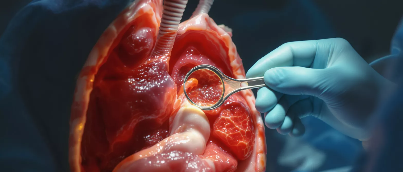
Innominate Vein
Learn about the innominate vein’s anatomy, function, role in cardiac surgery, and clinical significance, including thrombosis and stenosis.
The innominate vein, also known as the brachiocephalic vein, plays a vital role in your body's circulatory system—especially if you are facing heart surgery. Understanding this vessel helps patients and families feel more prepared and less anxious about procedures involving the chest and upper body. This glossary entry explains what the innominate vein is, where it sits in your anatomy, how it functions, and why cardiac surgeons like Dr Alejandro Molina pay close attention to it during operations.
What Is the Innominate Vein
The innominate vein is one of the major veins responsible for returning deoxygenated blood from your upper body back toward your heart. It collects blood from your head, neck, arms, and upper chest, channeling it into the superior vena cava—the large vein that empties directly into the right atrium of your heart. Without this critical pathway, blood from your brain and upper limbs would have no direct route to complete its journey through the circulatory system.
"Why is it called 'innominate'?" The term comes from Latin, meaning "nameless" or "without a name." Early anatomists found this vessel difficult to categorize simply, so the descriptive label stuck. Today, medical professionals often use both "innominate vein" and "brachiocephalic vein" interchangeably, though you will hear the former more frequently in cardiac surgical settings.
For patients preparing for heart surgery, knowing that your surgical team maps these vessels carefully before any procedure can bring reassurance. The innominate vein sits in a crowded space near the sternum, surrounded by the thymus gland, the aortic arch, and numerous nerves—making precision essential.
Innominate Vein Anatomy & Location
The innominate vein anatomy reveals two distinct vessels: one on the left side and one on the right. Both begin deep within the root of your neck, formed by the union of two smaller veins—the internal jugular vein (draining the brain and face) and the subclavian vein (draining the arm and shoulder). From this junction, each innominate vein travels downward and inward toward the center of your chest.
"Where exactly does the innominate vein sit in relation to my sternum?" The right innominate vein descends almost vertically behind the right edge of the sternum, measuring only about two tothree centimeters before joining its partner. The left innominate vein takes a longer, more oblique path—crossing from left to right behind the manubrium (the upper part of your breastbone), passing in front of the aortic arch and its three major branches. This longer course makes the left side more vulnerable during certain surgical approaches.
Both veins lie within the mediastinum, the central compartment of your thoracic cavity. Their proximity to the trachea, esophagus, and major arteries means that any swelling, clot, or external compression in this region can affect blood flow significantly. Surgeons must navigate this delicate terrain with extreme care during median sternotomy and other chest-opening procedures.
How the Innominate Vein Works: Function
The primary innominate vein function is to serve as a collecting chamber and transport highway for venous blood from the upper half of your body. Unlike arteries, which carry blood away from the heart under high pressure, veins operate under low pressure and rely on surrounding muscle contractions, one-way valves, and respiratory movements to keep blood moving forward.
"What happens if innominate vein function becomes compromised?" When this vein narrows, clots, or faces external compression, blood backs up into the territories it normally drains. Patients may experience facial swelling, arm heaviness, distended neck veins, or headaches—symptoms that often worsen when lying flat or raising the affected arm. These signs alert clinicians that venous outflow obstruction may be present and warrants immediate evaluation.
The innominate vein also serves as an important access point during cardiac surgery. Surgeons may cannulate it directly or use nearby tributaries to establish cardiopulmonary bypass, the temporary system that oxygenates and circulates your blood while your heart is stopped for repair. The vein's generous diameter and relatively thin wall make it suitable for this purpose, though its fragility demands gentle handling.
Left Innominate Vein vs Right Innominate Vein
Understanding the differences between the left and right innominate vein helps explain why certain complications arise more frequently on one side. The right innominate vein is shorter, straighter, and somewhat protected as it descends behind the right sternoclavicular joint. The left innominate vein, by contrast, arches across the midline in a longer traverse, making it more exposed to pressure from adjacent structures.
Several clinical scenarios highlight this asymmetry:
- Left-sided compression: The left innominate vein passes between the sternum anteriorly and the aortic arch posteriorly—an anatomical pinch point where swelling, aneurysm, or mass effect can narrow the channel
- Right-sided cannulation preference: Due to its straighter course, the right innominate vein and its tributaries are often preferred for central venous catheter placement and certain bypass configurations
- Variations in surgical exposure: During redo cardiac surgery or operations requiring deep sternal retraction, the left innominate vein requires particular attention to avoid avulsion or inadvertent injury
Despite these differences, both vessels perform identical physiological roles. Your surgical team will account for side-specific anatomy in preoperative planning, often reviewing CT or MRI imaging to map the exact relationship between your innominate veins and neighboring structures.
Innominate Vein in Cardiac Surgery
The innominate vein occupies center stage in many cardiac and thoracic operations. During coronary artery bypass grafting, valve replacement, or aortic surgery, surgeons must preserve venous integrity while gaining adequate exposure of the heart. The left innominate vein especially can obstruct visualization of the aortic arch and its branches, sometimes requiring temporary mobilization or gentle retraction.
"How do surgeons protect the innominate vein during complex operations?" Meticulous dissection and constant awareness of depth constitute the foundation of safe practice. Surgeons work within millimeters of this vessel when dividing the thymus, opening the pericardium, or placing cannulae for cardiopulmonary bypass. A small tear in the innominate vein can bleed profusely due to its thin wall and the negative intrathoracic pressure that draws air into venous injuries—creating a serious but manageable situation for experienced teams.
In some congenital heart operations and reoperative procedures, the innominate vein may be deliberately divided and later reconstructed, or bypassed using a graft. These advanced techniques demonstrate the vein's importance: surgeons go to considerable lengths to preserve or restore its patency rather than sacrifice it permanently.
Innominate Vein Compression Syndrome
Innominate vein compression syndrome occurs when external structures squeeze the vein and impede blood flow. The left innominate vein is far more commonly affected due to its longer transverse course and passage through the narrow space between sternum and aorta. Patients with this condition may develop progressive facial and arm swelling, visible collateral veins across the chest wall, and symptoms worsened by certain positions or arm elevation.
"What causes innominate vein compression besides anatomical factors?" Several conditions can narrow this space: substernal thyroid goiter, mediastinal tumors, enlarging aortic aneurysm, lymphadenopathy, or fibrosis from prior surgery or radiation. Even severe pectus excavatum (a sunken breastbone) has been reported to compress the vein in susceptible individuals. Diagnosis typically requires cross-sectional imaging—CT venography or MR venography—to visualize both the narrowing and any collateral pathways your body has developed to bypass the obstruction.
Treatment depends on severity. Mild cases may be observed with positional modifications. Significant compression often responds to stent placement by interventional radiologists or surgical decompression by removing the offending mass or repositioning structures. Your cardiac surgeon collaborates closely with these specialists when innominate vein compression complicates heart conditions.
Innominate Vein Thrombosis: Causes & Risks
Innominate vein thrombosis refers to clot formation within this major vessel, a serious condition that can propagate to the superior vena cava or embolize to the lungs. Unlike deep vein thrombosis in the legs, upper extremity and central thoracic vein clots often relate to specific triggers rather than spontaneous clotting tendencies.
Common risk factors include:
- Central venous catheters: Dialysis lines, chemotherapy ports, and pacemaker leads dwelling in or near the innominate vein can irritate the lining and promote clot formation
- Thoracic outlet obstruction: Compression between the first rib and clavavian structures slows flow and creates stasis, a key element of Virchow's triad for thrombosis
- Hypercoagulable states: Inherited or acquired tendencies toward clotting affect all veins, including the innominate, particularly when other risk factors coexist
Symptoms of innominate vein thrombosis overlap with compression syndrome but often include more acute pain, warmth, and pronounced discoloration of the affected arm. Anticoagulation remains the cornerstone of treatment, with catheter removal when feasible and endovascular or surgical intervention reserved for severe cases threatening limb viability or causing significant symptoms.
Innominate Vein Stenosis: Symptoms & Detection
Innominate vein stenosis describes abnormal narrowing of the vessel, distinct from external compression in that the vessel wall itself has become restrictive. This may follow prior surgery, radiation therapy, chronic inflammation, or successful thrombosis treatment where organization and scarring narrow the lumen. Some patients develop stenosis after prolonged central venous access, particularly in the oncology or dialysis populations.
"What symptoms suggest innominate vein stenosis rather than simple compression?" Stenosis typically produces gradual, progressive symptoms without the positional variation seen in pure compression. Patients report persistent arm fatigue, swelling that does not resolve overnight, and increasingly prominent superficial veins across the shoulder and anterior chest as collateral circulation develops. Unlike arterial disease, venous stenosis rarely causes the resting pain or tissue loss that signals critical limb ischemia—instead, the burden is one of chronic congestion and functional limitation.
Detection relies on careful physical examination and confirmatory imaging. Duplex ultrasound can assess flow patterns, though bony structures limit direct visualization. CT and MR venography provide detailed anatomic mapping, while conventional venography with pressure gradients across narrow segments helps quantify functional significance. For symptomatic patients, balloon venoplasty with or without stenting offers durable relief in experienced centers.
Innominate Vein Clinical Significance for Patients
The innominate vein clinical significance extends beyond its role as a passive conduit. For cardiac surgery patients, this vessel represents both opportunity and vulnerability—a structure that enables critical access yet demands respect for its fragility. Preoperative identification of variants, such as a particularly tortuous left innominate vein or anomalous tributary pattern, allows surgical teams to modify their approach and avoid surprises.
"Why does innominate vein health matter for long-term cardiovascular wellness?" Although not itself a site of atherosclerotic disease like coronary arteries, the innominate vein influences overall venous return and cardiac preload. Chronic obstruction forces the right side of your heart to work against elevated upstream pressures, potentially contributing to right heart strain over time. Moreover, thrombotic complications in central veins carry substantial morbidity and can complicate future cardiac interventions by eliminating preferred vascular access routes.
Patients with prior innominate vein interventions require coordinated follow-up. Stents need surveillance for restenosis or fracture; surgical reconstructions demand periodic assessment of graft patency. Your cardiac care team will integrate this surveillance into broader heart health monitoring, ensuring that venous and arterial concerns are addressed together.
Recovering After Innominate Vein Procedures
Recovery following surgery or intervention involving the innominate vein follows general cardiac surgical principles with a few specific considerations. Patients who have undergone stenting may receive dual antiplatelet therapy alongside any other blood thinners, requiring careful coordination with heart medications. Those with surgical reconstruction need monitoring for graft function and watchfulness for recurrent compression.
"What should patients watch for during recovery?" New or worsening facial swelling, arm discoloration, or unexplained shortness of breath warrant prompt communication with your care team. These symptoms could indicate early stent thrombosis, progressive stenosis, or a new compression issue. Most patients, however, recover uneventfully and find that previously troubling symptoms resolve substantially once venous outflow is restored.
Physical rehabilitation proceeds gradually, with attention to avoiding positions that stress fresh repairs or recently placed stents. Your surgical team will provide individualized guidance on lifting restrictions, recommended sleeping positions, and timelines for return to normal activities. The goal is complete restoration of venous function while protecting the investment made in your cardiovascular health.
The innominate vein, though "nameless" in origin, deserves full recognition for its essential contribution to circulatory physiology and cardiac surgical practice. Understanding this vessel empowers patients to participate meaningfully in their care and to recognize when symptoms suggest venous outflow problems. Under the guidance of experienced cardiac surgical teams, conditions affecting the innominate vein are eminently treatable, allowing patients to return to full, active lives.






