

J-incision
Learn how the j-incision technique enables minimally invasive heart valve surgery with faster recovery and less trauma.
What is a j-incision in cardiac surgery?
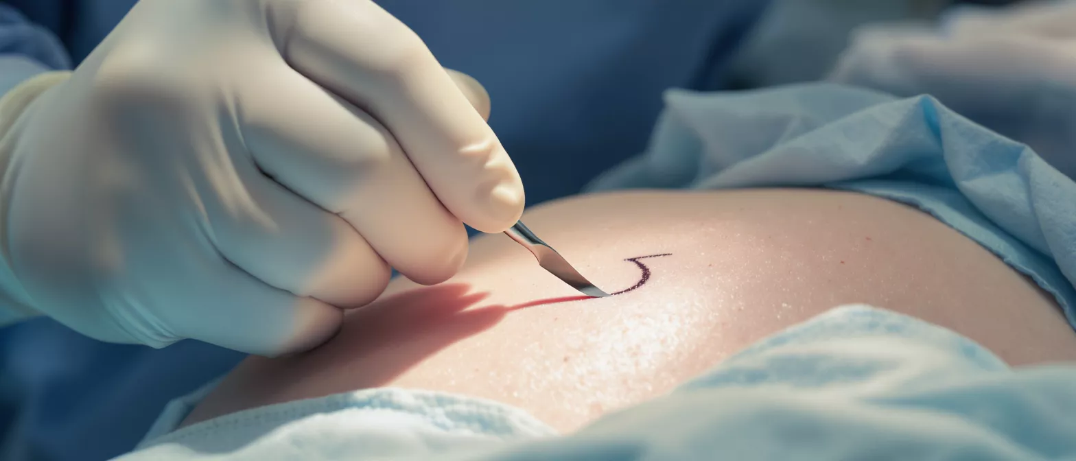
A j-incision is a specialized surgical technique used in minimally invasive cardiac surgery to access the heart. Instead of cutting directly down the entire length of the sternum, the surgeon makes a curving cut on the upper part of the breastbone, closely resembling the shape of the letter J. This approach allows the surgical team to reach vital structures while preserving the structural integrity of the lower chest cavity.
Lower surgical impact: By avoiding a full break of the breastbone, patients experience significantly less trauma to the chest wall. This smaller, targeted entry point forms the foundation of modern minimally invasive cardiac procedures, reducing both blood loss and the need for extensive postoperative pain management.
This method is most commonly employed when surgeons need to operate on the aortic or mitral valves. By lifting only a specific portion of the sternum, the surgeon can safely navigate specialized instruments to repair or replace faulty valves while maintaining a highly controlled and stable surgical environment.
How does the j-incision technique work?
During a j-incision procedure, the surgeon begins by making a precise skin incision over the upper portion of the sternum. Using specialized surgical saws, the bone is then carefully divided in a J-shape, extending from a specific point near the top and curving outward to the side. This technique creates a small, hinged bone flap that can be gently retracted rather than completely separated.
Retraction and access: Once the bone flap is lifted, the surgeon gains clear visibility of the pericardium, the protective sac surrounding the heart. The surgical team can then open the pericardium and introduce the necessary tools to perform complex valve repairs or replacements without needing to manipulate the entire chest cavity.
Throughout the operation, the patient is connected to a heart-lung bypass machine. This machine temporarily takes over the job of pumping blood and oxygenating it, allowing the heart to be safely stopped and operated on. The controlled window provided by the incision ensures the surgeon has enough room to execute these delicate maneuvers successfully.
J-incision vs traditional full sternotomy
A traditional full sternotomy involves making a straight cut down the entire length of the breastbone to fully expose the heart. While this approach provides excellent visibility and is the standard approach for major coronary bypass operations, it is highly invasive and requires a prolonged healing period. The j-incision was developed as an alternative for patients who require specific valve interventions but do not need total chest exposure.
Understanding the differences between these two approaches can help alleviate anxiety before surgery. Consider these structural and practical differences:
- Incision size: A full sternotomy requires a long central chest cut, whereas the j-incision is noticeably smaller and confined to the upper chest.
- Bone division: The traditional method splits the entire sternum, while the minimally invasive approach leaves the lower half of the breastbone fully intact.
- Initial healing: Because the lower bone remains unbroken, the structural stability of the chest is preserved, leading to a faster initial recovery phase.
Choosing between a full sternotomy and a partial incision depends entirely on the specific cardiac condition being treated. Complex coronary bypasses or major aortic aneurysms often require the full traditional approach, whereas isolated valve repairs are frequently excellent candidates for the targeted j-incision method.
Which heart valves can be repaired with a j-incision?
The j-incision provides excellent access to the aortic valve, which regulates blood flow from the heart to the rest of the body. Surgeons frequently use this approach to perform aortic valve replacements or repairs, treating conditions like aortic stenosis or regurgitation. The angle of the bone opening aligns perfectly with the natural anatomical position of this vital structure.
Accessing the mitral valve: The mitral valve, located between the left atrium and left ventricle, can also be effectively reached using this technique. Surgeons may opt for a right-sided version of the incision or use specialized instruments to navigate through the upper chest to repair mitral valve prolapse or stenosis.
While the aortic and mitral valves are the primary targets for this approach, the specific surgical route is always determined by the patient's unique anatomy. During preoperative imaging, the surgeon will map out the exact trajectory needed to safely access the malfunctioning valve while avoiding surrounding critical tissues.
J-incision recovery and healing timeline
Recovery from j-incision heart surgery generally begins in the intensive care unit, where patients are closely monitored for the first twenty-four hours. Because the chest wall retains much of its structural integrity, patients often experience less pain and are able to begin breathing without mechanical assistance much sooner than those who undergo a full sternotomy.
Returning to daily activities: Most patients are able to leave the hospital within four to six days following the procedure. While heavy lifting and strenuous activities are restricted for several weeks to allow the bone to knit back together, patients can typically resume gentle walking and basic self-care shortly after returning home.
Complete bone healing takes approximately six to eight weeks, similar to any broken bone. Patients will have follow-up appointments with their cardiologist to monitor heart function and ensure the surgical site is healing cleanly, gradually easing them back into their normal routines with confidence.
Benefits of a minimally invasive j-incision
The primary advantage of the minimally invasive j-incision is the significant reduction in surgical trauma. By leaving the lower portion of the sternum intact, the chest remains structurally rigid, which helps patients breathe more comfortably immediately after surgery and reduces the risk of deep sternal wound infections.
Patients often report a noticeable decrease in postoperative pain, which translates to a reduced need for strong pain medications. This allows individuals to participate more actively in their physical therapy and early mobilization, two critical components of a successful cardiac recovery.
Furthermore, the smaller incision results in a much more cosmetically favorable scar. While aesthetics are secondary to clinical outcomes, the psychological benefit of a smaller, easily hidden chest scar should not be underestimated for patients navigating the emotional journey of heart surgery.
Are j-incision procedures safe?
Extensive clinical experience has shown that the j-incision technique is a highly safe and effective approach for appropriately selected patients. The technique has been refined over many years by specialized cardiac surgeons, and the outcomes for valve repairs using this method are entirely comparable to traditional full sternotomy procedures.
Surgical monitoring: Throughout the operation, advanced transesophageal echocardiography is used to continuously monitor heart function and ensure the surgical repair is successful before the patient leaves the operating room. This high level of intraoperative oversight ensures that the limited visibility of a smaller incision is completely compensated for by specialized imaging technology.
As with any cardiac surgery involving general anesthesia, risks still exist, including bleeding, infection, or adverse reactions to medications. However, the preservation of chest stability and the minimization of tissue damage inherently reduce the likelihood of many common postoperative complications.
Potential complications of j-incision heart surgery
While generally safe, it is important for patients to understand the potential complications associated with any heart procedure. Bleeding during or after surgery is a risk, though the minimized tissue disruption of the j-incision often results in lower overall blood loss compared to larger incisions. In rare cases, additional transfusions may be required.
Infections at the incision site are another possibility, but because the bone is not completely split, the risk of deep sternal wound instability is significantly lowered. Patients with diabetes or compromised immune systems receive special attention and prophylactic antibiotics to ensure the soft tissue and bone heal cleanly without infectious complications.
Arrhythmias, or irregular heartbeats, can sometimes occur as the heart heals from the physical manipulation of surgery. These are usually temporary and managed effectively with medications before the patient leaves the hospital. Understanding cardiac arrhythmia monitoring equipment can help patients feel more at ease with the continuous supervision they receive during their hospital stay.
Who is a good candidate for a j-incision?
Ideal candidates for a j-incision procedure are patients requiring isolated aortic or mitral valve surgery who do not require concurrent coronary artery bypass grafting. Individuals with a relatively healthy body mass index and standard chest anatomy often provide the most straightforward surgical access for this targeted approach.
Preoperative assessment: A thorough evaluation including echocardiograms, CT scans, and cardiac catheterization is necessary to determine candidacy. These tests allow the surgeon to precisely visualize the size and position of the heart structures and ensure there is enough physical room to maneuver instruments through the limited opening.
Patients who have undergone previous heart surgeries may not be suitable candidates due to the presence of scar tissue and adhesions inside the chest cavity. In these complex reoperative cases, a traditional full sternotomy is often preferred to provide the maximum amount of visibility and control required to safely separate the heart from existing scar tissue.
Preparing for your j-incision procedure
Preparation for a j-incision procedure mirrors that of traditional open-heart surgery. Patients will receive detailed instructions from the surgical team regarding fasting, medication adjustments, and hygiene protocols to follow the night before the operation. It is vital to stop certain blood-thinning medications as directed to minimize intraoperative bleeding risks.
Mental and physical readiness: Engaging in light physical activity, such as walking, in the weeks leading up to surgery can help improve respiratory function. Equally important is preparing the home environment by placing frequently used items at waist level to avoid unnecessary reaching or straining during the initial recovery period.
Finally, arranging for a supportive caregiver to assist with daily tasks for the first week after discharge is essential. Knowing that a trusted family member or friend will be present to help with mobility and household chores provides immense peace of mind, allowing the patient to focus entirely on resting and healing their heart.






