

Mediastinal Drain
Learn about mediastinal drain placement, management, and removal after cardiac surgery.
What is a Mediastinal Drain
Understanding the mediastinum after surgery: The mediastinum is the central compartment of the chest cavity, housing the heart, major blood vessels, and other vital structures. During cardiac surgery, the surgeon opens the breastbone to access the heart. Consequently, some bleeding and fluid accumulation are natural in this space. It is a standard part of the postoperative recovery protocol.
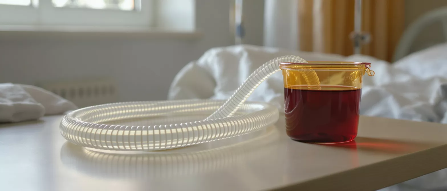
Purpose of the mediastinal drain: A mediastinal drain is a flexible, transparent tube placed inside the chest at the conclusion of the operation. It acts as an exit route for blood and fluids that collect around the heart. Without this drainage, accumulating fluid could compress the heart muscle. This condition, known as cardiac tamponade, would severely impair the heart's ability to pump effectively.
Family members seeing the tube for the first time should know it is a routine measure. The presence of the tube indicates the surgical team is actively monitoring the surgical site. It remains in place only as long as clinically necessary. Understanding its function helps alleviate the anxiety of seeing a loved one attached to medical equipment.
How Mediastinal Drain Placement Works
Surgical placement technique: Mediastinal drain placement occurs just before closing the chest incision. The surgeon carefully positions one or more flexible tubes beneath the sternum. The other end of the tube is threaded through a small skin incision, usually just below the main surgical cut. This approach keeps the drainage pathway separate from the primary wound closure.
Once positioned, the tube is secured to the skin with sutures. This prevents accidental dislodgement during patient transport from the operating room to the intensive care unit. The surgical team verifies tube patency before completing the procedure. Secure placement is critical for effective postoperative mediastinal drainage.
Patients remain under anesthesia during the placement. Consequently, there is no discomfort associated with the insertion phase. The drainage system is connected immediately, establishing negative pressure to assist fluid removal. This closed system protects the surgical site from external contaminants.
Mediastinal Drain Management and Care
Routine monitoring protocols: Mediastinal drain management is a highly structured process handled by specialized nursing staff. Nurses document the volume and color of the fluid at regular intervals. Fresh, bright red blood gradually transitions to a darker, serous fluid as healing progresses. This visual data provides real-time insight into the patient's recovery status.
Preventing tube blockages: Blood clots can form within the drainage tube. To prevent blockages, nurses routinely squeeze or milk the tubing. This technique maintains an open pathway for continuous fluid evacuation.
Proper care also involves monitoring the connection sites. The junction between the tube and the collection canister must remain sealed. Any air leak could compromise the negative pressure system. The nursing team is trained to identify and resolve these issues immediately.
Mediastinal Drain Output Explained
Normal drainage patterns: Mediastinal drain output is expected to be highest in the immediate hours following surgery. As the surgical site heals, the volume decreases significantly. The color also changes from bright red to a straw-colored fluid. This transition indicates bleeding has stopped and normal serous fluid is clearing the space.
Tracking this output guides the medical team's decisions. Excessive drainage over several hours may prompt the team to review clotting factors or blood products. Minimal drainage, conversely, could indicate a blockage in the tube. The medical team balances these data points against the patient's vital signs.
- Initial Phase: High volume, bright red blood in the first few hours.
- Transition Phase: Decreasing volume, darker fluid as bleeding subsides.
- Final Phase: Minimal output, serous fluid before removal.
What is a Mediastinal Drainage System
Components of the closed system: The mediastinal drainage system comprises three main parts: the chest tube, the connecting tubing, and the collection chamber. The collection chamber often uses a water seal mechanism. This mechanism allows air and fluid to exit the chest while preventing atmospheric air from entering the chest cavity.
How suction regulates pressure: Gentle suction is often applied to the system. This negative pressure helps pull fluid out of the chest and promotes lung re-expansion. The regulator ensures the pressure remains constant and safe. Any disruption to this pressure regulation is immediately addressed by the clinical staff.
The transparency of the entire system allows continuous visual inspection. Bubbling in the water seal chamber can indicate an air leak from the lung tissue. Quiet movements in the chamber, conversely, suggest the lung has sealed against the chest wall.
Mediastinal Drain Removal Process
Criteria for safe removal: Mediastinal drain removal occurs only when specific criteria are met. The primary requirement is a sufficiently low volume of output over a 24-hour period. Additionally, the fluid must have transitioned from bloody to serous. The patient must also demonstrate stable blood counts and hemodynamic stability.
Removal is a brief procedure performed at the bedside. The nurse or physician cuts the securing sutures. The patient is instructed to take a deep breath and hold it. This maneuver expands the lungs and prevents air from entering the chest cavity during the seconds the tube is extracted.
A gauze dressing is immediately applied to the exit site. A few sutures may be used to close the small opening if necessary. Most patients feel a brief sensation of pulling or slight pressure. Discomfort resolves rapidly once the tube is out, marking a significant milestone in recovery.
Mediastinal Drain Complications
Identifying potential risks: While generally safe, mediastinal drain complications can occur. The most common issue is tube blockage from clotted blood. If a clot blocks the tube, fluid can build up inside the chest. Nurses actively monitor for this by checking for firmness in the tubing and watching output levels.
Mediastinal drain infection signs: Any foreign object in the body carries a risk of infection. The exit site is monitored for redness, swelling, or purulent drainage. Strict sterile technique during placement and handling minimizes this risk. Prophylactic antibiotics are typically administered during the perioperative period.
- Blockage: Clotted blood preventing fluid exit.
- Infection: Localized redness or systemic fever.
- Dislodgement: Accidental pulling of the tube.
Are Mediastinal Drains Safe
Standard safety protocols: Are mediastinal drains safe? Yes, they are a routine and essential safety measure following cardiac surgery. The risks of not using a drain—such as cardiac tamponade—far outweigh the risks of having one. Modern cardiac surgery mediastinal drain protocols are refined to ensure maximum patient safety.
The closed drainage system acts as a barrier against environmental pathogens. Secure suturing and careful patient handling prevent accidental removal. Furthermore, the continuous monitoring by trained intensive care staff ensures any deviation from normal function is caught early. This vigilance is the cornerstone of postoperative safety.
Patient mobility is encouraged even with the drains in place. The tubes are long enough to allow patients to sit up and walk short distances. This early mobilization is crucial for preventing pneumonia and blood clots in the legs. The drains are integrated into the recovery process safely.
Mediastinal Drain Nursing Guidelines
Frequency of assessments: Mediastinal drain nursing protocols require hourly assessments in the immediate postoperative period. Nurses check the volume, color, and consistency of the output. They also assess the patency of the tubing and ensure all connections are tight. This high-frequency monitoring tapers off as the patient stabilizes.
Handling emergency situations: Nurses are trained for emergent scenarios. If a tube accidentally dislodges, the nurse immediately applies pressure to the site with a sterile dressing. If output suddenly stops, the nurse investigates for internal clots. These guidelines ensure swift action, protecting the patient from adverse events.
Communication is a key component of these guidelines. The nursing team relays output data to the surgical team. If drainage exceeds predetermined thresholds, the surgeon is notified immediately. This collaborative approach ensures the patient receives comprehensive care.
Postoperative Mediastinal Drainage Recovery
Transitioning off drainage support: Postoperative mediastinal drainage recovery culminates in the removal of the tubes. This event signals that the surgical site has stabilized. The body has resumed its natural ability to manage fluid reabsorption. Patients often feel more comfortable and mobile immediately after removal.
Healing after drain removal: The small incision where the tube was placed heals quickly. Patients are instructed on how to care for the site at home. Keeping the area clean and dry is usually sufficient. A small scar is the only visible remnant of the drain.
Understanding the entire process helps demystify the intensive care experience. The drain is a temporary but vital companion on the journey to heart health. By evacuating fluid and monitoring internal healing, it provides a critical safety net. Patients can focus on their cardiac rehabilitation knowing this phase of recovery is complete.






