

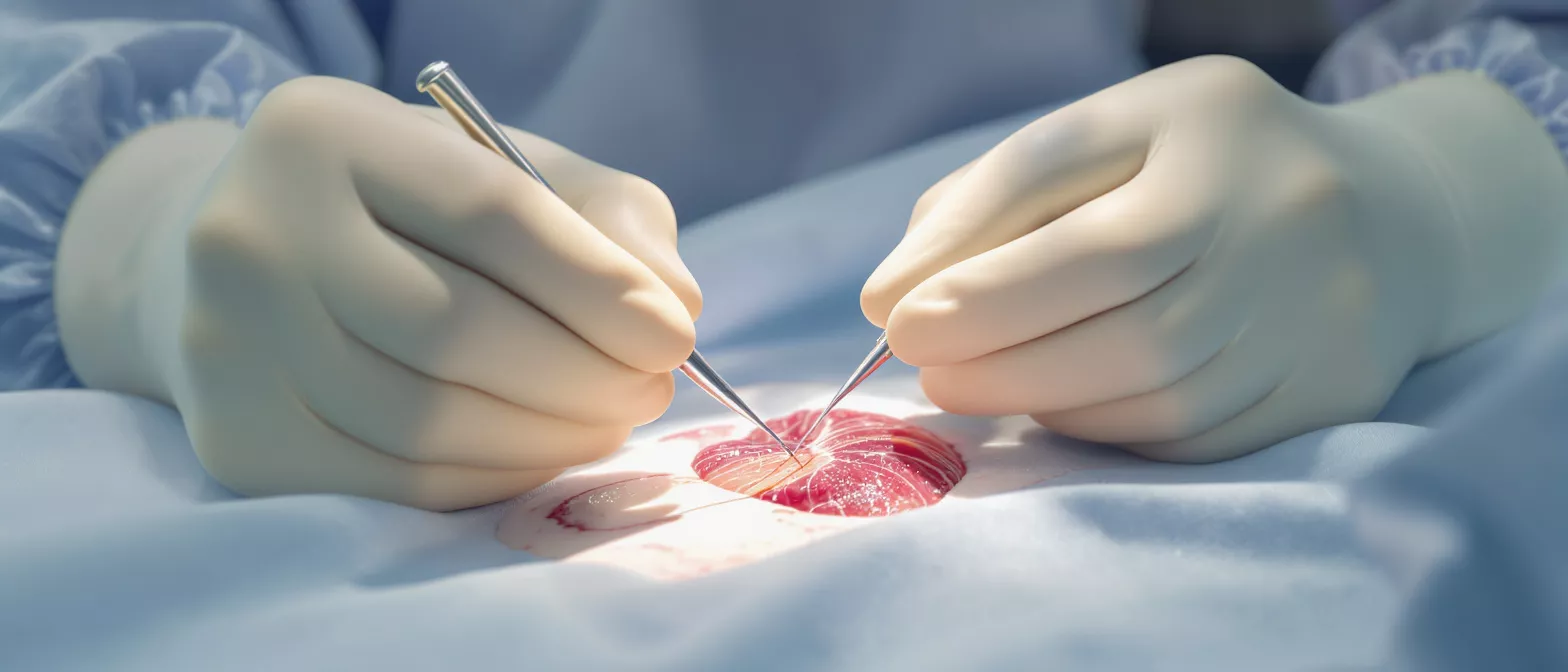
Septal Defect Closure
Septal defect closure repairs holes in the heart wall.
What is a septal defect closure?
Understanding a hole in heart repair begins with understanding the heart's structure. The human heart is divided into four chambers separated by walls called septa. Sometimes, a baby is born with an opening in one of these walls—a congenital anomaly known as a septal defect. Septal defect closure is the medical procedure used to safely patch this opening, restoring normal blood flow and preventing long-term strain on the heart and lungs. For families hearing this term for the first time, the diagnosis can feel overwhelming, but modern cardiac care offers highly effective solutions.
Why does this hole matter to a heart surgery patient? When a gap remains in the heart's wall, blood can travel abnormally between the chambers. Oxygen-rich blood may mix with oxygen-poor blood, forcing the heart to work harder than it should. Over time, if left untreated, this extra workload can lead to fatigue, high blood pressure in the lungs, and irregular heartbeats. Closing the defect alleviates this burden immediately.
The goal of the procedure is straightforward: to seal the opening and allow the heart to function efficiently. Whether the defect is small and asymptomatic or large and causing noticeable issues, a septal defect closure is a well-established intervention. Dr Molina and experienced surgical teams perform these repairs with precision, ensuring the heart heals correctly so patients can return to their normal lives without the constant fatigue associated with a heart murmur or shunting blood.
How septal defect closure works
The exact approach to septal defect closure depends largely on the size and location of the opening. Surgeons generally choose between two primary methods: a minimally invasive catheter-based approach or a traditional surgical repair. Both methods aim to permanently seal the gap, but they achieve this through different pathways. The cardiac team evaluates the patient's specific anatomy to determine which technique offers the safest and most effective outcome.
Catheter-based ASD closure procedure is often used for atrial defects. In this method, a thin, flexible tube is guided through a blood vessel directly to the heart. Once the catheter reaches the defect, a specialized device—resembling a small umbrella—is deployed to plug the hole. The heart tissue eventually grows over this device, making it a permanent part of the heart structure. This approach does not require opening the chest, allowing for a significantly shorter recovery time.
For more complex cases or larger defects, open-heart surgery may be necessary. During this operation, the surgeon makes a small incision in the chest to access the heart directly. The defect is then stitched closed or covered with a special patch made from the patient's own tissue or a synthetic material. This method provides the surgeon with a direct, clear view of the heart, ensuring precise placement and a durable, long-lasting repair that stands the test of time.
Types of septal defect repair
Septal defects are categorized by their location within the heart, which dictates the specific repair required. The two most common variations are atrial septal defects and ventricular septal defects. An atrial defect occurs in the wall between the upper two chambers of the heart. This opening allows freshly oxygenated blood to flow back into the lungs, creating inefficiency in the circulatory system over time.
VSD closure surgery addresses openings in the wall between the two lower pumping chambers. Because the ventricles handle the heart's most forceful pumping action, a hole here can cause significant stress on the organ. Blood is pushed from the left ventricle into the right, increasing the volume of blood the lungs must handle. Surgical repair is often recommended early in life to prevent permanent damage to the lung vessels.
Understanding the difference helps patients and families grasp why their surgical plan looks a certain way. The specific location influences the surgeon's approach and the expected timeline for intervention.
- Atrial Septal Defect (ASD): Occurs in the upper chambers, often repaired with a minimally invasive catheter device.
- Ventricular Septal Defect (VSD): Occurs in the lower pumping chambers, frequently requiring surgical patching.
- Complete Septal Repair: Sometimes involves a combination of patches to address multiple structural issues at once.
Atrial septal defect closure procedure
The atrial septal defect closure procedure is a remarkable intervention that has transformed cardiac care. When the defect is in the upper chambers and meets specific size and anatomical criteria, cardiologists often prefer the transcatheter approach. This method avoids the need for a sternotomy—opening the chest—and relies instead on sophisticated imaging to guide a closure device through the body's vascular system directly into the heart.
What happens during the ASD closure procedure? Under general anesthesia, the medical team threads a catheter through a vein, usually in the leg. They guide it up to the heart and across the defect. Once positioned, a self-expanding disc is released on either side of the hole, clamping the tissue in place. This device acts as scaffolding, allowing the heart's natural cells to grow over it and seal the gap permanently within a few months.
Patients typically spend only one night in the hospital following a catheter-based closure. The lack of a large chest incision means less post-operative pain and a faster return to daily activities. Regular follow-up echocardiograms ensure the device remains properly seated and that the heart is adapting beautifully to its newly efficient circulatory path.
Ventricular septal defect closure surgery
When the hole is located in the lower, more muscular chambers of the heart, surgical intervention is typically required. Ventricular septal defect closure surgery is a highly effective procedure performed to halt the abnormal flow of blood between the ventricles. The strength of the ventricular muscle and the high pressure in these chambers make catheter-based devices less reliable, necessitating a direct surgical approach for a definitive fix.
The surgical approach to VSD closure involves placing the patient on a heart-lung machine, which takes over the pumping of blood and oxygenation while the heart is still. The surgeon opens the right atrium or the ventricle itself to access the defect. Depending on the size of the hole, it may be closed with strong sutures or covered with a patch crafted from synthetic material or the patient's own pericardial tissue.
Once the patch is secured and the heart is beating normally again, the surgical team closely monitors the repair. This operation provides a complete and permanent closure, drastically reducing the workload on the heart and lungs. While it requires a slightly longer hospital stay than a catheter procedure, the long-term outcomes for patients undergoing ventricular repair are excellent and allow for completely normal heart function.
Septal defect symptoms and diagnosis
Identifying a septal defect early is crucial for optimal heart health. In many infants, a ventricular septal defect is discovered shortly after birth during a routine checkup because it causes a distinct heart murmur. The sound occurs because blood is shunting through the opening, creating a turbulence that the doctor can hear through a stethoscope. Pediatricians often refer patients to a cardiologist immediately upon detecting this sound.
Recognizing heart murmur treatment needs depends on the symptoms present. Small defects may cause no symptoms at all and sometimes close on their own as the child grows. However, larger defects can cause poor weight gain in infants, rapid breathing, and extreme fatigue during feeding. Adults with undiagnosed atrial defects might experience shortness of breath, palpitations, or frequent respiratory infections.
Diagnosis is confirmed using an echocardiogram, which uses sound waves to create a detailed moving picture of the heart. This allows the cardiologist to see the exact size and location of the opening and measure the amount of blood flowing across it. Additional tests, such as an electrocardiogram (ECG) or a chest X-ray, help assess the overall size and function of the heart before a surgical plan is finalized.
Are septal defect closure procedures safe?
The safety of modern septal defect closure is incredibly high, thanks to decades of medical advancement. Like all surgical interventions, it carries some risks, but the likelihood of a successful, uncomplicated outcome far outweighs the dangers of leaving a significant heart defect untreated. Surgeons and specialized cardiac teams perform these operations using rigorous protocols designed to protect the patient at every stage.
Understanding the risks of septal defect repair involves looking at both catheter and surgical methods. Catheter closures carry minimal risk of bleeding or device dislodgement, while open surgeries carry standard surgical risks such as infection or reaction to anesthesia. The surgical team will discuss these possibilities openly, providing families with a clear, honest picture of what to expect.
It is important to weigh these surgical risks against the severe complications that arise from ignoring a defect. Untreated holes in the heart can lead to pulmonary hypertension, heart failure, and irregular rhythms that become harder to treat over time. By choosing to undergo a cardiac defect closure device placement or surgical repair, patients drastically improve their life expectancy and quality of life. The consensus in the medical community is that the benefits of closure heavily outweigh the procedural risks for most patients.
Septal defect recovery time and hospital stay
Recovery from a septal defect closure depends entirely on the method used and the patient's age. For a transcatheter atrial repair, the recovery is remarkably swift. Most patients are up and walking within a few hours of the procedure and return home the following day. Full recovery, where the heart tissue has completely healed over the device, generally takes about six months, but patients resume normal activities within a week or two.
What is the typical septal defect recovery time for surgery? Open-heart ventricular repairs require a more extended hospital stay, usually ranging from four to seven days. During this time, the medical staff monitors the heart's rhythm and ensures the patch is functioning perfectly. Pain management is a priority, and physical therapists assist the patient in getting out of bed and moving safely to prevent complications.
Once home, surgical patients gradually increase their activity over four to six weeks. The surgical team provides clear guidelines on wound care, activity restrictions, and when it is safe to return to work or school. Following these instructions carefully ensures the chest bone heals properly and the heart fully adapts to its new, efficient workload.
- Catheter Procedure: 1 night in the hospital, 1-2 weeks of light activity before returning to normal routines.
- Open-Heart Surgery: Up to a week in the hospital, 4-6 weeks of restricted activity at home.
- Long-Term Healing: Heart tissue fully grows over patches or devices over 3 to 6 months.
Pediatric septal defect closure considerations
When an infant or child requires pediatric septal defect closure, the considerations are highly specialized. Children's hearts are smaller and still growing, which influences both the timing of the surgery and the materials used. Cardiac surgeons who specialize in congenital defects tailor their approach to ensure the repair supports the child's development and allows the heart to grow normally alongside the repaired septum.
Timing the intervention for children is a critical decision. Some small ventricular defects close spontaneously during the first year of life, so doctors may choose a watch-and-wait approach with regular checkups. However, if the defect is large or causing breathing and feeding difficulties, surgical closure is often scheduled within the first few months to prevent lung damage and promote healthy weight gain.
The patch material used in pediatric cases is chosen to accommodate future growth. Synthetic patches do not grow, but surgeons place them in a way that allows the surrounding heart muscle to expand naturally. Parents can feel reassured knowing that once a child recovers from this procedure, their heart typically functions perfectly, allowing them to participate in sports and all normal childhood activities without restriction.
Congenital heart defect treatment in Guadalajara
Accessing high-quality congenital heart defect treatment is vital for families facing a new diagnosis. In Guadalajara and the broader Jalisco region, patients have access to advanced cardiac care that rivals international standards. Dr Alejandro Molina and his specialized team bring extensive experience to the diagnosis and surgical management of these structural anomalies, offering a reassuring presence to frightened families.
Why choose septal defect surgery in Guadalajara? The region offers state-of-the-art facilities equipped with the latest imaging and surgical technology. Families benefit from receiving world-class cardiac intervention without the need to travel abroad, keeping their support networks intact during a stressful time. The medical team prioritizes clear communication, ensuring parents and adult patients understand every step of the treatment plan.
From the initial echocardiogram to the final post-operative checkup, the standard of care is meticulous. Patients considering a custom heart surgery care package will find comprehensive support tailored to their specific medical and logistical needs. If you or a loved one has been diagnosed with a hole in the heart, seeking a consultation with an expert cardiac surgeon is the most important first step toward a healthy, active future.






