

The Atrial Appendage and Clot Formation in AFib
Atrial appendage clots AFib patients face 5x higher stroke risk.
What Is the Atrial Appendage and Why Does It Matter?
The left atrial appendage is a small, ear-shaped pouch that extends from the left atrium — the upper-left chamber of your heart that receives oxygenated blood from the lungs and pumps it into the left ventricle. Though this structure is tiny compared to the rest of the heart, it plays an outsized role in stroke prevention for patients living with atrial fibrillation. Understanding its anatomy and function helps explain why AFib patients face a dramatically higher stroke risk and what treatment options exist to address it.
In a normally beating heart, the atrial appendage contracts along with the surrounding atrium muscle, pushing blood efficiently through the chamber with each heartbeat. The interior surface contains ridges called pectinate muscles, which give the pouch a comb-like texture. During atrial fibrillation, the heart's upper chambers quiver chaotically instead of squeezing in an organized fashion, and this irregular movement means blood can pool inside the appendage rather than flowing forward with each cycle.
Why this pouch becomes a clot factory: When blood sits stagnant inside the appendage's crevices, the body's natural clotting system activates — platelets clump together and clotting factors form a mesh that traps blood cells. If a clot breaks free from this pouch, it can travel through the aorta and into the arteries supplying the brain, causing an ischemic stroke. Roughly 90% of AFib-related clots originate in the left atrial appendage, which is why this small structure has become central to modern stroke prevention strategies. The ascending aorta serves as the highway these clots travel to reach the brain, making the appendage's role in stroke formation remarkably direct.
How Does AFib Cause Blood Clots in the Left Atrium?
Atrial fibrillation disrupts the normal electrical signals that coordinate your heartbeat. Instead of a steady, organized contraction driven by the heart's natural pacemaker, the atrium quivers rapidly and erratically. This means the blood that normally flows smoothly through the chamber begins to stagnate, particularly in the atrial appendage where the pouch-like shape creates natural pockets where flow is slowest. The condition known as atrial flutter, a related rhythm disorder, can produce similar pooling effects, though AFib tends to cause more chaotic and unpredictable blood stasis.
What happens inside the atrium during fibrillation: The quivering muscle cannot generate enough force to empty the appendage completely with each cycle. Blood that entered on the previous beat remains, and fresh blood layers on top of it. Over hours and days, this stagnant blood becomes increasingly likely to clot. The appendage's internal ridges further slow blood flow, creating micro-environments where clotting proteins concentrate. The longer blood remains motionless, the larger and more organized a clot can become.
Not every AFib episode produces a dangerous clot, but the risk accumulates over time, especially in people who have persistent or permanent AFib rather than occasional episodes. According to Verywell Health, patients with atrial fibrillation face a stroke risk roughly four to five times higher than people with normal heart rhythm. This elevated risk makes proactive prevention essential rather than optional, and it is the reason your cardiac team takes AFib seriously even when symptoms feel mild or intermittent.
What Are the Risk Factors for AFib Clot Formation?
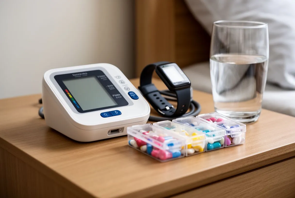
Several factors beyond AFib itself raise the likelihood that clots will form in the atrial appendage. Age is a major driver — patients over 75 face significantly higher risk than younger patients with the same rhythm pattern. High blood pressure, diabetes, and heart failure all damage the heart's structure over time, making the atrium wall stiffer and less efficient at moving blood, which increases clot formation during episodes of fibrillation.
A clinical scoring system called CHA2DS2-VASc helps doctors estimate individual stroke risk based on validated criteria. The acronym represents Congestive heart failure, Hypertension, Age over 75, Diabetes, prior Stroke or transient ischemic attack, Vascular disease, Age 65 to 74, and Sex category (female sex adds one point). A score of 2 or higher typically warrants formal anticoagulation therapy. The presence of atheromatous plaque in major arteries, measured by markers such as Apolipoprotein B levels, can also contribute to the vascular disease component of this score.
- High blood pressure: Damages atrial tissue over years, creating a stiff chamber where blood pools more easily
- Age over 75: Structural heart changes accumulate, reducing contractility and increasing stasis risk
- Prior stroke or TIA: The strongest single predictor that the appendage has already produced dangerous clots
- Heart failure: Reduced pumping efficiency means blood moves more slowly through all chambers, including the atrium
How Is Stroke Risk Assessed in Atrial Fibrillation?
Your cardiac team evaluates stroke risk using more than just a numerical score. They combine clinical history, imaging studies, and rhythm monitoring to build a complete picture of your individual risk. A transthoracic echocardiogram — which uses sound waves against the chest wall to create images of your heart — helps assess the left atrium's size and pumping function. An enlarged or poorly contracting atrium suggests higher clot risk. During the initial physical examination, auscultation, or careful listening to the heart with a stethoscope, may reveal an irregular rhythm that prompts further investigation.
Doctors may also use transesophageal echocardiography to look directly inside the left atrial appendage for existing clots before any procedure. This involves a thin probe passed down the esophagus to capture detailed images from behind the heart, where the left atrium sits closest to the esophagus. If a clot is already present, your team must manage it with medication before considering closure procedures or electrical cardioversion. Pre-procedure imaging can also reveal other cardiovascular conditions that need attention — for example, detailed aortic imaging sometimes identifies an intramural haematoma of the thoracic aorta that requires separate management, as discussed in research published in the Journal of Cardiothoracic Surgery.
Why rhythm pattern matters for risk stratification: Some patients have paroxysmal AFib, meaning episodes start and stop on their own, while others have persistent or permanent AFib. The pattern matters because continuous AFib may carry different clot management implications than intermittent episodes. Your electrophysiologist may recommend a Holter monitor worn for 24 to 48 hours, or an implantable loop recorder that monitors rhythm for months, to capture data that guides your prevention strategy. Atrial flutter, which some patients have alongside AFib, produces a more organized but still abnormally fast atrial rhythm and may require its own specific management approach.
What Medications Help Prevent AFib-Related Strokes?
Blood thinners, or anticoagulants, are the cornerstone of stroke prevention in atrial fibrillation. These medications do not dissolve existing clots but prevent new ones from forming by interfering with different steps in the clotting cascade. Two main categories exist: warfarin, a traditional option that requires regular blood testing and dietary management, and direct oral anticoagulants such as apixaban and rivaroxaban, which have largely replaced warfarin for most AFib patients due to their predictable dosing and fewer interactions.
The landmark ARISTOTLE trial demonstrated that apixaban reduced stroke and systemic embolism while causing fewer major bleeding complications compared to warfarin. This study fundamentally changed how cardiologists approach AFib stroke prevention worldwide. Direct oral anticoagulants do not require the dietary restrictions and frequent blood-level monitoring that warfarin demands, making them more convenient for most patients. However, warfarin remains important for patients with mechanical heart valves or severe kidney disease where newer agents have limitations.

Anticoagulants carry inherent bleeding risk, and some patients cannot tolerate long-term blood thinners due to prior gastrointestinal bleeding, intracranial hemorrhage, or conditions that increase fall risk. In these cases, doctors explore procedural alternatives. Aspirin alone is generally insufficient for AFib stroke prevention and is no longer recommended as a primary strategy by most cardiology guidelines. For patients who cannot safely take anticoagulants, left atrial appendage closure offers a mechanical alternative that addresses the source of clots directly rather than thinning the blood systemically.
What Is Left Atrial Appendage Occlusion?
Left atrial appendage occlusion is a procedure that physically seals off the appendage from the rest of the heart's circulation. By closing this pouch, blood can no longer pool inside it, eliminating the primary source of AFib-related clots. This approach offers an alternative for patients who cannot safely take long-term blood thinners but still need effective stroke prevention. The concept is elegantly simple: if the structure where clots form is sealed, the clots cannot escape to the brain.
The procedure is typically performed under general anesthesia with transesophageal echocardiography providing real-time imaging guidance. A cardiologist threads a catheter through a vein in the leg, advancing it to the right atrium, then crossing to the left atrium through a small puncture in the interatrial septum — the wall between the heart's two upper chambers. Once inside the left atrium, measurements are taken of the appendage opening, and the closure device is deployed precisely at the mouth of the pouch. This catheter-based approach means no chest incision is needed, and recovery is measured in days rather than weeks.
What happens after the device is placed: Over the following weeks, heart tissue grows over the device surface, completely sealing the appendage. During this healing period, patients typically remain on a short course of anticoagulation to prevent clots from forming on the device itself before tissue coverage is complete. After confirmation that the appendage is fully sealed — usually via a follow-up imaging study at approximately 45 days — many patients can reduce or stop taking blood thinners entirely. The decision to discontinue anticoagulation is made individually based on your overall stroke risk and bleeding history.
How Does a Left Atrial Appendage Occlusion Device Work for AFib?
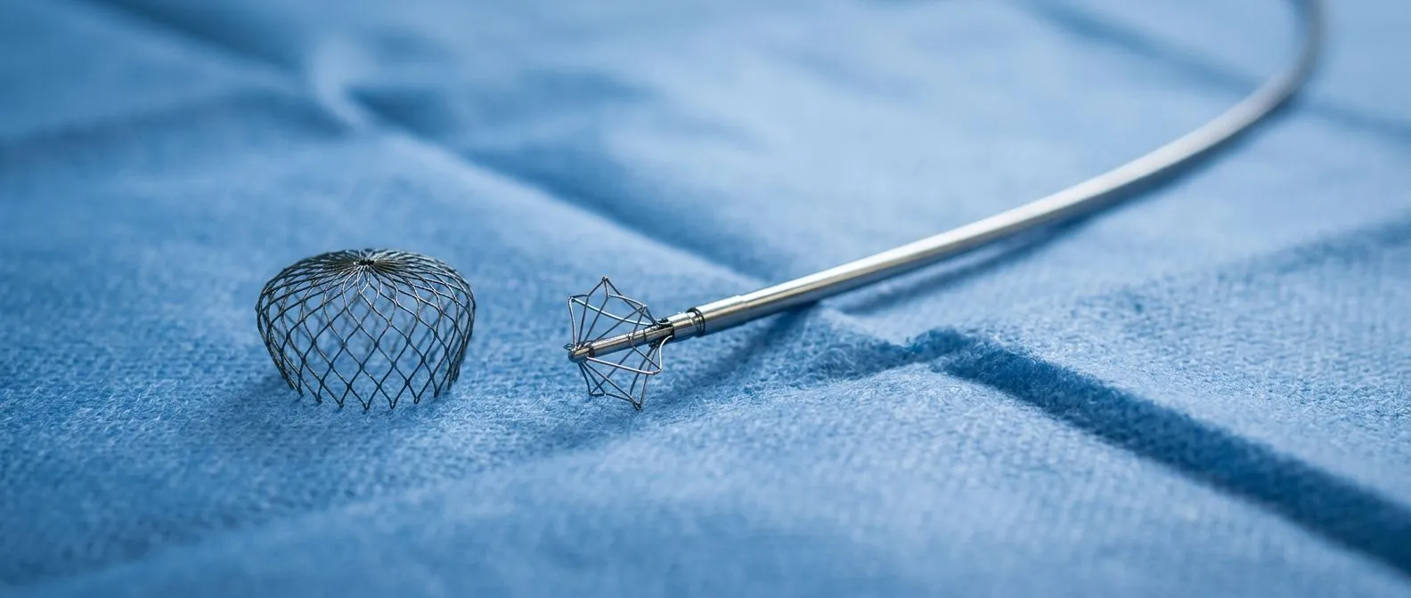
Left atrial appendage occlusion devices are among the most widely studied structural heart implants. It consists of a self-expanding nitinol frame — a metal alloy that returns to its pre-shaped form when released from the catheter — covered with a fabric membrane. The device is compressed into a delivery catheter, guided into position, and then expanded to fill the opening of the appendage like an umbrella opened in a doorway.
Clinical evidence supporting left atrial appendage occlusion comes from multiple randomized trials comparing it to warfarin therapy. These studies demonstrated that the device provides comparable stroke protection while significantly reducing hemorrhagic stroke and major bleeding events over long-term follow-up. For patients who have experienced serious bleeding episodes on blood thinners, this reduction in bleeding risk represents a meaningful improvement in both safety and quality of life. The procedure typically takes about an hour, and most patients stay overnight for observation before returning home the next morning.
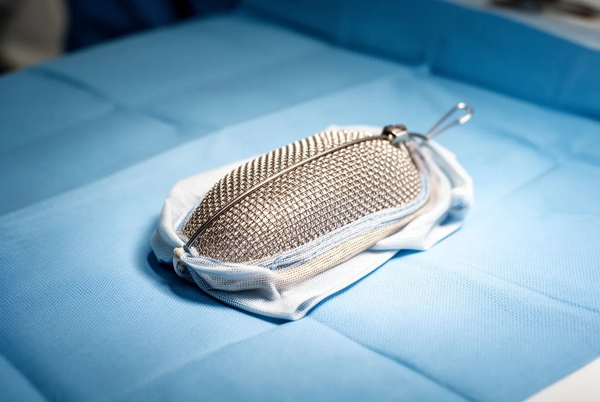
Recovery and follow-up after left atrial appendage occlusion: Most patients return to normal daily activities within a week, though your cardiologist may recommend avoiding strenuous exercise for a short period. The critical follow-up milestone occurs around 45 days after implantation, when a transesophageal echocardiogram confirms whether the device has sealed the appendage completely. If residual flow is detected around the device edges, your doctor may extend anticoagulation temporarily and re-evaluate later. The vast majority of implants achieve complete sealing, but the follow-up process is essential because incomplete closure could leave a pathway for clot migration. Patients who also have vascular conditions requiring complex interventions — such as those described in a case report on physician-modified endografts for aortic arch ulcers — may need coordinated planning between their cardiac and vascular teams.
Is Atrial Appendage Closure Surgery Right for You?
Not every AFib patient needs or qualifies for appendage closure. The ideal candidate has a high stroke risk — meaning an elevated CHA2DS2-VASc score — but cannot tolerate long-term anticoagulation due to bleeding complications. This includes patients who have experienced gastrointestinal bleeding, intracranial hemorrhage, or those with medical conditions or living situations that increase fall risk and bleeding danger. The procedure addresses a specific problem: clot formation in the appendage of a patient who cannot safely take blood thinners.
The decision requires a thorough evaluation by a cardiac team experienced in structural heart procedures. Your cardiologist will assess the shape and size of your appendage using imaging, because anatomical variations can affect whether a standard device fits properly. Some appendages are too large, too small, or have unusual configurations such as multiple lobes that make catheter-based closure challenging. In these cases, alternative device sizes, surgical closure performed during another planned heart operation, or continued medical therapy may be recommended instead.
- Good candidate: High stroke risk plus a documented bleeding event on anticoagulants
- Good candidate: High fall risk with CHA2DS2-VASc score of 2 or more
- Needs further evaluation: Appendage anatomy outside standard device size range
- Not indicated: Low stroke risk without anticoagulation contraindications
It is important to understand that appendage closure does not treat atrial fibrillation itself. You may still need rate-control medication, rhythm management with antiarrhythmic drugs, or catheter ablation to address the underlying arrhythmia. The closure only addresses the stroke risk associated with the appendage. Patients should view it as one component of a comprehensive AFib management plan, not a standalone cure for the rhythm disorder. In rare cases, surgical closure of the appendage may be performed during open-heart surgery for other conditions, such as coronary artery bypass or valve repair, when the surgical team already has direct access to the heart.
Catheter Ablation vs LAA Closure: Which Is Better?
Catheter ablation and left atrial appendage closure address different aspects of atrial fibrillation and are not competing procedures. Ablation targets the electrical signals causing the irregular rhythm, attempting to restore normal conduction by isolating the areas where abnormal impulses originate. LAA closure targets the mechanical risk of clot formation in the appendage. Many patients benefit from both approaches at different stages of their treatment, depending on whether the priority is rhythm control or stroke prevention.
Catheter ablation uses radiofrequency energy or cryotherapy to create small scars in heart tissue that block abnormal electrical pathways. For selected patients — particularly those with paroxysmal AFib and structurally normal hearts — successful ablation can reduce AFib burden significantly, which may lower clot risk by restoring more organized atrial contraction. However, ablation does not eliminate stroke risk entirely, especially in patients with persistent risk factors such as hypertension or heart failure. Current cardiology guidelines recommend continuing stroke prevention therapy based on CHA2DS2-VASc score, regardless of whether ablation is successful, because the appendage can still harbor stagnant blood even when rhythm improves.
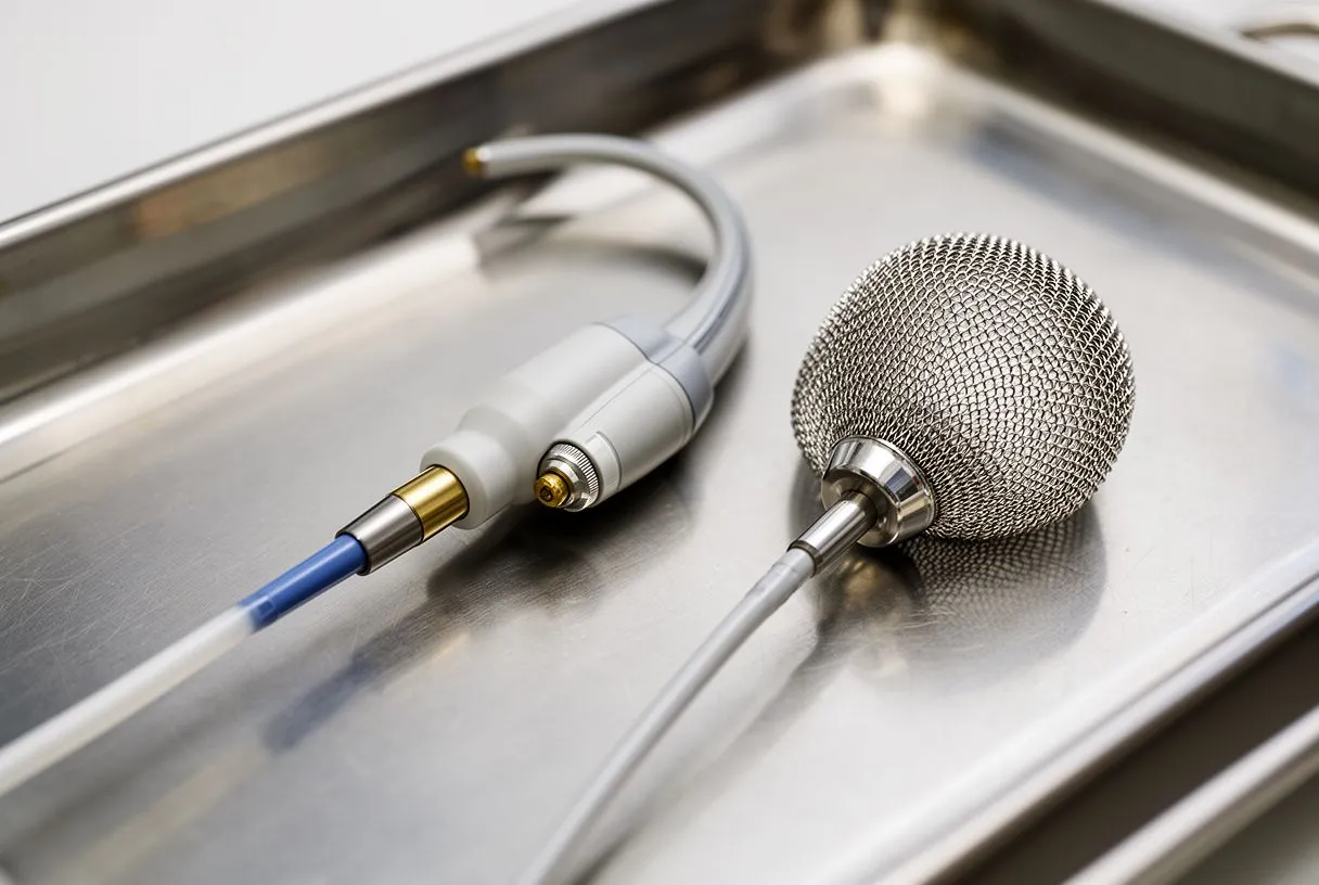
Can both procedures be done together? In experienced centers, both procedures can sometimes be performed during the same catheter-based session. The ablation addresses the rhythm disorder while the closure device addresses the mechanical clot risk, offering a comprehensive approach in a single anesthetic event. Whether a combined approach is appropriate depends on your specific anatomy, AFib pattern, procedure duration, and overall cardiac health. Atrial flutter, which some patients have concurrently with AFib, can also be treated during the same ablation session using targeted energy at the cavotricuspid isthmus, the tissue corridor where flutter circuits typically propagate.
Living With AFib: Long-Term Stroke Prevention
Managing atrial fibrillation is a lifelong commitment that extends well beyond any single procedure or medication change. Regular follow-up with your cardiac team ensures that your stroke prevention strategy remains appropriate as your health evolves over time. Blood pressure control, diabetes management, and maintaining a healthy weight all reduce AFib burden and clot risk independently of any specific treatment. Annual or semi-annual checkups allow your team to adjust medications, reassess your risk score, and determine whether your prevention plan still fits your current situation.
Lifestyle modifications play an underappreciated but well-documented role in AFib management. Reducing alcohol intake, treating sleep apnea with continuous positive airway pressure when indicated, and maintaining regular physical activity can decrease AFib episode frequency and may improve the effectiveness of other treatments. Some patients find that addressing these factors reduces their reliance on medications. However, lifestyle changes complement — they do not replace — formal stroke prevention strategies, and discontinuing anticoagulation or postponing a recommended closure procedure based solely on lifestyle improvements should only happen after careful discussion with your cardiologist.
If you have atrial fibrillation and concerns about your stroke risk, the most important step is to have an honest conversation with a qualified cardiac team. Every patient's anatomy, risk profile, and tolerance for treatment differs, and what works well for one person may not be appropriate for another. We encourage you to schedule a consultation with our team at Cirugía de Corazón to discuss your specific situation and build a prevention plan tailored to your needs rather than self-diagnosing or delaying care. In cardiac emergencies, an automated external defibrillator may be life-saving for rhythm-related cardiac arrest, but preventing the stroke that AFib threatens requires planning ahead — not emergency response after the fact.






