

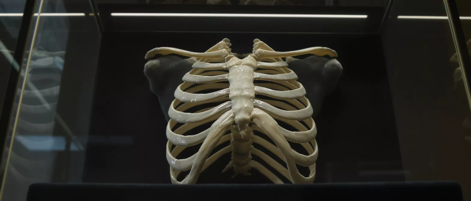
Thoracic Cavity
Explore the thoracic cavity, its anatomy, boundaries, and vital organs.
What is the thoracic cavity?
When preparing for cardiac surgery, understanding the basic anatomy of your chest can bring enormous peace of mind. The thoracic cavity is the medical term for the protected chamber inside your chest that houses your heart and lungs. By learning the thoracic cavity medical glossary terms your surgical team uses, you can better follow your treatment plan and feel more confident heading into your procedure.
Understanding the thoracic cavity definition: In straightforward terms, the thoracic cavity is a bony box formed primarily by your ribs, sternum, and spine. It acts as a sturdy shield for some of the most vital structures in your body, defending delicate tissues from external impact while allowing them the space they need to function continuously.
Knowing this foundational anatomy helps demystify the surgical process. Your surgical team must carefully navigate this space to access your heart, and understanding what they are working around can help you visualize your own recovery journey.
Thoracic cavity anatomy explained
To understand how your surgeon protects your heart, it helps to look at how this space is organized. The thoracic cavity anatomy is divided into three distinct compartments, ensuring that your heart and lungs operate without crowding one another.
Central mediastinum and surrounding spaces: The central section, called the mediastinum, houses the heart, esophagus, and trachea. On either side of the mediastinum sit the pleural cavities, which are sealed, airtight compartments that separately contain the left and right lungs. This separation ensures that a change in pressure on one side does not collapse the other.
This precise organization is what allows cardiac surgeons to operate safely. By clearly mapping these distinct compartments, your surgeon can isolate the specific structures they need to repair without disturbing the surrounding environment.
What organs are in the thoracic cavity?
The thoracic cavity organs are essential for circulation and breathing. Alongside the heart and lungs, this space contains major blood vessels, the trachea, and the esophagus. Understanding the thoracic cavity contents: These structures work in tight coordination, with the heart continuously pumping blood to the lungs for oxygenation before sending it out to the rest of your body.
Because these organs are packed so efficiently, any procedure involving the heart requires meticulous planning. The heart sits right in the middle of these critical pathways, meaning your surgeon must expertly manage surrounding structures to maintain stable circulation throughout the operation.
For families awaiting news during a procedure, knowing that these organs are in a dedicated, well-protected space explains why operating takes time. The surgeon is carefully navigating a network of vital structures to ensure everything functions perfectly when the chest is closed.
Thoracic cavity vs chest cavity
Patients often hear doctors use both terms, leading to confusion at the bedside. Comparing the two terms: The thoracic cavity is the precise medical term for the internal anatomical space, whereas the chest cavity is the broader, everyday term most people use to describe the same general area.
Think of the chest as the outer structure—muscles, ribs, and skin—and the thoracic cavity as the deeply protected inner room. By making this distinction, it becomes easier to understand how your surgical team plans their approach to the heart.
What is the thoracic cavity function?
The primary thoracic cavity function is to provide a protected, pressurized environment where the heart and lungs can perform without interruption. The ribs create a rigid cage, but the flexible cartilage between them allows the cavity to expand and contract with every breath you take.
Supporting respiration and circulation: By maintaining a vacuum-like environment in the pleural spaces, the cavity allows the lungs to inflate effortlessly. Simultaneously, it holds the heart securely in the mediastinum, giving it a stable anchor point to pump blood efficiently.
Any disruption to this delicate pressure system can affect your breathing or heart rhythm. This is why, after cardiac surgery, your care team closely monitors the pressure inside your chest to ensure the space is healing correctly and supporting normal function.
What are the thoracic cavity boundaries?
To safely access the heart, surgeons must understand exactly where the cavity begins and ends. Anatomical borders explained: The thoracic cavity boundaries are defined by the rib cage at the front, sides, and back, with the diaphragm muscle forming the floor that separates it from the abdominal cavity.
- Superior boundary: The upper thoracic aperture, where vital nerves and vessels exit to the neck and arms.
- Lateral and anterior boundaries: The ribs and sternum, providing rigid structural protection.
- Inferior boundary: The diaphragm, a muscular dome separating the heart and lungs from the digestive organs.
These borders are critical for surgical planning. When a cardiothoracic surgeon plans an incision, they rely on these boundaries to avoid damaging neighboring regions while gaining the clearest possible route to the heart.
What is the thoracic cavity medical definition?
If you are reading medical reports or consent forms, you may encounter clinical jargon that can feel overwhelming. Decoding clinical terms: The thoracic cavity medical definition refers specifically to the hollow space enclosed by the thoracic wall, extending from the neck down to the diaphragm.
This clear-cut definition helps medical professionals communicate exactly which part of the body they are discussing, eliminating ambiguity during complex multi-step procedures. When your doctor says they are operating within the thoracic cavity, they are referring to entering this specific, enclosed anatomical zone.
Understanding this terminology empowers you to ask informed questions. Recognizing that this space is clearly mapped by anatomists can reassure you that there is a structured, highly predictable plan for your operation.
How the thoracic cavity houses the heart
The heart is the centerpiece of the thoracic cavity, carefully suspended within a protective sac. The pericardium and its role: This double-walled sac anchors the heart in the mediastinum, preventing it from shifting as you move, while allowing it to beat freely without friction.
Because the heart is so securely housed, conditions like fluid buildup around the heart can become dangerous, as the restricted space limits how much the heart can expand. Understanding this physical constraint highlights why cardiac surgeons are so precise in their work, carefully managing this tight space to restore healthy blood flow.
How the thoracic cavity houses the lungs
The lungs flank the heart on either side, completely separated by the central mediastinum. The pleural space mechanics: Each lung sits within its own pleural cavity, surrounded by a thin membrane that secretes fluid, allowing the lungs to glide smoothly against the chest wall during every breath.
This physical separation is a brilliant evolutionary safeguard. Because the left and right lungs occupy entirely separate compartments, an issue affecting one lung—such as a collapsed lung—does not directly impact the other, ensuring you maintain adequate oxygenation.
- Independent compartments: Each pleural cavity operates under its own pressure.
- Lubrication: Pleural fluid prevents friction during continuous respiratory motion.
- Protection: The rib cage shields the fragile lung tissue from physical trauma.
During heart surgery, the surgical team temporarily manages how your lungs function, sometimes using a bypass machine to oxygenate your blood. Knowing how securely the lungs are housed in their own compartments makes it easier to visualize how the team safely resumes normal breathing after the operation.
Thoracic cavity structures and surgery recovery
After your operation, the way your thoracic cavity heals is directly related to its structure. The ribs, cartilage, and protective membranes must recover together to restore the stable environment your heart and lungs depend on.
Recovery involves managing pain, protecting the sternum as it mends, and gradually re-expanding the lungs to their full capacity. By understanding the intricate anatomy of your chest, you can approach your rehabilitation with patience, knowing that you are healing the very structure that protects your heart.
Always remember that your surgical team is deeply familiar with every boundary and compartment inside your chest. Their expertise ensures that from the first incision to the final stitch, your thoracic cavity is treated with the utmost care to restore your quality of life.






