

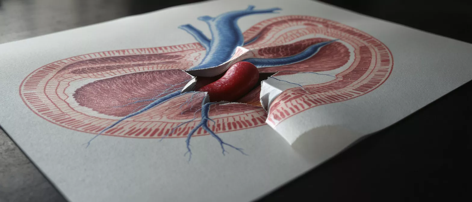
Acute Aortic Syndrome
Acute aortic syndrome is a life-threatening emergency.
What is acute aortic syndrome?
Acute aortic syndrome is a life-threatening medical emergency involving a sudden tear or rupture within the aorta, the main artery carrying blood from the heart to the rest of the body. When patients hear this diagnosis, it is natural to feel frightened, but understanding the condition is the first step toward effective cardiac care. This syndrome requires immediate surgical intervention to restore blood flow and prevent fatal complications.
Difference between aortic dissection and a heart attack: While both cause sudden, severe chest pain, a heart attack results from a blockage in the coronary arteries supplying the heart muscle, whereas acute aortic syndrome involves damage to the wall of the aorta itself. Distinguishing between the two rapidly is critical, as their treatments are entirely different.
Intramural hematoma and penetrating aortic ulcer explained: Acute aortic syndrome is an umbrella term covering three distinct pathologies. Type A aortic dissection involves the inner layer of the aorta tearing away, while an intramural hematoma is a bleed into the aortic wall without a tear, and a penetrating aortic ulcer occurs when plaque erodes through the artery's inner lining. Each variant presents unique challenges for surgical planning.
How aortic dissection symptoms present
Recognizing the signs of sudden chest pain aorta is crucial for survival. The most classic symptom of aortic dissection is abrupt, severe, tearing or ripping pain in the chest or upper back. Unlike a heart attack, which often builds in intensity, this pain is typically maximal at the moment of onset.
Sudden chest pain aorta warning signs: The location of the pain can shift as the dissection progresses along the aorta, moving from the chest to the back or abdomen. Patients may experience differences in pulse or blood pressure between the left and right arms. Recognizing these specific aortic emergency symptoms ensures faster transport to a specialized unit.
In some cases, patients might not present with classic chest pain but rather experience neurological symptoms, such as fainting or stroke-like effects, if the dissection interrupts blood supply to the brain. This complexity is why immediate medical evaluation and imaging are essential for anyone with severe, unexplained chest or back pain.
Types of aortic dissection
Medical professionals classify acute aortic syndrome using the Stanford classification aortic system to determine the urgency and type of intervention required. Understanding these classifications helps families comprehend the treatment plan and why a specific surgical approach was chosen.
Type A aortic dissection: This type involves a tear in the ascending aorta, the portion of the aorta directly attached to the heart. It is a critical emergency that requires immediate open-heart surgery to repair the tear and prevent rupture into the pericardium, the sac surrounding the heart.
- Type A: Involves the ascending aorta, requiring immediate surgical repair.
- Type B: Involves the descending aorta, often managed medically unless complications arise.
Type B aortic dissection: This occurs in the descending aorta, away from the heart. Initial treatment often focuses on strict blood pressure control, but surgery may be necessary if the dissection restricts blood flow to vital organs or threatens to rupture.
What causes acute aortic syndrome?
Understanding aortic dissection causes involves looking at underlying conditions that weaken the aortic wall. Chronic high blood pressure is the most significant risk factor, as it continuously stresses the aortic tissue. Over time, this relentless pressure can lead to structural fatigue, making patients susceptible to specialized cardiovascular support equipment.
Connective tissue disorders and high blood pressure: Genetic conditions such as Marfan syndrome or Ehlers-Danlos syndrome directly compromise the structural integrity of the aorta, predisposing patients to acute events. When coupled with unmanaged hypertension, the risk of aortic disease treatment necessity increases significantly.
Other risk factors include advanced age, atherosclerosis (hardening of the arteries), and a history of cardiac surgery or trauma. While some factors are genetic, managing modifiable risks through medication and lifestyle adjustments is highly effective in preventing acute aortic syndrome.
Acute aortic syndrome diagnosis
A prompt acute aortic syndrome diagnosis is vital for survival. Because initial symptoms mimic other cardiac events, emergency physicians rely heavily on advanced imaging techniques to confirm the presence and extent of an aortic emergency. Rapid diagnostic protocols are standard in emergency departments.
Imaging techniques used in thoracic aorta syndrome: The gold standard for diagnosing this condition is a CT angiogram, a specialized scan that provides detailed images of blood flow within the aorta. This scan allows the surgical team to pinpoint the exact location and severity of the dissection, guiding the surgical approach.
Other diagnostic tools include a transesophageal echocardiogram (TEE), which uses sound waves from a probe placed in the esophagus to capture close-up images of the heart and aorta. This is particularly useful if a patient is unstable and cannot easily move to a CT scanner, ensuring rapid diagnostic clarity.
Aortic dissection treatment options
Aortic dissection treatment varies significantly based on the type and location of the tear. The primary goal of any intervention is to prevent aortic rupture and restore normal blood flow. For Type A dissections, emergency aortic repair is universally required.
Medical management for uncomplicated Type B dissection: When the descending aorta is involved without life-threatening complications, treatment focuses on aggressive blood pressure control to lower the stress on the aortic wall. Intravenous medications are used in intensive care to keep the heart rate and blood pressure in a safe range.
If a Type B dissection becomes complicated—meaning it blocks blood flow to the kidneys, intestines, or limbs—interventional aortic syndrome management is necessary. This often involves endovascular stent grafts or open surgical repair to secure the damaged aorta and re-establish healthy circulation.
Emergency aortic repair procedures
Emergency aortic repair is a highly specialized procedure requiring a dedicated cardiac surgical team. For Type A aortic dissection, the surgeon must open the chest to directly access the ascending aorta, replace the torn segment with a synthetic tube, and ensure blood flow to the heart is preserved.
Endovascular stent grafting techniques: For Type B dissections requiring intervention, surgeons often use a minimally invasive approach. A catheter is threaded through the femoral artery to deploy a stent graft at the site of the tear, reinforcing the aortic wall without the need for a large chest incision, which can speed up recovery.
The choice between open surgery and endovascular repair depends on the patient's anatomy, overall health, and the extent of the damage. Both techniques aim to stabilize the aorta, and surgeons often recommend utilizing advanced hemodynamic monitoring systems to ensure optimal outcomes during the critical post-operative phase.
Aortic syndrome management and recovery
Effective aortic syndrome management extends well beyond the operating room. Survivors of acute aortic syndrome require lifelong medical follow-up to monitor the aorta and prevent future complications. This includes routine imaging to ensure the repaired aorta remains stable over time.
Long-term blood pressure control and lifestyle adjustments: Strict blood pressure management is the cornerstone of ongoing care. Patients must adhere closely to their medication regimens and frequently monitor their blood pressure at home, often integrating portable post-operative cardiac monitors into their daily routine to catch irregularities early.
Physical activity should be resumed gradually and under medical supervision. Cardiac rehabilitation programs are highly beneficial, providing structured, monitored exercise and education to help patients return to a healthy lifestyle safely and confidently.
Are acute aortic syndrome patients safe post-surgery?
Patients and families frequently ask, "Are acute aortic syndrome patients safe post-surgery?" While the initial event is terrifying, modern cardiac surgery offers excellent survival rates for those who reach the hospital in time. Safety post-surgery relies heavily on meticulous adherence to a lifelong management plan.
Monitoring for future aortic complications: Because patients are at risk for subsequent aortic issues, regular CT or MRI scans are mandatory. The surgical team will schedule these follow-ups at specific intervals to catch any signs of aortic enlargement or new tears long before they become symptomatic.
Psychological support is also an essential component of safety and recovery. The trauma of experiencing an aortic emergency can leave lasting emotional impacts, and accessing support groups or counseling helps patients process the event and regain a sense of normalcy in their lives.
Key takeaways for aortic emergency preparedness
Understanding acute aortic syndrome transforms fear into preparedness. Recognizing that sudden, severe tearing chest pain requires immediate emergency room evaluation is the most critical step a patient or family member can take to improve survival rates.
Importance of specialized cardiac care: Receiving care at a specialized cardiac center like Cirugía de Corazón provides the multidisciplinary expertise required for this complex condition. Our dedicated team utilizes advanced imaging and surgical techniques to ensure the highest standard of care from diagnosis through recovery.
In summary, while acute aortic syndrome is a profound medical challenge, outcomes continue to improve with rapid diagnosis and intervention. By maintaining strict blood pressure control and attending regular follow-ups, patients can lead full, healthy lives after aortic surgery.






