

Adventitia
Learn what the adventitia is, how it protects blood vessels, and why it matters in heart surgery.
What is the adventitia
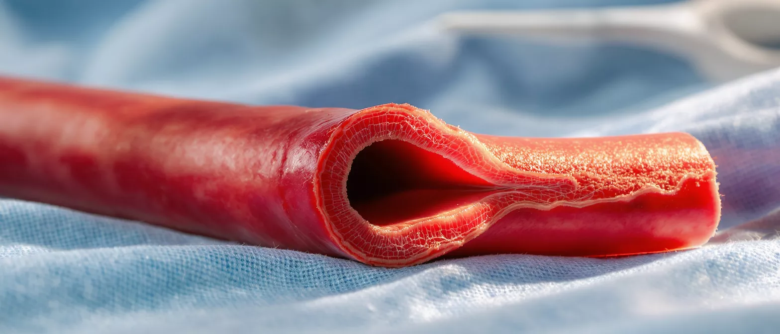
The adventitia is the tough, outermost layer of connective tissue that surrounds your blood vessels. When a cardiac surgeon explains the structure of an artery or a vein, the adventitia serves as the protective outer tunic. It is primarily composed of strong collagen fibers, which give it both durability and flexibility, allowing the vessel to withstand the continuous pressure of blood flow without tearing or collapsing.
Where is the adventitia located: In the arterial wall, it sits just outside the media, which is the middle smooth muscle layer. The transition between the media and the adventitia is marked by the external elastic lamina, a thin sheet of elastic tissue. In some larger veins, the adventitia is actually the thickest layer of the vessel wall, providing essential structural support against external pressures.
For patients preparing for heart surgery, understanding the architecture of a blood vessel can provide peace of mind. During procedures like coronary artery bypass grafting, a surgeon must handle the adventitia with extreme care. This layer is gently dissected to access the vessel lumen for attaching a healthy graft, but preserving its integrity helps the vessel maintain its strength long after the procedure is complete.
How does adventitia vs media differ
Patients often hear medical terms that describe vascular anatomy, and distinguishing these layers helps clarify surgical discussions. The key difference in the adventitia vs media comparison lies in their tissue composition and primary role. The media consists of smooth muscle cells and elastic tissue, which actively controls the diameter of the vessel, while the adventitia is predominantly loose connective tissue that anchors the vessel securely in place.
Adventitia vena y arteria structural differences: The thickness of the adventitia varies significantly depending on whether it is covering an artery or a vein. In arteries, the media is usually the thickest layer to manage high-pressure blood flow. In veins, where blood pressure is naturally lower, the adventitia often becomes the dominant layer, providing the bulk of the vessel's overall wall structure.
Cardiac surgeons must recognize these subtle tissue differences when evaluating and harvesting grafts. A saphenous vein graft, which is commonly used to bypass blocked coronary arteries, features a robust adventitia that is carefully managed to prevent spasms or surgical damage during the bypass operation.
What is the function of the adventitia
The primary function of the adventitia is to provide structural support and firmly anchor the blood vessel to surrounding tissues. Without this protective outer sheath, vessels would be highly prone to collapsing, tearing, or shifting under the continuous stress of pulsatile blood flow. The collagen matrix acts like a durable, flexible sleeve, keeping the vessel patent and properly aligned within the body.
Adventicia tejido conectivo roles: Beyond mere mechanical support, this connective tissue houses the vasa vasorum, which are tiny blood vessels that supply oxygen and nutrients to the walls of larger arteries and veins. It also contains the nervi vasorum, a network of nerves that help regulate vascular tone and transmit sensory signals back to the brain.
During heart surgery, preserving the integrity of this layer is vital. When a surgeon creates an anastomosis, which is the connection between a graft and a native coronary artery, the tissue must hold fine sutures securely. A strong adventitia prevents tearing at the suture line, ensuring a leak-free and durable connection that will last for years.
Adventitia collagen fibers and structural support
The mechanical strength of the vessel wall comes from the densely packed collagen fibers found within the outer tunic. These fibers are woven together in a specialized matrix that provides high tensile strength. This structural support ensures that a coronary artery can expand slightly during systole and recoil during diastole, maintaining a steady, controlled flow of oxygenated blood to the heart muscle.
Adventicia grosor vaso implications: The thickness of the adventitia correlates directly with the amount of vascular stress a particular vessel endures. Larger arteries, such as the aorta, have a substantial adventitial layer to prevent aneurysms from forming. When disease weakens the vessel wall, maintaining a healthy collagen framework is the body's primary defense against vascular rupture.
Patients recovering from cardiac surgery often ask about the long-term durability of their vascular grafts. The natural resilience of the adventitia is a major reason why vascular repairs, once fully healed, can last for many years. Surgeons rely on collagen-rich vascular repair products alongside the patient's own tissue to ensure the highest possible long-term success rate.
How adventitia heart surgery relates to grafts
Coronary artery bypass surgery relies entirely on the precise manipulation of blood vessel layers. The adventitia heart surgery relationship is critical because a graft must be attached to a healthy, intact portion of the native artery. If the native coronary artery's outer layer is diseased or heavily calcified, suturing becomes incredibly difficult and the risk of postoperative bleeding increases.
Adventitia coronary artery surgical planning: A cardiac surgeon evaluates the physical quality of the adventitia coronary artery before making any incisions. The tissue must be pliable enough to accept fine surgical sutures without fraying. If the outer layer is heavily scarred, the surgeon will search for a healthier segment of the artery to ensure the bypass graft heals properly and securely.
For those undergoing bypass surgery, selecting the right graft is a crucial step. Surgeons often prefer using an internal thoracic artery because its robust adventitia handles surgical stress exceptionally well. In certain cases, synthetic vascular graft materials are utilized when native veins are unsuitable, though managing the interface between artificial materials and natural tissue requires meticulous surgical skill.
Adventitia vascular nerves and blood supply
The outer layer of a blood vessel is not just a static wrapper; it is a living, dynamic tissue that actively participates in circulatory health. The adventitia vascular networks include the vasa vasorum, which are microscopic vessels that deliver oxygen to the thick walls of larger arteries, preventing the tissue from becoming ischemic and dying.
Nervi vasorum and vascular tone: The nerves that reside in the adventitia play a vital role in regulating blood pressure and systemic flow. They respond to chemical and mechanical signals, instructing the smooth muscle in the media to constrict or relax as needed to maintain circulatory homeostasis throughout the body.
- Vasa vasorum: Tiny vessels supplying oxygen to the outer vascular walls.
- Nervi vasorum: Sympathetic nerves regulating vessel dilation and tone.
- Immune surveillance: Resident macrophages guarding against inflammation.
Understanding these networks is crucial because inflammation in the adventitia can trigger plaque instability. Cardiovascular researchers are increasingly focusing on the outer layers of vessels to understand exactly how diseases like atherosclerosis progress and ultimately rupture.
What are adventitia pathological conditions
When the outer layer of a blood vessel becomes diseased, serious complications can arise. Adventitia pathological conditions include inflammation, fibrosis, and structural degradation. These issues can severely weaken the arterial wall, leading to life-threatening aneurysms or spontaneous dissections, where blood tears through the layers of the vessel wall.
Adventitia pathological conditions and inflammation: Conditions like vasculitis can directly target the vasa vasorum, reducing oxygen supply to the vessel wall and causing tissue necrosis. When the tissue becomes inflamed, the collagen matrix weakens significantly, making the artery vulnerable to rupture even under normal blood pressure.
Cardiologists monitor patients closely for signs of vascular weakening, especially those with a history of connective tissue disorders.
Adventitia arterial health and recovery
Postoperative recovery after heart surgery involves the careful healing of all vascular layers. Adventitia arterial health is vital for proper wound healing at the anastomosis site where the graft is attached. The collagen in the outer layer provides the necessary scaffolding for new tissue growth, ensuring the graft connection remains sealed and strong over time.
Adventicia significado in patient recovery: Understanding the meaning of this outer layer helps patients appreciate why they must avoid sudden, strenuous movements immediately after surgery. Straining can increase intra-thoracic pressure, putting unnecessary stress on freshly sutured vascular tissue. Gradual, supervised increases in physical activity allow the collagen to heal and remodel safely.
Patients are encouraged to support their vascular health through prescribed medications and controlled cardiac rehabilitation. A well-healed adventitia is a primary indicator of a successful bypass recovery and long-term surgical success.
Adventitia tissue healing after cardiac procedures
Healing is a complex biological cascade, and the outer vascular layer plays a highly significant role in postoperative recovery. Adventitia tissue healing begins with inflammation, followed by cellular proliferation, and ends with matrix remodeling. Fibroblasts within the connective tissue produce new collagen, sealing the surgical incisions and reinforcing the arterial wall against future stress.
Adventicia capa externa vaso sanguíneo remodeling: The remodeling phase can take several weeks to months. During this time, the external elastic lamina and the surrounding connective tissue adapt to the new hemodynamic forces created by the bypass graft. Proper blood pressure control is absolutely essential to prevent excess stress on the healing tissue.
Surgeons monitor this healing process through regular follow-up appointments. Ensuring the graft and native vessel are integrating properly requires patience and strict adherence to medical guidelines. Patients must prioritize sleep and stress management during this critical healing window.
Key takeaways about the layers of the vessel wall
Understanding the layers of a blood vessel provides clarity and confidence for patients navigating cardiac surgery. The layers of the vessel wall work together to ensure blood reaches the heart muscle efficiently and safely. The outer layer, with its durable collagen matrix, acts as the ultimate structural safeguard for the entire vascular system.
- Layered Protection: Each layer has a specific role in overall vascular health.
- Surgical Importance: The outer tunic holds sutures and prevents tearing.
- Healing Dynamics: Collagen remodeling is essential for postoperative recovery.
When facing heart surgery, knowledge is a powerful tool for reducing anxiety. Knowing how your body is structured and how it heals builds confidence in the surgical process. Trust in your medical team and the remarkable regenerative capabilities of your own vascular system.






