

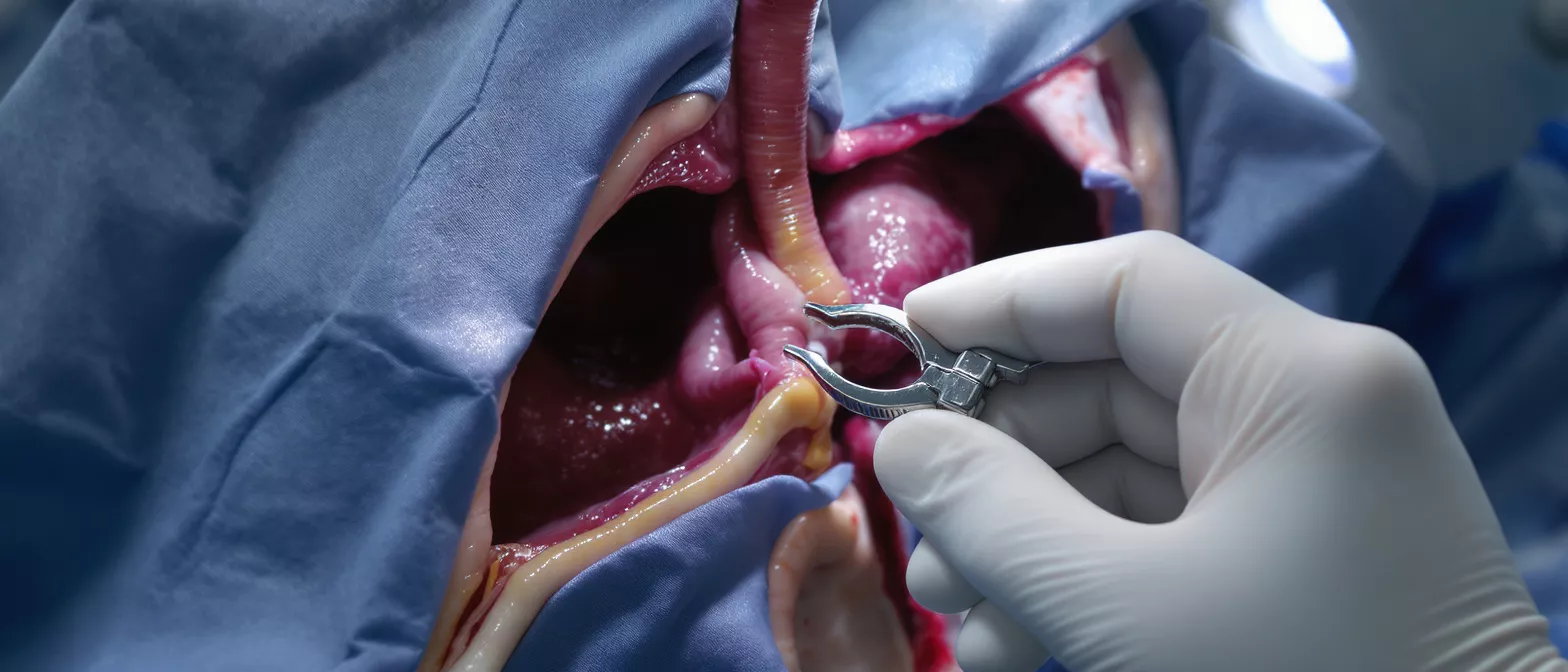
Cannulation
Learn how cannulation works in cardiac surgery: techniques, types, risks, and ECMO.
What Is Cannulation in Cardiac Surgery?
Cannulation is a foundational step in modern heart surgery where thin, specialized tubes called cannulas are carefully inserted into specific blood vessels. These tubes act as secure pathways that connect the patient's circulatory system to the cardiopulmonary bypass machine, often referred to as the heart-lung machine. This connection allows the surgical team to temporarily take over the work of the heart and lungs, providing a still, bloodless operative field.
Why cannulation matters for heart surgery patients: This step is critical because it ensures the rest of the body continues to receive oxygen-rich blood while the surgical team repairs the heart. By diverting blood flow away from the heart, the surgeon can operate with high precision. Understanding this process helps patients and families feel more confident about the operating room workflow.
For families watching a loved one prepare for surgery, the technical aspects can feel overwhelming. However, cannulation is a standard, meticulously practiced procedure. The surgical team uses real-time imaging and decades of refined technique to place these tubes safely, ensuring a stable circulation throughout the operation.
How Does Cardiopulmonary Bypass Cannulation Work?
Cardiopulmonary bypass cannulation works by establishing an alternate route for blood circulation. Once the cannulas are securely placed, venous blood is drained from the body, typically from the right side of the heart or the major veins. This blood is then routed into the heart-lung machine where it is oxygenated, cleared of carbon dioxide, and gently warmed or cooled to the required temperature before being returned to the body.
The mechanics of arterial perfusion cannulation: The oxygenated blood is returned to the body through an arterial cannula, usually placed in the aorta. This high-pressure return line is carefully secured by the surgeon to ensure a tight seal, preventing any air from entering the bloodstream while maintaining steady perfusion to vital organs.
The entire bypass circuit is continuously monitored by a perfusionist, a specialist who manages the heart-lung machine. They adjust the flow rate and pressure to match the patient's specific needs. The cannulas are designed to be highly efficient, allowing maximal blood flow through relatively small, carefully engineered tubes.
What Is the Difference Between Arterial and Venous Cannulation?
Understanding arterial and venous cannulation requires looking at the direction of blood flow. Venous cannulation involves placing tubes into the body's major veins or the right atrium of the heart to drain deoxygenated blood away from the tissues and into the bypass machine. Arterial cannulation is the process of returning that newly oxygenated blood back into the arterial system, supplying the body with the oxygen it needs to survive during the operation.
Arterial perfusion vs venous drainage: Arterial lines handle high-pressure flow, meaning the placement must withstand the force of blood being actively pumped back into the body. Venous lines operate on lower pressure and rely on gravity and gentle suction to draw blood out of the body. Both are equally vital for a successful bypass run.
The surgeon's approach to each type of cannulation is tailored to the patient's anatomy. In some cases, a single venous cannula is sufficient, while in complex cases, multiple venous tubes are used to ensure complete drainage. The choice of arterial cannulation site is equally varied, depending on the specific operation being performed.
Types of Cannulation: Central vs Peripheral Approaches
Central cannulation sites: In traditional open-heart surgery, the surgeon places cannulas directly into the chest, typically into the aorta and the right atrium. This central approach provides the most direct and efficient pathway for blood flow. It is the standard approach for major procedures like coronary artery bypass grafting or valve replacements, where a full incision provides complete access to the heart.
Peripheral cannulation approaches: When minimally invasive surgery is planned, the surgeon may use peripheral cannulation, placing the tubes in blood vessels outside the chest, such as in the groin or neck. This approach allows the surgical team to establish bypass support without making a large central incision, which can lead to smaller scars and potentially faster recoveries.
The choice between central and peripheral cannulation depends on the specific surgical goals, the patient's vascular health, and the surgeon's preference. Both methods effectively maintain circulation, but they require different types of cannulas and specialized techniques to ensure safe placement and stable flow.
What Is Femoral Cannulation in Heart Surgery?
Femoral cannulation is a specific type of peripheral approach where the tubes are inserted into the femoral artery and vein in the groin. This technique is frequently used in minimally invasive cardiac surgery, endovascular aortic repairs, and emergency situations where rapid access to the circulation is needed. It allows the surgical team to establish bypass support without opening the chest.
Minimally invasive aortic cannulation alternatives: While femoral access is common, surgeons may also utilize the axillary artery in the shoulder for arterial return. This high-flow arterial perfusion cannula is particularly useful when the surgeon needs to protect the brain and ensure blood flow is distributed evenly during complex aortic arch surgeries.
Using the femoral vessels requires careful preoperative assessment to ensure the blood vessels are wide enough and free of disease. The surgical team uses ultrasound guidance to safely access the vessels, confirming that the cannula is seated properly and that blood is flowing without obstruction.
How Are Cannulation Sites Chosen for Surgery?
Choosing the right cannulation sites is a decision tailored entirely to the individual patient. The surgeon evaluates the specific heart condition, the planned surgical procedure, and the patient's overall vascular health. The goal is to provide the safest, most efficient pathway for blood circulation while avoiding areas of vascular disease.
Preoperative vascular assessment: Before surgery, the medical team may use ultrasound or CT scans to evaluate the size and quality of the blood vessels. This step is crucial for identifying calcifications or plaque buildup that could complicate cannula placement. Selecting a healthy vessel reduces the risk of complications and ensures smooth blood flow during bypass.
The selected sites must provide enough room for the cannula to deliver the required blood volume without causing damage to the vessel wall. The surgeon prioritizes locations that are easy to access and repair once the procedure is complete. If specialized peripheral access surgical tray equipment is needed for minimally invasive cases, the team confirms its availability beforehand.
What Are Cannulation Complications and Risks?
While cannulation is a standard procedure, it carries potential complications that surgical teams actively work to prevent. One of the primary concerns is the risk of dislodging plaque from an artery during tube insertion, which could travel to the brain and cause a stroke. Surgeons mitigate this risk through careful vessel selection, preoperative imaging, and gentle surgical technique.
Vascular injury and bleeding prevention: Placing cannulas into delicate blood vessels can cause tears or excessive bleeding. To minimize this, surgeons use specialized sutures to secure the insertion site, known as purse-string sutures, which tighten around the cannula. This creates a secure seal that prevents leakage while the tube is in place and allows for easy repair when it is removed.
Other potential risks include air entering the bloodstream, infection, or the formation of blood clots. The surgical team takes extensive precautions to prevent these events, including flushing the bypass circuit to remove air, maintaining strict sterile fields, and administering anticoagulant medications. Continuous monitoring ensures any minor issue is immediately addressed.
How Does ECMO Cannulation Differ From Bypass?
Extracorporeal Membrane Oxygenation (ECMO) is a life support system that uses cannulation to bypass the heart and lungs for prolonged periods, sometimes days or weeks. While the cannulas are similar in function to those used in standard cardiac surgery, they are designed for extended use. A durable long-term ECMO cannulation kit is utilized to ensure tissues do not become irritated over time.
Prolonged life support via ECMO: Unlike surgical bypass, which lasts for a few hours during an operation, ECMO is used in intensive care settings to allow failing hearts or lungs to rest and recover. The cannulas used for this purpose are often larger and feature specialized coatings to prevent blood clots from forming during extended circulation.
The insertion technique for ECMO is often performed percutaneously, meaning through the skin without a large surgical incision, using a needle and a guidewire. This allows critical care physicians to place the cannulas at the bedside in an emergency. The management of these cannulas is intensive, requiring constant monitoring to prevent infection.
What Happens During Cannula Placement?
During cannula placement, the patient is fully anesthetized and monitored. The surgeon exposes the target blood vessel, whether through a chest incision or a peripheral approach. Once the vessel is prepared, the surgeon places specialized purse-string sutures around the insertion point, which will hold the tube securely in place.
Securing the bypass circuit: A small incision is made in the blood vessel, and the cannula is gently inserted. The sutures are tightened to create a seal, and the tube is connected to the bypass circuit. The perfusionist then gradually starts the flow of blood, checking the system for air and ensuring pressures remain stable before full bypass is initiated.
This phase of the operation requires seamless teamwork. The surgeon, anesthesiologist, and perfusionist communicate continuously to ensure the patient's hemodynamics remain stable. The following list summarizes the core steps of the process:
- Preparation: Vessel exposure and ultrasound confirmation.
- Suturing: Placement of purse-string sutures for a secure seal.
- Insertion: Careful advancement of the cannula into the vessel.
- Connection: Attaching to the bypass machine and initiating flow.
Is Cannulation Safe During Heart Surgery?
Cannulation is an exceptionally safe and refined procedure in modern cardiac surgery. Decades of medical innovation have led to advanced materials, better imaging, and highly trained surgical teams. The techniques used to place these tubes have been standardized globally, making complications rare. Surgeons undergo rigorous training to master these steps before operating independently.
The safety of modern cannulation techniques: Continuous improvements in cannula design, such as softer materials and specialized coatings, have significantly reduced the risk of blood vessel trauma. Real-time monitoring allows the surgical team to detect and address any issues instantly. The use of transesophageal echocardiography ensures precise placement and secure flow.
In conclusion, cannulation is the critical bridge that makes modern heart surgery possible. By safely connecting the patient to the heart-lung machine, it provides the still, bloodless environment surgeons need to repair the heart while keeping the rest of the body oxygenated. Understanding this process can help patients approach surgery with greater confidence and clarity. The following list highlights the key takeaways regarding cannulation safety:
- Proven safety: Standardized techniques minimize risks.
- Advanced tech: Modern materials protect delicate vessels.
- Expert teams: Highly trained surgeons and perfusionists.






