

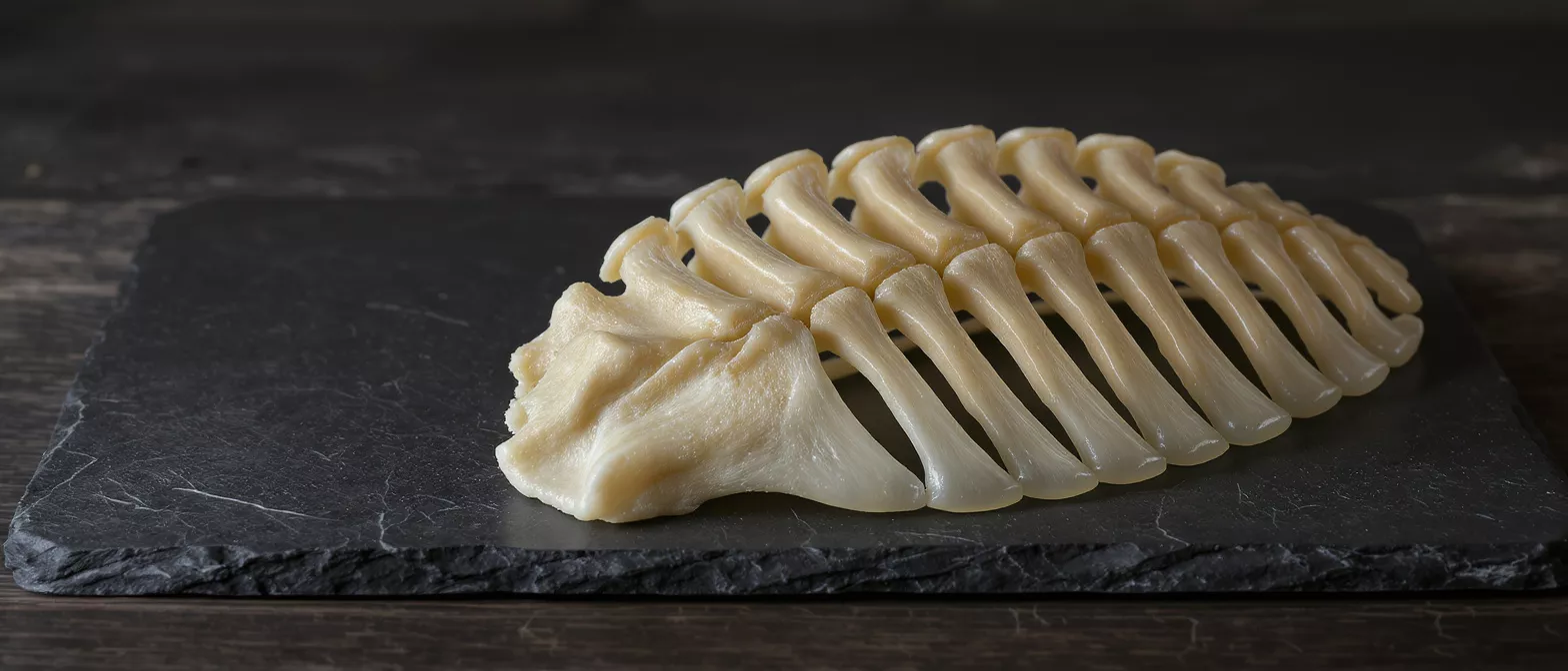
Costal Cartilage
Explore costal cartilage anatomy, function, and role in thoracic procedures, and learn how it impacts heart surgery.
What Is Costal Cartilage?
The costal cartilage is a tough, flexible connective tissue that attaches the front ends of your ribs to the sternum, or breastbone. If you are preparing for heart surgery or caring for someone who is, understanding this structure can help ease anxiety about the chest wall. The term costal cartilage refers to the smooth, resilient bars of tissue that allow the rib cage to be both a protective shield and a flexible framework for breathing. You can read more in-depth details about the thoracic cartilage support structures in our comprehensive anatomy guide.
Role in the skeletal system: This tissue acts as a crucial bridge between bone and the central chest structure. It creates a semi-rigid connection that absorbs shock and movement. Without this connection, the rib cage would be entirely solid bone, making breathing difficult and the chest highly vulnerable to fractures.
From a surgical standpoint, this anatomy matters deeply. When a cardiac surgeon operates, they need to access the heart by managing the chest wall. Knowing how this tissue behaves helps surgeons plan their approach, minimize trauma, and ensure the chest heals securely afterward.
How Costal Cartilage Function Supports the Rib Cage
The costal cartilage function is primarily about elasticity and structural support. Every time you breathe, your ribs expand and contract. The cartilage at the front of the chest allows the lower ribs to flare outward and upward, accommodating lung expansion without tearing or breaking. For a patient recovering from surgery, preserving this flexibility is vital for restoring normal respiration.
Why the rib cage needs cartilage: Pure bone is strong but brittle. The rib cartilage structure provides the necessary give, preventing the ribs from snapping during sudden impacts or intense physical exertion. It essentially functions as a natural shock absorber for the upper body.
- Resilience: Absorbs impact forces during chest compressions or minor trauma.
- Expansion: Enables rib cage flexibility required for deep breathing.
- Protection: Maintains the structural integrity of the thoracic cavity to shield vital organs.
Understanding this dual role helps patients appreciate why surgical techniques are designed to protect or carefully reconstruct these areas. A well-healed chest wall ensures the heart remains protected while the lungs recover their full capacity.
Costal Cartilage Anatomy and the Costochondral Junction
Examining costal cartilage anatomy reveals how the chest wall is assembled. The costochondral junction anatomy is the specific region where the hard rib bone meets the softer, flexible cartilage. This transition zone is uniquely important because it acts as a growth plate in younger patients and a stress distributor in adults.
Anatomical positioning: The first seven ribs connect directly to the sternum via their own costal cartilage segments, forming the true ribs. The next three ribs share a connection to the cartilage of the rib above them. This layered network provides comprehensive, overlapping protection for the heart and lungs.
For a heart surgeon, navigating this anatomy is a daily reality. The way the cartilage articulates with the sternum dictates how the chest is opened and how it is closed. A thorough understanding of the costochondral junction ensures that when wires or sutures are placed to close the chest, they hold firmly in both bone and cartilage.
What Causes Costal Cartilage Pain and Injury?
Experiencing costal cartilage pain can be frightening, especially when it occurs near the heart. The most common cause of this discomfort is costochondritis, an inflammation of the cartilage connecting the ribs to the breastbone. While the sharp, aching pain can mimic cardiac symptoms, it is typically musculoskeletal and treatable.
A costal cartilage injury can occur from direct chest trauma, severe coughing, or the physical stress of surgery. Surgical impact on cartilage: During a sternotomy, the surgeon must cut through the sternum and separate the surrounding tissues, which inevitably stresses the costal cartilage. Postoperative pain often stems from the inflammation of these manipulated tissues as they heal.
It is crucial to differentiate between surgical recovery pain and a new cartilage injury. Patients should communicate any new or worsening chest pain to their medical team. Proper diagnosis ensures that pain management is targeted effectively, allowing for a smooth recovery without unnecessary complications.
How Costal Cartilage Imaging Helps Diagnose Thoracic Issues
Diagnosing chest wall problems requires advanced techniques. Costal cartilage imaging involves specialized medical scans to visualize the soft tissues of the rib cage. Because standard X-rays are designed to highlight bone, they often miss cartilage issues entirely.
Distinguishing cartilage from bone on scans: Doctors typically rely on CT scans or MRIs to evaluate the cartilage. An MRI can reveal inflammation, tears, or a subtle costal cartilage fracture that would remain invisible on a standard chest X-ray. For postoperative patients, these scans help verify that the chest wall is healing correctly and that no structural gaps have developed.
Effective imaging provides peace of mind. When a patient reports lingering chest discomfort months after surgery, a detailed scan can confirm whether the issue is simply slow-healing cartilage or something requiring further intervention. Clear pictures lead to precise diagnoses and better outcomes.
Costal Cartilage Biomechanics and Flexible Thoracic Protection
The study of costal cartilage biomechanics explores how this tissue handles physical forces. Every breath, cough, or upper body movement generates mechanical stress. The cartilage’s unique cellular composition allows it to bend and twist without losing its original shape, a property known as elastic recoil. You can learn about the science behind flexible chest wall grafts through our advanced clinical resources.
Stress distribution during movement: When you lift something heavy, the force transfers from your arms to your shoulders, and ultimately to your rib cage. The cartilage distributes this pressure evenly, preventing localized stress from cracking the sternum or ribs. This mechanical buffer is essential for everyday physical function.
In the context of cardiac surgery, preserving biomechanical integrity is a top priority. Surgeons aim to restore the chest to its original dynamic state. A rigid chest wall limits breathing and physical activity, while a properly repaired one restores the natural, protective flexibility of the thorax.
Pediatric Costal Cartilage and Developing Chest Structures
When children require heart surgery, their anatomy requires special consideration. Pediatric costal cartilage is highly elastic and contains active growth centers. Because children’s chest walls are still developing, the way surgeons manage these tissues can impact future growth.
Growth plate importance: The costochondral junction acts as a growth plate, allowing the rib to lengthen as the child grows. Disrupting this area during early childhood surgeries must be done with extreme care. Surgeons use techniques that minimize trauma to these zones, ensuring the chest expands normally as the child ages.
Parents should know that pediatric cardiac surgeons are specially trained to navigate these growing structures. The priority is not just fixing the immediate heart condition, but ensuring the child develops a strong, functional chest wall that accommodates a growing heart and expanding lungs.
Costal Cartilage Transplantation in Cardiothoracic Surgery
In complex reconstructive cases, surgeons sometimes use costal cartilage transplantation. This involves harvesting a patient’s own rib cartilage to rebuild or support structures within the chest or airway. Because it is the patient’s own tissue, the risk of rejection is incredibly low.
Reconstructive applications: While less common in standard adult coronary bypasses, cartilage grafting is highly valuable in pediatric cardiac surgery. Surgeons may use a patient's own cartilage to reconstruct damaged or underdeveloped airways, or to provide structural support to a malformed chest wall.
Understanding these advanced techniques can be reassuring for families facing complex diagnoses. It highlights the versatility of the body’s tissues and the innovative ways cardiac surgeons use a patient’s own anatomy to heal and rebuild vital structures safely.
Are Costal Cartilage Deformities a Concern After Heart Surgery?
Patients often wonder if their chest will look or feel different after surgery. Costal cartilage deformities can occur if the healing process is disrupted, leading to a sunken chest or a protruding breastbone. These changes are more common in pediatric patients but can happen in adults.
Recognizing postoperative changes: If the cartilage heals asymmetrically, or if the sternum shifts during recovery, the chest wall can change shape. Careful monitoring, especially in the first few months post-surgery, is essential. Surgeons use supportive measures to ensure the bones and cartilage knit together perfectly flat.
Addressing deformities early is the best approach. Minor irregularities often resolve with physical therapy and time, while more significant shifts might require corrective bracing or a minor follow-up procedure. Patients should discuss any visible changes with their cardiologist immediately.
Costal Cartilage Regeneration and Long-Term Healing
The healing process after chest surgery is a marathon, not a sprint. Costal cartilage regeneration and repair occur slowly because cartilage lacks a rich blood supply. Unlike skin, which heals in weeks, cartilage heals over months, relying on surrounding tissues to deliver nutrients.
Timeline for recovery: Most patients feel significantly better six to eight weeks after surgery, but the deep structural healing continues for up to a year. The cartilage slowly remodels and strengthens, ensuring the chest wall becomes entirely stable. During this time, following restrictions on heavy lifting is critical to prevent setbacks.
Ultimately, understanding the resilience and healing capacity of the thorax provides immense comfort. The body is remarkably adept at rebuilding these critical structures. With proper care and patience, the chest wall will return to full strength, protecting the heart and allowing for a full, active life.






