

Doppler Echocardiography
Doppler echocardiography shows blood flow through your heart’s valves and chambers.
What is Doppler Echocardiography
Understanding the doppler echocardiogram meaning: Doppler echocardiography is a specialized, completely non-invasive ultrasound test used to evaluate how blood flows through your heart's chambers and valves. By using high-frequency sound waves, this imaging technique creates detailed, real-time pictures of the heart's internal structures and captures the speed and direction of circulating blood. For a patient preparing for or recovering from cardiac surgery, it provides your surgical team with critical information about your heart's mechanical function without making a single incision.
The basic principle of the test: The technology relies on the Doppler effect, a phenomenon where sound waves change pitch as they bounce off moving objects. When ultrasound waves reflect off red blood cells moving through the heart, the machine calculates their velocity. Faster blood flow produces a higher frequency shift, while slower flow produces a lower shift, allowing cardiologists to determine exactly how well your heart is pumping.
Understanding what this test measures helps alleviate anxiety when approaching cardiac diagnostics.
How does a Doppler Heart Test Work
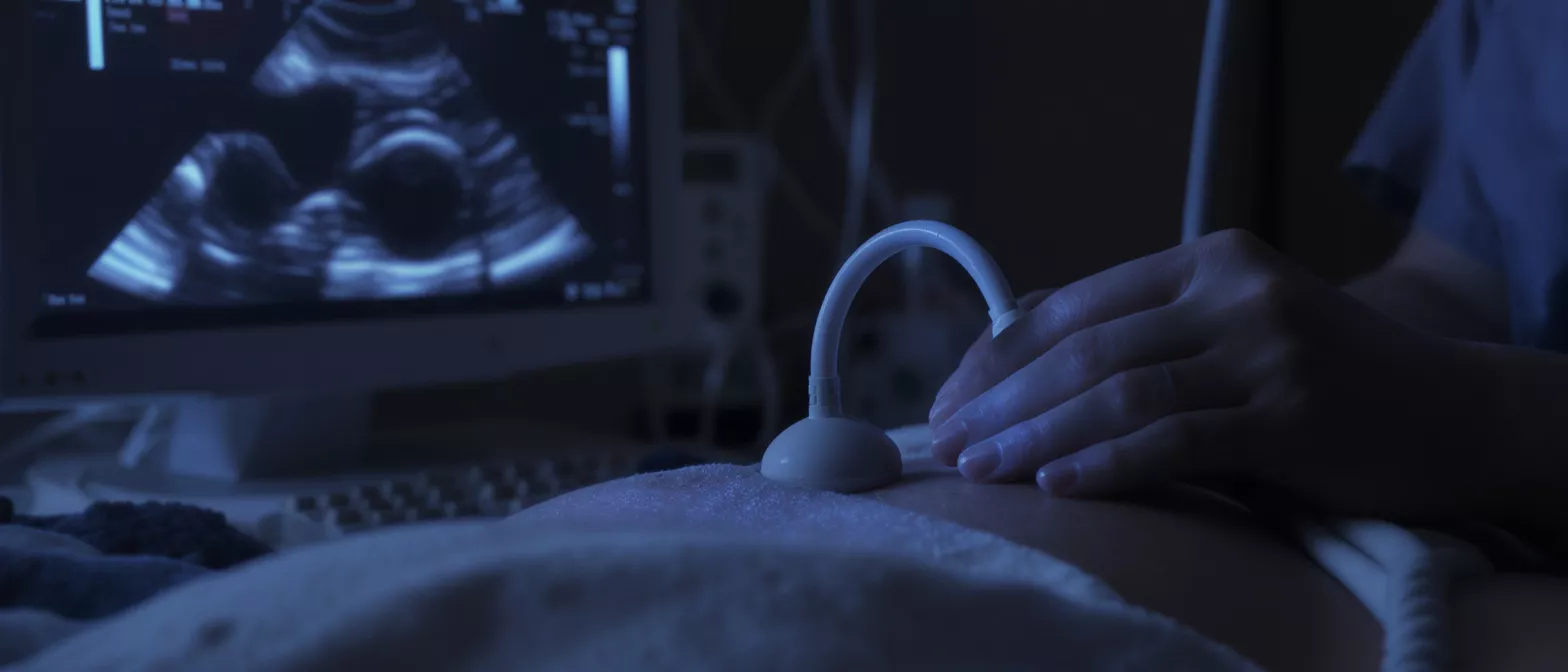
The role of sound waves: During a Doppler heart test, a trained sonographer places a small handheld device called a transducer against your chest. This device emits brief pulses of ultrasound that travel harmlessly through the skin and ribcage. When these waves encounter solid structures like heart muscle or moving blood cells, they bounce back to the transducer, which translates the returning echoes into dynamic visual images on a monitor.
Measuring blood flow velocity: The true strength of echocardiography doppler lies in its ability to measure the exact speed and turbulence of blood flow. By analyzing the frequency shifts of the returning sound waves, the system maps out areas of normal flow and regions where blood might be moving too quickly, which can indicate a narrowed valve or an obstructed vessel.
The procedure relies entirely on sound energy rather than ionizing radiation, making it remarkably safe for patients of all ages. The real-time nature of the imaging allows your medical team to watch the heart cycle through its pumping and relaxing phases, noting any structural abnormalities immediately as they occur.
Why is Cardiac Blood Flow Imaging Important
Identifying valve disease: Cardiac blood flow imaging is a critical tool for diagnosing valvular stenosis, regurgitation, or structural defects. If a valve is too narrow, the blood must accelerate rapidly to push through, creating turbulent flow patterns that the Doppler test easily detects. Recognizing these patterns early allows your surgical team to determine if a valve repair or replacement is necessary.
Measuring chamber pressures: The speed of the blood flow is directly related to the pressure differences inside the heart's chambers. By calculating the peak velocity of the blood using Doppler ultrasound, cardiologists can accurately estimate pulmonary artery pressures and ventricular function, providing crucial data for pre-surgical risk assessment and post-surgical recovery monitoring.
Before any heart surgery, establishing a clear baseline of your cardiovascular function is essential. Using precision heart valve diagnostic tools ensures that your surgical team has exact measurements of your cardiac anatomy and hemodynamics, forming a reliable roadmap for the operating room and tracking your progress throughout recovery.
What to Expect During a Doppler Echo Procedure
Understanding the physical experience of the Doppler echo procedure helps significantly reduce patient anxiety before an appointment. The test typically takes place in a darkened, quiet room to help the technician see the monitor clearly. You will lie comfortably on an examination table, usually positioned on your left side to bring your heart closer to the chest wall, and you may be asked to change positions slightly or hold your breath briefly to capture the clearest images.
The ultrasound gel and transducer: The technician will apply a warm, water-based gel to your chest. This gel eliminates the tiny air gap between the transducer and your skin, allowing the sound waves to transmit efficiently. You will feel slight pressure as the technician presses the probe against your chest to get the correct angle on your heart, but the sensation is not painful.
- The test is completely painless and involves no needles or injections.
- A standard resting Doppler echocardiogram typically takes between 30 to 60 minutes.
- You can resume all normal daily activities immediately after the test is finished.
Color Doppler Echocardiography Explained
Visualizing flow direction: Color Doppler echocardiography takes the numerical data of blood flow velocity and translates it into a highly intuitive visual map. The computer assigns colors to represent the direction of the moving blood relative to the ultrasound probe. Traditionally, red indicates blood flowing toward the transducer, while blue indicates blood flowing away from it, providing an immediate, clear picture of circulatory dynamics.
Detecting abnormal turbulence: When blood flow becomes turbulent, such as when it leaks backward through a damaged valve, the smooth color pattern breaks down into a mosaic of mixed colors. This visual disturbance immediately alerts the cardiologist to potential anatomical problems that might require surgical correction, such as valve regurgitation or an unexpected hole in the heart wall.
This color mapping feature is what makes the test so powerful for rapid diagnostics. Instead of trying to interpret raw waveform data alone, surgeons and cardiologists can instantly see the exact location and severity of a leak or blockage, making the pre-operative planning process much more precise.
Heart Valve Doppler Assessments
Evaluating stenosis and regurgitation: Heart valve doppler assessments are specifically designed to measure the severity of valve disease. When a valve becomes stenotic, or narrowed, the Doppler probe records abnormally high blood velocities as the fluid is forced through a smaller opening. Conversely, if a valve fails to close properly, the test captures blood leaking backward, a condition known as regurgitation.
Calculating valve area: By applying specific mathematical formulas to the velocity data captured during the echocardiogram, your cardiology team can estimate the exact physical opening size of the valve. This calculation is vital for surgeons deciding whether a valve can be safely repaired or if it requires a complete replacement. This is why accurate cardiac doppler ultrasound is so heavily relied upon.
For patients presenting with heart murmurs or symptoms like shortness of breath, this specific assessment provides the definitive diagnosis. Utilizing non-invasive cardiac imaging technology eliminates the need for exploratory procedures, ensuring that patients only undergo surgical intervention when strictly necessary.
How to Read Doppler Echo Test Results
Interpreting Doppler echo test results requires specialized medical training, but understanding a few key terms helps patients actively participate in their own care. The report will typically describe the ejection fraction, which is the percentage of blood pushed out of the left ventricle with each heartbeat, along with specific measurements of blood velocity across each of the four heart valves. A normal ejection fraction generally falls between 50 and 70 percent, indicating a healthy, strong pumping function.
Recognizing peak gradients: The results will list pressure gradients, measured in millimeters of mercury (mmHg), which indicate the pressure difference across a heart valve. A higher peak gradient means the valve is significantly narrowed, forcing the heart to work much harder to pump blood through. Your surgeon will use these specific numbers to determine the urgency and type of intervention required.
Always review your final results directly with your cardiologist or cardiac surgeon. They can correlate the numerical data from the test with your physical symptoms, ensuring that the diagnostic picture accurately reflects your overall cardiac health and readiness for any proposed surgical treatment.
Benefits of Non-Invasive Cardiac Imaging
Zero radiation exposure: One of the most significant benefits of non-invasive cardiac imaging is the complete absence of ionizing radiation. Unlike CT scans or traditional angiograms that require X-rays, Doppler echocardiography relies entirely on harmless sound waves, making it safe for repeated use over time, which is ideal for closely monitoring a patient's recovery after heart surgery.
Real-time dynamic evaluation: Because Doppler imaging shows the heart actively beating and pumping blood in real time, it captures functional issues that static images would miss entirely. It allows surgeons to evaluate how well the cardiac muscle contracts, how the valves open and close dynamically, and how efficiently blood circulates with every single heartbeat cycle.
- Completely safe for patients of all ages, including pregnant women if absolutely necessary.
- Highly effective for diagnosing structural defects, valve failures, and chamber enlargement.
- Ideal for tracking long-term postoperative recovery without physical stress or risk.
Is a Doppler Echocardiogram Safe
No known biological risks: A Doppler echocardiogram is widely recognized as one of the safest medical imaging procedures available today. The ultrasound frequencies used for cardiac diagnostics have no known harmful biological effects on human tissue. There is no radiation, no contrast dye to stress the kidneys, and no invasive penetration of the skin, which virtually eliminates the risk of infection or bleeding complications.
Comfort and patient experience: The safety profile extends to patient comfort during the examination. The only physical sensation is the gentle pressure of the transducer probe and the warmth of the conductive gel on the chest. There are no side effects following the test, meaning you can drive yourself home, return to work, or continue your normal daily routine immediately after leaving the clinic.
For heart surgery patients who require multiple follow-up appointments, this exceptional safety record is invaluable. Your surgical team can perform repeated echocardiography checks at various stages of your recovery to ensure your heart is healing properly, all without exposing you to any cumulative physical risk.
Doppler Echocardiography in Cardiac Diagnostics
In the broader landscape of cardiac diagnostics, Doppler echocardiography serves as the foundational pillar for evaluating mechanical heart function. While electrocardiograms (ECGs) measure the electrical activity of the heart, an echocardiogram reveals exactly how well that electrical activity translates into physical pumping action. This distinction is crucial for surgical planning, as a heart might have a normal rhythm on an ECG but still suffer from severely compromised mechanical output due to valve disease.
Guiding surgical decisions: Before opening the chest, a surgeon needs a precise understanding of the internal landscape. Doppler imaging maps out the exact degree of valve damage, the size of the cardiac chambers, and the presence of any abnormal communications between the right and left sides of the heart, reducing unexpected surprises during the operation.
Monitoring long-term recovery: The utility of this technology extends far beyond the operating room. In the weeks and months following cardiac surgery, regular Doppler assessments allow your medical team to verify that repaired valves are functioning properly, artificial valves are opening fully, and overall heart function is steadily returning to a healthy baseline.






