

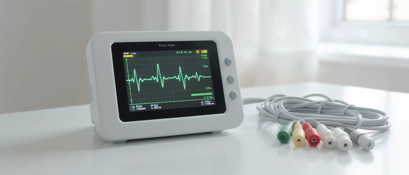
Electrocardiogram
Learn how the electrocardiogram works and why it matters for heart surgery prep.
What is an electrocardiogram?
An electrocardiogram, commonly referred to as an ECG test, is a straightforward and painless medical examination that records the electrical activity of your heart. Every time your heart beats, it generates small electrical signals that travel through the muscle tissue, causing it to contract and pump blood throughout your body. The electrocardiogram captures these signals and translates them into a visual graph displayed on a monitor or printed on paper. This allows your cardiac care team to assess how well your heart is functioning and to diagnose a wide variety of potential conditions with precise accuracy.
For patients preparing for cardiac procedures, understanding this diagnostic tool helps ease anxiety. What the heart electrical activity test reveals is a moment-by-moment picture of your heart's rhythm, showing how fast or slow it is beating and whether the electrical impulses are traveling through the correct pathways. It gives your surgeon a baseline understanding of your heart health before surgery, ensuring they can tailor their approach to your specific physiological needs.
This diagnostic tool is frequently used in routine physical exams, emergency departments, and pre-operative assessments. Because it provides immediate feedback regarding heart rhythm and can identify damage to the heart muscle, it remains an essential cornerstone of cardiology. Your surgeon relies on this quick snapshot to make informed decisions regarding your overall care plan.
How does an ECG test work?
The electrocardiogram operates on a simple principle: detecting the tiny electrical impulses that naturally trigger your heartbeats. During the test, a trained technician places small, flat patches called electrodes on the skin of your chest, arms, and legs. These sensors are connected by wires to an electrocardiogram machine, which measures the electrical signals produced with each heartbeat. The machine amplifies these signals and draws them as a series of waves on a moving strip of paper or a digital screen.
How cardiac monitoring test tracks heart waves is by measuring the timing and duration of each electrical phase. The first wave represents the upper chambers of the heart, known as the atria, squeezing to push blood into the lower chambers. The next, larger wave corresponds to the ventricles, which are the main pumping chambers, pushing blood out to the lungs and the rest of the body. A final wave shows the heart's electrical system resetting for the next beat.
By looking at the spacing between these waves, your cardiologist can determine if the electrical pathway is normal or if there is a delay. Different leads or sensors provide different vantage points of the heart, creating a comprehensive twelve-dimensional view of your heart's activity from just a few minutes of recording. If any patterns look unusual, further testing is often recommended to pinpoint the exact cause.
Why is an electrocardiogram important before heart surgery?
Before any cardiac procedure, your surgical team needs a complete and accurate picture of your heart's condition. An electrocardiogram is a critical first step in that evaluation process. It reveals silent issues such as arrhythmias, which are irregular heartbeats that might complicate anesthesia or surgery. It can also show evidence of a previous heart attack or inadequate blood flow to the heart muscle, factors that heavily influence surgical planning.
Identifying heart rhythm test risks early ensures your surgeon, such as Dr Alejandro Molina, can mitigate complications before they occur. If your heart is already struggling with an abnormal rhythm, additional medications or a different surgical approach might be necessary to keep you safe. Finding these details out beforehand allows the entire surgical team to be fully prepared.
Furthermore, this test serves as a baseline for monitoring your heart throughout your hospital stay. By comparing electrocardiogram results taken before, during, and after surgery, doctors can immediately detect if any new changes occur in your heart's electrical system. This continuous vigilance is a major reason why modern cardiac surgery is so remarkably safe and effective today.
ECG vs EKG: is there a difference?
Many patients feel confused when they hear both terms used interchangeably. The distinction is entirely based on language translation rather than medical function. ECG stands for electrocardiogram, derived from the original English spelling, while EKG is an abbreviation of the German word, elektrokardiogramm. In the early days of cardiology, medical professionals adopted the German abbreviation to avoid confusing it with another similar-sounding medical term. Both acronyms refer to the exact same heart electrical activity test.
Why medical teams use both abbreviations simply comes down to habit and regional preference. Whether your doctor writes ECG or EKG on your chart, the procedure, the equipment, and the resulting wave patterns are exactly identical. You do not need to prepare differently or worry about which abbreviation is used on your medical paperwork.
Knowing this distinction can help reduce unnecessary worry for patients researching their upcoming procedures. If you see either term on your pre-operative instructions or consent forms, you can rest assured that you are simply undergoing a standard, non-invasive cardiac monitoring test. When seeking an electrocardiogram near me, you can confidently use either abbreviation knowing you are requesting the right procedure.
What to expect during an electrocardiogram procedure
Knowing exactly what will happen during your electrocardiogram procedure can greatly reduce any pre-test anxiety. The test takes place in a quiet room, usually in a doctor's office or a hospital outpatient clinic. You will be asked to lie flat on an examination table while a technician places up to twelve small electrodes on your chest, arms, and legs. The patches use a light adhesive and sometimes a small amount of conductive gel to ensure a clear reading.
How electrocardiogram procedure is performed requires you to remain very still and breathe normally while the machine records. Even talking or shivering can interfere with the sensitive sensors and blur the wave patterns. The recording itself takes only a few seconds, although preparing the skin and positioning the electrodes takes about five to ten minutes from start to finish.
It is important to remember that no electricity is passed through your body during this test. The electrodes are strictly passive receivers that listen to your heart's natural electrical output. Once the recording is complete, the technician will remove the patches, wipe away the gel, and you can return to your normal activities immediately without any downtime.
Is the ECG test safe and painful?
Patients frequently ask if the electrocardiogram is safe and if it causes any discomfort. The test is completely safe for people of all ages, including pregnant women and elderly individuals. Because the machine only records the heart's electrical signals and does not emit any electrical current into the body, there is zero risk of electrical shock or harmful side effects. It is one of the safest diagnostic tools in modern medicine.
Are electrocardiogram tests uncomfortable? The examination is entirely painless. The most common sensation patients report is the cool feeling of the conductive gel on the skin and the slight pull of the sticky patches when they are removed, similar to peeling off a bandage. If you have sensitive skin, you might experience very mild redness where the patches were placed, but this fades quickly.
Because it carries no inherent risks, the test can be repeated as many times as necessary without causing any harm. This makes it an ideal tool for ongoing cardiac monitoring and routine postoperative checkups to ensure your recovery is progressing exactly as expected after heart surgery.
How to read electrocardiogram results
Electrocardiogram interpretation is a highly specialized skill that takes years of medical training to master. The results appear as a graph with a series of spikes and valleys, representing the different phases of your heartbeat. Each wave is labeled with a letter and corresponds to a specific electrical event in the heart muscle. While it might look like a chaotic line to the untrained eye, these patterns tell a very clear story to a trained cardiologist.
Understanding basic electrocardiogram interpretation involves looking at the height, length, and spacing of these waves. For instance, if the space between the waves is too long, it indicates a slow heart rate, while very narrow spaces indicate a fast heart rate. If a wave is unusually tall or flat, it might suggest that a specific part of the heart muscle is enlarged or lacking proper blood flow.
Your surgical team will review these findings with you in detail. They will explain what your specific electrocardiogram results mean in the context of your overall heart health and upcoming surgery. Having a basic understanding of the process helps you feel more involved and informed during your medical consultations.
What causes an abnormal electrocardiogram?
Receiving news of an abnormal electrocardiogram can sound frightening, but it does not always indicate a severe medical emergency. There are many benign and highly treatable reasons why a reading might deviate from the standard. Sometimes, an abnormal reading is simply the result of a harmless variation in your heart's anatomy, or it can be triggered by an electrolyte imbalance in your blood, such as low potassium levels.
Common reasons for abnormal electrocardiogram findings include rhythm irregularities like atrial fibrillation, where the upper chambers beat out of sync with the lower chambers. It can also detect signs of an enlarged heart, inflammation of the heart lining, or evidence of a past silent heart attack that occurred without obvious symptoms. Even certain medications can alter your heart's electrical patterns temporarily.
- Electrolyte imbalances: Low potassium or calcium levels can disrupt electrical signals.
- Arrhythmias: Extra beats or skipped beats are often harmless but require monitoring.
- Medication side effects: Some prescriptions alter heart rhythms without indicating permanent damage.
- Ischemia: Reduced blood flow to the heart muscle, which may require further imaging tests.
If your results are abnormal, your doctor will likely order additional testing, such as an echocardiogram or a stress test, to gather more information before making a final surgical decision. These can complement a prescribed regimen but should always be discussed with your cardiologist first.
ECG test cost and accessibility in Guadalajara
The ECG test cost in Guadalajara is highly competitive compared to many other international medical hubs. Because it is a quick, non-invasive procedure requiring minimal equipment overhead, clinics and hospitals can offer this essential screening at a very accessible price point. This financial accessibility makes it easier for patients to get the baseline evaluations they need before undergoing major heart surgery.
Finding an electrocardiogram specialist in Guadalajara ensures you are receiving top-tier medical care without the extreme financial burden. The city has become a leading destination for medical tourism, particularly for complex cardiology and cardiac surgery. You will find modern facilities equipped with the latest technology and staffed by bilingual, internationally trained professionals who understand the needs of international patients.
Taking a proactive approach to your health by scheduling your cardiac evaluation today is the first step toward a healthier, more active life.
Key takeaways about your heart rhythm test
Understanding the electrocardiogram helps demystify the cardiac surgery preparation process. It is a vital, painless, and incredibly fast diagnostic tool that paints a real-time picture of your heart's electrical system. Whether your doctor is looking for a baseline measurement before surgery or monitoring your recovery after a procedure, the ECG provides the essential data needed to ensure your heart is beating strongly and safely.
What to remember about your heart rhythm test is that it is a tool for safety, not something to fear. It allows your surgical team to catch hidden irregularities, tailor your anesthesia plan, and monitor your heart health with precise detail. If you or a loved one is facing heart surgery, knowing what to expect from this test can bring immense peace of mind.
- Painless and fast: The entire procedure takes under ten minutes and causes no discomfort.
- Essential for surgery: Provides a critical baseline for your cardiac team to ensure surgical safety.
- Safe for everyone: No electrical currents are put into the body; it only listens to your heart.
- Early detection: Identifies silent arrhythmias, previous attacks, or blood flow issues before they become emergencies.
If you are seeking expert cardiac care, finding an experienced heart surgeon in Guadalajara is the most important step you can take. If you have symptoms, discuss them with your doctor, who can arrange the right evaluation. Your heart deserves expert attention, and an ECG is the perfect starting point.






