

In-stent Restenosis
Learn about in-stent restenosis, why arteries re-narrow after stent placement, symptoms, and treatment options.
What is in-stent restenosis
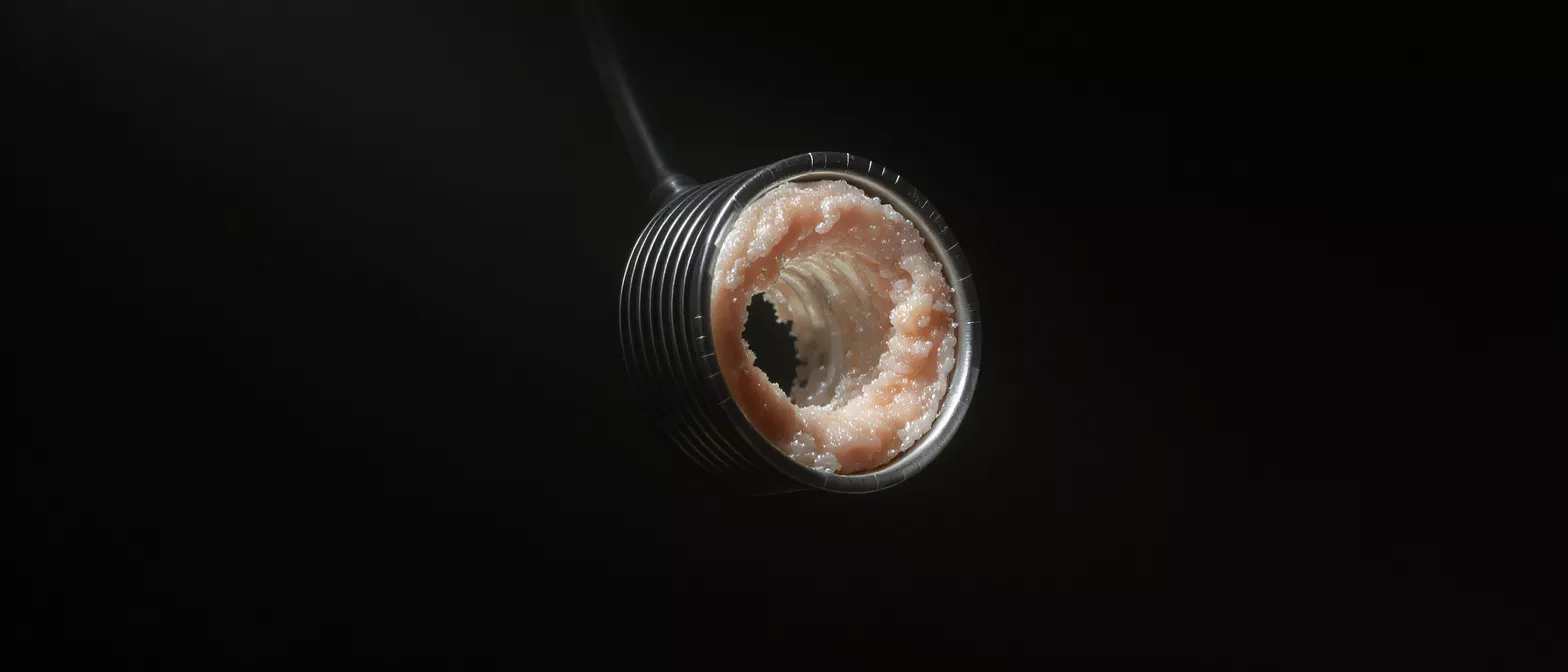
When a patient undergoes an angioplasty to open a blocked heart artery, a tiny metal mesh tube called a stent is almost always placed to keep the vessel propped open. In-stent restenosis refers to the re-narrowing of the artery inside that implanted stent. This occurs when tissue growth builds up within the stent over time, restricting blood flow to the heart muscle again. For patients recovering from heart surgery or an interventional procedure, understanding this condition is crucial because it is the most common reason a previously treated artery might fail.
Why scar tissue forms inside a stent: The body views a stent as a foreign object. In response to the stent's presence and the pressure of the balloon angioplasty that placed it, the arterial wall triggers a healing response. This involves white blood cells and muscle cells migrating to the area, creating a layer of tissue. If this tissue overgrows, it narrows the channel through the stent.
It is natural for patients and families to feel anxious when they hear that a stent might re-block. The condition is generally slow-growing rather than a sudden, life-threatening event. Recognizing the signs early makes it highly manageable, and cardiologists have excellent tools to resolve the narrowing and restore healthy blood flow.
How restenosis after stent placement happens
The biological process behind stent reblockage is called neointimal hyperplasia. When the balloon catheter expands the stent against the artery wall, it inevitably stretches the tissue and causes microscopic injury. The body responds by trying to heal that injury. Smooth muscle cells from the middle layer of the artery wall multiply and migrate outward into the stent struts. As they collect, they form a thick layer of fibrous tissue that gradually fills the empty space inside the metal mesh.
Healing response gone into overdrive: Think of this healing process like a thick scar forming on the skin after a deep cut. Inside the heart artery, a thin, smooth layer of new tissue is normal and helps the artery heal smoothly over the metal. However, if the healing mechanism overcompensates, the resulting internal scar becomes too thick, physically blocking the path for blood.
Unlike atherosclerosis, which is driven by fatty cholesterol plaques building up over decades, restenosis is entirely a tissue-healing phenomenon. It is not caused by poor diet or a sudden rupture of a plaque. Because the cause is mechanical and biological rather than purely dietary, the management approach requires distinct interventions.
Causes of coronary in-stent restenosis
Several distinct factors can drive the development of coronary in-stent restenosis. The primary cause is the arterial wall's exaggerated healing response to the physical stretch of the stent. When the balloon expands the stent to flatten a cholesterol blockage, it causes controlled trauma to the vessel. The body attempts to repair this trauma by growing smooth muscle cells to cover the exposed metal struts.
Inflammation and mechanical stress: The body's immune system reacts to the stent as a foreign material, creating chronic, low-grade inflammation. Additionally, if the stent is undersized for the artery, or if it expands unevenly against the vessel wall, the turbulence of blood flowing past the irregular surface can stimulate further tissue growth.
Patient-specific biological factors also weigh heavily on the risk of stent reblockage. People with diabetes, for instance, often have an overactive tissue healing response that makes cellular overgrowth more aggressive. Similarly, patients with complex anatomy, such as very small arteries or long, branching blockages, face a higher probability of restenosis due to the technical difficulty of achieving perfect stent apposition against the vessel walls.
Restenosis symptoms to watch for
Identifying restenosis symptoms early is essential for timely treatment. Because the re-narrowing develops gradually as tissue grows, symptoms typically return slowly rather than striking suddenly. Patients often notice a familiar sensation they experienced before their original procedure: chest pressure or angina, especially during physical exertion or emotional stress.
Returning angina and chest pressure: The most common warning sign is exertional chest pain that stops when resting. You might feel a squeezing tightness in the center of the chest or pain radiating to the jaw, shoulders, or left arm. This occurs because the heart muscle is starved of oxygen when the narrowed stent cannot deliver enough blood during increased demand.
Other indicators include unexplained shortness of breath or a noticeable decrease in exercise tolerance. If you could comfortably walk a mile a few months after your procedure but suddenly find yourself winded after half a mile, it is a signal to contact your cardiologist. It is worth noting that severe, crushing chest pain at rest is not typical of restenosis and points instead to acute plaque rupture, requiring emergency medical attention.
Drug-eluting stent restenosis vs bare metal stent restenosis
The type of stent originally placed significantly influences the likelihood of narrowing. Years ago, cardiologists used bare metal stents, which acted simply as scaffolds. These older bare metal stent restenosis rates were notoriously high, often affecting up to a quarter of all patients. To combat this, engineers developed drug-eluting stents coated with slow-release medications designed to stop the local tissue from overgrowing.
How modern coatings suppress tissue growth: Drug-eluting stents release anti-proliferative medications directly into the arterial wall over several weeks. These drugs safely pause the rapid multiplication of smooth muscle cells, allowing the artery to heal with only a thin, healthy tissue layer. Today, drug-eluting stent restenosis is rare, occurring in less than ten percent of cases.
If you are preparing for an intervention, it is natural to worry about which hardware will provide the safest, longest-lasting result for your heart.
Restenosis risk factors
Understanding restenosis risk factors helps patients and their care teams take preventive action. While the mechanical stretch of the artery is the trigger, certain biological conditions make the healing response more aggressive. Identifying these risks early allows cardiologists to tailor their approach during the initial procedure.
Diabetes and vascular resistance: Diabetic patients have higher insulin levels and vascular inflammation, which act as powerful fertilizers for smooth muscle cell growth inside the stent. Because of this, diabetic vessels are highly prone to re-narrowing, requiring specialized stent strategies and meticulous medication management.
- Diabetes: Promotes aggressive tissue growth inside the stent struts.
- Small vessel size: Arteries less than 3mm limit blood flow and increase reblockage risk.
- Complex lesions: Long blockages or branching vessels create uneven stent expansion.
- Smoking: Ongoing tobacco use severely impairs arterial healing and promotes inflammation.
Other factors include long overlapping stent segments, stents that are under-expanded during the initial placement, or a patient's failure to take prescribed blood thinners. Each of these elements alters the local blood dynamics, creating the perfect environment for scar tissue to accumulate and narrow the newly opened channel.
How stent restenosis treatment works
If restenosis does occur, cardiologists have highly effective interventions to restore flow without needing open-heart surgery. The approach for stent reblockage treatment usually involves a repeat angiogram to visualize the narrowed stent. Once the area is located, specialized wires and balloons are threaded through the arteries to physically compress the built-up tissue against the stent walls.
Techniques to clear reblocked stents: Doctors use cutting balloons or rotational atherectomy devices to gently shave away the excess tissue inside the old stent. Once cleared, a new drug-eluting stent is often placed inside the old one to provide a fresh dose of tissue-suppressing medication. In some complex cases, drug-coated balloons are expanded inside the blockage, delivering anti-growth medicine before being removed.
For patients researching options, specialized intracoronary drug-coated balloons represent a modern, hardware-free option that leaves behind medication to prevent scar regrowth without adding extra metal layers inside the artery. Your surgeon will decide which tool best fits the specific anatomy of your narrowed stent.
How to prevent stent restenosis
While you cannot change the biological reality of how arteries heal, you can take meaningful steps to prevent stent restenosis. The most critical action a patient can take is strictly adhering to their prescribed dual antiplatelet therapy. These blood thinners ensure blood flows smoothly through the new stent, preventing clots and reducing the turbulent friction that triggers tissue overgrowth.
Medication adherence and follow-up: Stopping aspirin or prescribed blood thinners prematurely disrupts the delicate healing process of the endothelium. Patients must take these medications for the exact duration prescribed by their cardiologist. Attending all scheduled follow-up appointments ensures any silent narrowing is caught before symptoms develop.
Controlling systemic conditions is equally vital. By strictly managing these vascular risk factors, patients reduce the inflammatory burden on their arteries, creating a much healthier environment for the stent to integrate permanently.
Is recurrent stenosis dangerous
It is understandable that patients worry about the risks of recurrent stenosis. The reassuring truth is that in-stent restenosis is generally a mechanical and biological issue, not an acute emergency. Because it involves the slow growth of scar tissue rather than the sudden rupture of a cholesterol plaque, it does not typically cause a massive heart attack without warning.
Gradual narrowing versus sudden blockage: Unlike a thrombosis, where a clot instantly seals an artery, restenosis acts like a slowly closing valve. The heart muscle gets gradually deprived of oxygen, causing predictable exertional angina. This slow progression gives patients and doctors ample time to schedule a planned repeat angioplasty to fix the issue safely.
However, ignoring the symptoms is dangerous. If left untreated, the restricted blood flow can eventually lead to a serious heart event or cause damage to the heart muscle from chronic oxygen deprivation. Any return of chest pain or breathlessness in a patient with a known stent must be evaluated by a medical professional immediately.
Living with stent narrowing long term
Recovering from a heart intervention and living with a stent requires a lifelong partnership with your cardiology team. The placement of a stent is not a permanent cure for coronary artery disease, but rather a mechanical fix for a localized blockage. The systemic condition driving the disease must be managed daily through a combination of medication, diet, and exercise.
Building heart-healthy habits: Patients who thrive long-term after a stent embrace their role as active participants in their care. This means tracking blood pressure, maintaining a heart-healthy diet, and participating in supervised exercise programs. It also involves reporting any unusual fatigue or chest sensations immediately rather than waiting for the next scheduled appointment.
Understanding the signs, risks, and treatments of in-stent restenosis empowers you to face your recovery with confidence. Knowing what to watch for removes the fear of the unknown. With modern drug-eluting technology and excellent treatment options available, even if a stent narrows, your cardiology team has the tools to keep your heart beating strongly for years to come.






