

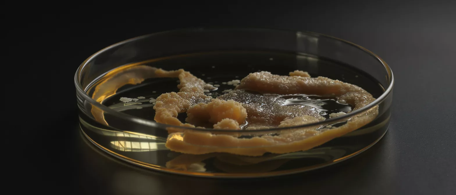
Infective Endocarditis
Learn about infective endocarditis, a serious heart valve infection, including symptoms, causes, diagnosis, and treatment options from cardiac specialists.
What is infective endocarditis?
Understanding the basics of a heart valve infection: Infective endocarditis is an infection of the endocardium, which is the smooth inner lining of the heart chambers and valves. When bacteria enter the bloodstream and settle on this delicate tissue, they can multiply and cause severe inflammation. This condition is a serious medical event that requires immediate attention and careful management by a specialized cardiac team.
For patients preparing for or recovering from heart surgery, understanding this condition is vital. The heart valves are mechanical gateways that ensure blood flows in one direction. When a valve becomes infected, it may develop lesions or growths—often called vegetations—that disrupt normal blood flow and compromise cardiac function.
Receiving a diagnosis can feel overwhelming, but modern cardiac care offers highly effective treatments. Dr Alejandro Molina and surgical teams specializing in heart conditions approach this infection with precision, using a combination of targeted antibiotic therapies and, when necessary, advanced surgical repair. The goal is always to clear the infection completely while preserving your heart's natural function and structural integrity.
What are endocarditis symptoms?
Recognizing acute endocarditis versus subacute endocarditis: The presentation of endocarditis symptoms largely depends on the specific bacteria involved and the patient's overall health. Acute endocarditis develops rapidly over a few days with high fever, chills, and severe fatigue. Subacute endocarditis progresses more slowly over weeks, presenting with mild fever, night sweats, and a general feeling of being unwell.
Symptoms can sometimes mimic common illnesses like the flu, making early detection challenging. A patient might experience joint pain, unexplained weight loss, or a new heart murmur. In some cases, tiny broken blood vessels may appear on the whites of the eyes or under the fingernails. If you or a loved one experience persistent fevers alongside a known heart condition, seeking prompt medical evaluation is essential.
- Fever and chills: Often the earliest and most common indicator of a systemic bloodstream infection.
- New heart murmur: A sound heard by a physician indicating altered blood flow due to valve damage.
- Vegetations: Bacterial growths on the heart valve that can break off and travel through the bloodstream.
What are endocarditis causes?
How daily bacteria reach the heart valves: The primary causes of this condition are bacteria that enter the bloodstream during everyday activities or medical procedures. Normally, your immune system clears these transient bacteria quickly. However, if a heart valve has a slight structural abnormality, the bacteria can attach to the roughened surface and form a protective microbial layer.
Once this layer is established, the bacteria multiply rapidly, effectively hiding from the body's natural defenses. The immune response causes severe local inflammation, which further damages the valve tissue. Over time, this cycle of infection and inflammation destroys the delicate valve leaflets, leading to leakage or stenosis, which requires cardiac surgical intervention.
The link between dental procedures and endocarditis: Routine dental work is a well-known entry point for oral bacteria. Simple actions like brushing, flossing, or professional cleanings can push streptococcus bacteria into the bloodstream. For patients with high-risk heart conditions, maintaining excellent oral hygiene and antibiotic prophylaxis before dental work are critical preventative steps.
How does bacterial endocarditis develop?
The formation of valve vegetation endocarditis: When bacteria adhere to the endocardium, they rapidly multiply and attract platelets and fibrin. This creates a mesh-like structure known as a vegetation. These growths are not just biological debris; they are active bacterial colonies encased in a protective shield, making them remarkably resilient to standard immune responses.
The physical presence of these vegetations alters the mechanical movement of the heart valve. As the mass grows, it prevents the valve from closing tightly or opening fully. This mechanical disruption forces the heart to work harder to pump blood, eventually leading to heart failure if the infection and structural damage are not addressed urgently by cardiac specialists.
Furthermore, pieces of these fragile vegetations can break off. When this happens, the emboli travel through the arteries, potentially blocking blood flow to vital organs such as the brain, kidneys, or lungs. This risk of embolization is why immediate diagnosis and treatment are critical to preventing permanent organ damage.
What are endocarditis risk factors?
Identifying congenital and acquired cardiac vulnerabilities: Patients with pre-existing heart conditions carry the highest endocarditis risk factors. This includes individuals with congenital heart defects, prosthetic heart valves, or a previous history of endocarditis. Damaged or artificial materials provide an ideal surface for bacteria to latch onto and multiply.
While structural heart issues are the primary concern, certain lifestyle factors and medical interventions also increase risk. Intravenous drug use is a major cause of acute right-sided heart infections, as needles introduce aggressive bacteria directly into the venous system. Additionally, having a central venous catheter or undergoing procedures involving the urinary or respiratory tracts can introduce bacteria into the bloodstream.
- Prosthetic valves: Artificial valves are highly susceptible to bacterial colonization within the first year after surgery.
- Congenital defects: Structural abnormalities, particularly unrepaired cyanotic heart disease, increase colonization risk.
- Healthcare exposure: Prolonged hospital stays and intravenous lines can serve as entry points for opportunistic bacteria.
How is heart valve infection diagnosed?
Confirming endocarditis blood culture results: The cornerstone of endocarditis diagnosis is taking multiple blood cultures. By drawing blood samples over a short period, the laboratory can identify the exact bacteria causing the infection. This step is crucial because it allows your medical team to select the most effective antibiotic rather than guessing blindly.
Using echocardiography for heart valve infection diagnosis: Blood tests alone cannot determine the extent of physical damage. An echocardiogram uses sound waves to create detailed images of the heart. This non-invasive test allows the cardiologist to visualize the valves, measure the size of any vegetations, and assess how much the infection is interfering with cardiac function.
In complex cases, a transesophageal echocardiogram (TEE) may be necessary. This involves passing a small probe down the esophagus to get a clearer, close-up view of the heart structures. Combining positive blood cultures with echocardiogram evidence gives Dr Alejandro Molina and the surgical team the precise anatomical information needed to plan medical or surgical treatment.
What is endocarditis treatment?
Administering endocarditis antibiotics: The first line of defense against this infection is high-dose, intravenous antibiotics administered in a hospital setting. Because the bacteria are protected within their vegetations, oral medications are not strong enough to penetrate the infection. Patients typically require several weeks of dedicated intravenous therapy to eradicate the bacteria completely.
During this time, the medical team monitors blood cultures daily to ensure the infection is clearing. They also perform follow-up echocardiograms to check the size of the vegetations. If the fever resolves, blood cultures become sterile, and the valve remains structurally sound, the patient may complete the antibiotic course at home under careful supervision.
However, antibiotics alone are not always sufficient. Infections caused by aggressive bacteria, particularly staphylococcus endocarditis, can destroy valve tissue faster than antibiotics can kill the bacteria. In these instances, relying solely on medication would lead to irreversible heart failure, making surgical intervention necessary to remove the infected tissue and restore cardiac function.
When is heart surgery needed for endocarditis?
Addressing heart failure from valve destruction: When the infection severely damages a heart valve, causing significant leakage, surgery becomes inevitable. The pressure of blood flowing backward into the heart chambers causes fluid to back up into the lungs. In these cases, surgical repair or replacement is performed to restore forward blood flow and relieve the strain on the heart muscle.
Managing uncontrolled infection and embolism risks: If the infection does not respond to intravenous antibiotics within a week, or if large vegetations are at high risk of breaking off and causing a stroke, urgent surgery is required. The surgeon must physically remove the infected tissue, clear the area, and implant a new biological or mechanical valve to secure the heart's function.
Undergoing heart surgery while actively fighting a severe infection is complex, but it is a routine scenario for specialized cardiac surgeons. They employ advanced techniques, sometimes using bioprosthetic heart valves for infection repair, to ensure the best long-term outcome. Post-operative care includes a tailored antibiotic regimen to ensure the infection does not return once the physical source has been removed.
What are endocarditis complications?
Preventing embolic stroke from endocarditis: One of the most feared endocarditis complications is an embolic event. When a piece of the valve vegetation breaks away, it can travel to the brain and block an artery, causing a stroke. Patients experiencing this require immediate multidisciplinary care involving both cardiology and neurology teams to minimize permanent damage.
Beyond the brain, these emboli can travel to other critical areas. If they reach the kidneys, they cause localized damage and blood in the urine. If they travel to the lungs, which is common in right-sided heart infections, they can cause pulmonary infarctions and localized pneumonia. These secondary infections complicate the patient's recovery timeline significantly.
Another severe complication is the formation of a heart abscess. The infection can extend beyond the valve into the surrounding heart muscle, creating a localized pocket of pus that is difficult to treat with antibiotics alone. This often requires emergency surgical drainage and aggressive debridement to prevent the spread of infection into the cardiac electrical pathways.
How is infective endocarditis prophylaxis used?
Implementing infective endocarditis prophylaxis: Prophylaxis means taking preventative antibiotics before a procedure to stop an infection from ever starting. For high-risk patients—specifically those with artificial valves, previous endocarditis, or complex congenital heart conditions—a single dose of antibiotics taken an hour before dental cleanings or surgical procedures can prevent bacteria from colonizing the heart valve.
While prophylaxis is highly effective, it is no longer recommended for everyone. Guidelines have been refined to focus only on patients with the highest risk factors. Taking antibiotics unnecessarily can lead to bacterial resistance and adverse reactions. It is crucial to discuss your specific cardiac history with your dentist and cardiologist to determine if you require prophylactic coverage.
Ultimately, endocarditis prevention is a shared responsibility. Maintaining excellent oral hygiene, seeking prompt treatment for skin infections, and avoiding unnecessary invasive procedures are all powerful preventative measures. By working closely with your healthcare providers and understanding your personal risk factors, you can protect your heart health and avoid the need for emergency intervention.






