

Pectus excavatum: understanding and treating “funnel chest”
Pectus excavatum causes, symptoms, diagnosis, and treatment options.
What is pectus excavatum?
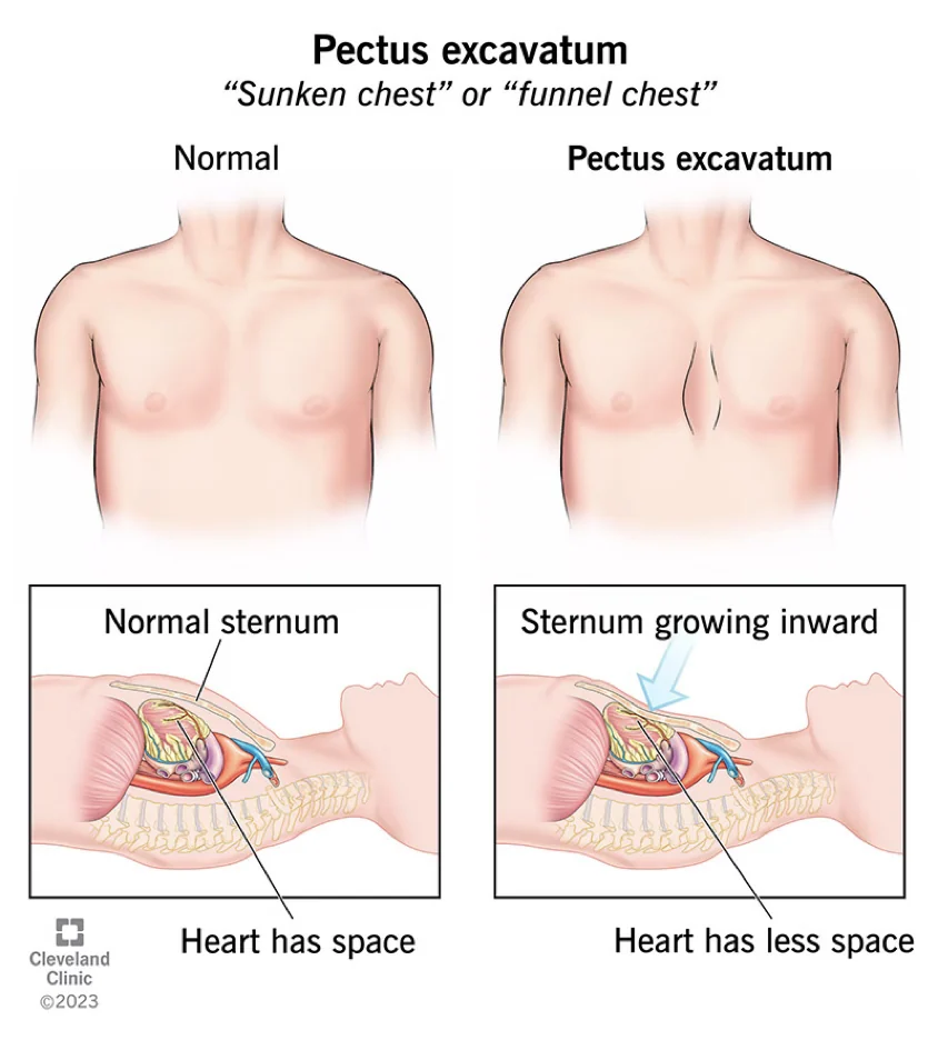
Pectus excavatum is a congenital chest wall anomaly where the sternum and adjacent ribs grow inward, creating a sunken appearance in the front of the chest. Often noticed for the first time in early childhood or during the rapid growth phases of adolescence, this structural deformity varies significantly from person to person. For some, the depression is mild and barely noticeable; for others, it creates a deep hollow that visibly alters the shape of the torso.
Understanding the funnel chest deformity: The term "funnel chest" describes exactly how this anomaly looks from the outside. The chest wall functions not only as a protective cage for vital organs like the heart and lungs but also as an anchor for the muscles that help us breathe. When the structural support of this cage is compromised, it can cause a cascade of physical and psychological effects that need proper evaluation.
How common is a sunken chest deformity: This is the most frequent type of pediatric chest deformity. Families often worry that their child's chest looks different, especially when comparing them to their peers. Recognizing that this is a documented medical condition, rather than a simple cosmetic quirk, is the first step toward finding effective, reassuring medical support and exploring appropriate corrective options.
What causes pectus excavatum?
Genetic connective tissue syndromes link: The exact origin of this condition is still being researched, but specialists know it involves the overgrowth of the costal cartilage that connects the ribs to the sternum. Instead of growing straight, this cartilage pushes the breastbone inward. There is a well-documented connection between this condition and connective tissue disorders, which can sometimes warrant a screening for conditions that affect the body's structural integrity.
When examining pectus excavatum causes, medical professionals often look for a family history of chest wall anomalies. Up to forty percent of patients have a relative with a similar structural presentation. If someone in your family has Marfan syndrome, their chest wall might be particularly flexible, allowing the deformity to develop as they grow. A thorough clinical assessment helps identify these patterns early.
It is important to remember that no specific action during pregnancy or childhood causes the sternum to sink. There is no fault to be found, and it is not a result of poor posture. The inward growth is simply a localized anomaly in how the rib cartilage develops alongside the rest of the skeleton.
Pectus excavatum symptoms
Many patients first discover they have a chest wall anomaly because they notice a physical change, but the actual pectus excavatum symptoms can vary widely. The most common physical sign is the visible depression itself. As children enter their teenage growth spurts, the chest may sink further, making the deformity much more pronounced and drawing attention to the area.
Cardiac and pulmonary assessment findings: Because the sternum sits directly in front of the heart and lungs, a severe depression can physically compress these vital structures. Patients may experience shortness of breath during exertion, unexplained fatigue, or a sensation of rapid heartbeat. When evaluating symptoms, doctors frequently use a pediatric chest wall deformity brace as a baseline reference in conservative management to see if structural support and posture can relieve mild physical compression.
Even when physical symptoms are minimal, the primary concern driving patients to seek medical help is often psychological. Adolescents and adults alike can feel significant self-consciousness about their chest appearance, which can severely impact their self-esteem and quality of life. Recognizing this emotional weight is just as important as measuring the physical depth of the depression.
Pectus excavatum diagnosis
What tests confirm pectus excavatum: A definitive pectus excavatum diagnosis always begins with a thorough physical examination by a specialist. The doctor will visually inspect the chest wall, assess the flexibility of the ribs, and listen to the heart and lungs. This initial clinical assessment provides immediate context regarding the severity of the sunken appearance.
How is pectus excavatum measured on imaging: To truly understand what is happening beneath the skin, advanced imaging is required. A chest CT or MRI scan provides a clear, three-dimensional view of the skeletal structure. This allows the surgical team to see exactly how much the sternum is compressing the heart or restricting lung expansion. Radiology is the only way to accurately measure the internal impact of the deformity.
Beyond structural imaging, a complete diagnosis requires functional testing. Doctors typically order an echocardiogram to evaluate heart function and check for valve compression, alongside pulmonary function tests like spirometry. These tests help determine if the chest wall anomaly is just a cosmetic issue or if it is actively limiting cardiopulmonary performance.
What is the Haller index?
The Haller index is the specific radiological measurement used to quantify the severity of funnel chest deformity. Calculated using data from a CT or MRI scan, it relies on a precise mathematical formula. By dividing the internal horizontal width of the chest by the anteroposterior distance from the sternum to the spine, doctors get a clear numerical value representing the depth of the depression.
Haller index pectus excavatum severity scale: A normal chest typically yields an index of 2.5 or lower. When the number rises above 3.25, the patient is classified as having a severe structural depression. This threshold is widely recognized by surgeons and is often a prerequisite for insurance approval when recommending pectus excavatum surgery.
Understanding this metric is vital for patients and families facing a potential diagnosis. The index transforms a subjective visual assessment into an objective, measurable fact.
Types of pectus excavatum treatment
There are several distinct types of pectus excavatum treatment, ranging from conservative monitoring to surgical repair. For young children with mild, asymptomatic cases, doctors often recommend simple observation. The chest wall is highly flexible at a young age, and careful monitoring allows the medical team to track the deformity's progression before deciding on further intervention.
Non-surgical funnel chest management options: Physical therapy is a mainstay of conservative care. Targeted postural and breathing exercises can strengthen the chest muscles, improve lung capacity, and help posture. For younger patients with flexible chests, non-surgical therapies aim to gradually lift the sternum over time through external suction, though this requires immense daily discipline.
Surgical intervention becomes an excellent therapeutic option when the deformity causes significant physical symptoms or emotional distress. Choosing the right path requires a collaborative decision between the patient, their family, and the surgical team, based on the severity of the measurements, the patient's age, and the impact on their daily life.
Best pectus excavatum surgery options
When exploring the best pectus excavatum surgery options, specialists primarily compare the Nuss and Ravitch procedures. The Nuss procedure is a minimally invasive technique where curved metal bars are inserted beneath the sternum and flipped to push the chest outward. This method is highly recommended for younger patients with highly flexible rib cages, offering a shorter operating time and excellent cosmetic results with small incisions.
Nuss procedure vs Ravitch technique comparison: The Ravitch procedure is an open surgical technique used more frequently for adults, asymmetric deformities, or rigid chest walls. It involves an anterior incision to remove the abnormal costal cartilage and elevate the sternum directly. While it requires a longer recovery period and leaves a more visible scar, it is unparalleled for complex structural corrections.
For patients seeking a modern, less invasive approach, there are newer techniques like taulinoplasty, which uses an external plate to progressively lift the deformity without entering the chest cavity. Regardless of the surgical method chosen, pain management has improved dramatically, with techniques like cryoanalgesia providing comfortable, smooth recoveries.
Pectus excavatum recovery guide
A comprehensive pectus excavatum recovery guide is essential for anyone undergoing surgical repair. The immediate postoperative phase requires a few days in the hospital to manage pain and monitor breathing. While the Nuss procedure is minimally invasive, it involves significant structural reshaping, and patients should expect initial discomfort that requires targeted medical management and rest.
- Initial 6 weeks: Focus on gentle walking and strict avoidance of heavy lifting or twisting motions to allow the initial healing of the chest wall.
- 3 to 6 months: Gradual reintroduction of cardiovascular exercise, though contact sports must be strictly avoided to protect the underlying hardware.
- 2 to 3 years later: The implanted bars are removed during a short, outpatient procedure, completing the full corrective cycle.
Costal cartilage deformity healing process: As the months pass, the ribs and cartilage adapt to their new, corrected position. Patients must sleep on their backs and practice specific breathing exercises to keep the lungs fully expanded. Adhering to these guidelines ensures the best possible structural outcome and a safe return to normal activities without complications.
Is pectus excavatum safe to leave untreated?
Many families ask if it is safe to leave a mild deformity untreated. The answer depends entirely on the severity of the structural depression and its impact on the body. If the Haller index is normal, and the patient experiences no shortness of breath or cardiac compression, careful observation is a perfectly safe and medically sound approach.
Risks of untreated severe funnel chest deformity: If imaging shows the sternum physically compressing the right ventricle of the heart or displacing the lungs, leaving it untreated can lead to progressive cardiopulmonary decline. Over time, this compression can severely limit exercise tolerance and overall stamina. In these cases, surgical intervention is strongly recommended to prevent long-term functional deterioration.
For those with moderate cases, a surgeon might suggest conservative management alongside the use of a posture correction support brace to assist with structural alignment. This is often combined with an active physical therapy regimen to support the chest wall and monitor any changes as the child grows.
Long-term outlook with pectus excavatum
The long-term outlook with pectus excavatum is highly positive. Whether managed through observation or surgically corrected, patients typically experience a profound improvement in their quality of life. Physical symptoms like shortness of breath and fatigue often resolve entirely once the chest wall is normalized and the heart and lungs have adequate space to function.
Following pectus excavatum repair, the psychological benefits are often just as significant as the physical ones. Restoring a typical chest contour dramatically improves self-esteem, allowing patients to participate confidently in sports and social activities without the burden of self-consciousness. This boost in confidence is a critical component of the healing journey.
Continuous medical follow-up remains necessary to monitor the healing process and ensure the stability of the chest wall, especially after the removal of any surgical bars. With modern, less invasive solutions and highly effective pain management techniques, a healthy, active future is the standard outcome for those living with this congenital condition.






