Pectus Excavatum Surgery: Nuss and Ravitch Options
Pectus excavatum surgery can correct funnel chest with the Nuss or Ravitch procedure.
As a cardiothoracic surgeon, I have accompanied many patients and their families through the management of pectus excavatum, a condition that, although common, can raise concerns that are both cosmetic and functional. My aim in this article is to give clear, understandable information about the condition, what it means and the treatment options available.
What is pectus excavatum?
Pectus excavatum, also known as “funnel chest”, is a congenital deformity in which the sternum and neighbouring ribs sink into the chest, creating a visible depression. It is the most common abnormality of the front of the chest wall, affecting roughly 1 in every 1,000 children, and is four times more common in boys than in girls.
What causes pectus excavatum?
The exact cause of pectus excavatum is still not fully understood, but it is considered a congenital abnormality in the development of the costal cartilage. In most cases, the cartilage that joins the ribs to the sternum grows abnormally and pushes the sternum inwards.
Although there is not always a family history, a hereditary pattern is found in up to 40% of patients, which suggests a genetic basis. A higher incidence of pectus excavatum has also been seen in people with certain genetic connective tissue syndromes, such as Marfan syndrome or Ehlers-Danlos syndrome. This link suggests that a structural weakness of the connective tissue may contribute to the deformity.
How it presents
How pectus excavatum shows itself varies with age and with the severity of the deformity:
- Early childhood: it may be barely noticeable, or appear as a slight dip in the chest.
- Adolescence: during growth spurts, the deformity can become more pronounced and more obvious.
- Associated symptoms: some patients experience tiredness, shortness of breath on exercise, palpitations or chest pain. In many cases, however, the main concern is cosmetic.
Diagnosis
The diagnosis of pectus excavatum is based on clinical assessment and additional tests:
- Physical examination: shows the depth and extent of the chest depression.
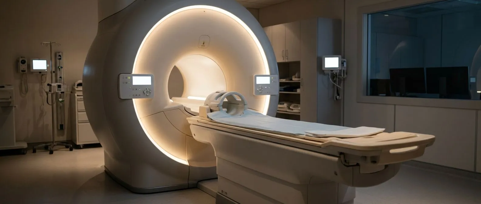
- Imaging: a CT scan or MRI of the chest helps quantify the severity using the Haller index.
- Heart and lung assessment: tests such as an echocardiogram and spirometry show whether the deformity is affecting how the heart or lungs work.
What is the Haller index?
The Haller index is a measurement used in radiology to assess the severity of pectus excavatum, a chest deformity in which the sternum is sunken into the chest.
How is it calculated?
It is calculated from a CT scan or MRI of the chest, taking two measurements on an axial (horizontal) slice:
- Internal transverse diameter of the chest: the horizontal width of the chest, from rib to rib.
- Anteroposterior distance: from the sternum to the spine, at the deepest point of the sternal depression.
Formula: Haller index = internal transverse diameter ÷ anteroposterior distance.
Normal and abnormal values
- Normal index: around 2.5 or less.
- Above 3.25: considered to indicate severe pectus excavatum, and may be a criterion for corrective surgery, especially if there are breathing or heart symptoms.
Clinical importance
A high index suggests the chest is so depressed that it could affect how the heart and lungs work.

Treatment options
The choice of treatment depends on how severe the deformity is, whether there are symptoms, and the patient's preferences.
1. Observation and exercise
In mild cases without symptoms, especially in young children, observation together with posture and breathing exercises may be chosen to improve the appearance and function of the chest.
2. Non-surgical therapies
- Vacuum bell: a device that uses suction to lift the sternum gradually. It works best in young patients whose chest is still flexible.
- Physiotherapy: specific exercises can improve posture and lung capacity, although they do not correct the structural deformity.
3. Surgical treatment: techniques and details
When pectus excavatum causes significant physical symptoms or a significant emotional impact, surgery becomes an excellent treatment option. Over the years, different surgical techniques have been developed, from open approaches to minimally invasive procedures, transforming patients' outlook and quality of life.
The Nuss procedure (minimally invasive technique)
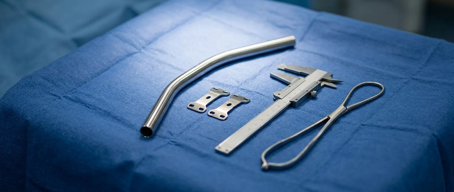
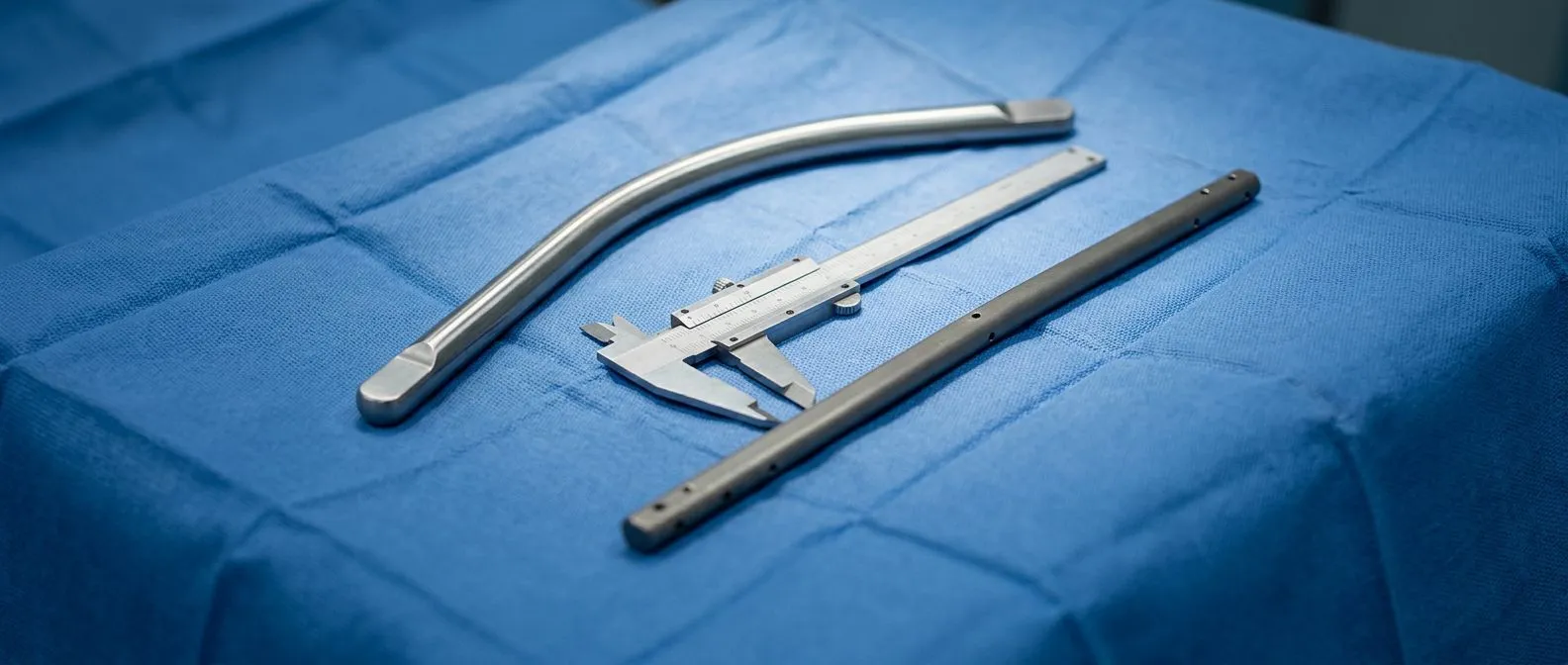
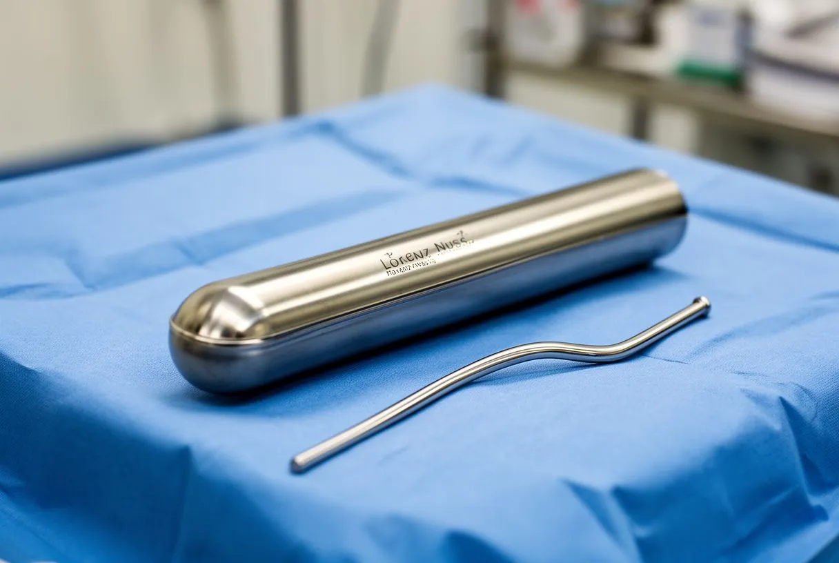
This is currently the most widely used surgical treatment in the world and is recommended for young patients whose chest is still flexible. One or more curved metal bars are passed beneath the sternum and, when rotated, push the depression outwards. The bar is secured with stabilisers and left in place for 2 to 3 years.
- Advantages: a minimally invasive approach, an excellent cosmetic result and a shorter operation.
- Considerations: it may cause discomfort at first, and contact sports should be avoided while the bar is in place.
The Ravitch procedure (open technique)
This is indicated in adults, asymmetrical cases or rigid deformities. It involves an incision on the front of the chest, removal of the deformed costal cartilages and elevation of the sternum, which is stabilised with a bar or mesh.
- Advantages: it allows direct structural correction and gives better results in complex deformities.
- Considerations: a longer recovery and a more visible scar.
Comparative results in the medical literature
Comparative studies have shown that both the Nuss and the Ravitch procedures have success rates above 90% in correcting the defect. The Nuss procedure, being less invasive, is better accepted cosmetically and involves shorter operations, but may be associated with more pain early after surgery; cryoanalgesia has helped reduce this. The Ravitch procedure, for its part, shows good long-term results, especially in adults or in cases with calcified ribs, although it needs longer hospital stays and carries a slightly higher surgical risk. The choice between the two should be based on individual factors such as age, severity, the symmetry of the deformity and the patient's preferences.
Pectus Up (taulinoplasty)
A recent, less invasive technique developed in Spain. It uses an external metal plate attached to the sternum that gradually lifts the deformity, and it does not require entering the chest cavity.
- Advantages: less postoperative pain and a quick recovery.
- Considerations: it is indicated in children and adolescents with a flexible chest, and its availability is limited.
Innovations: intercostal cryoanalgesia
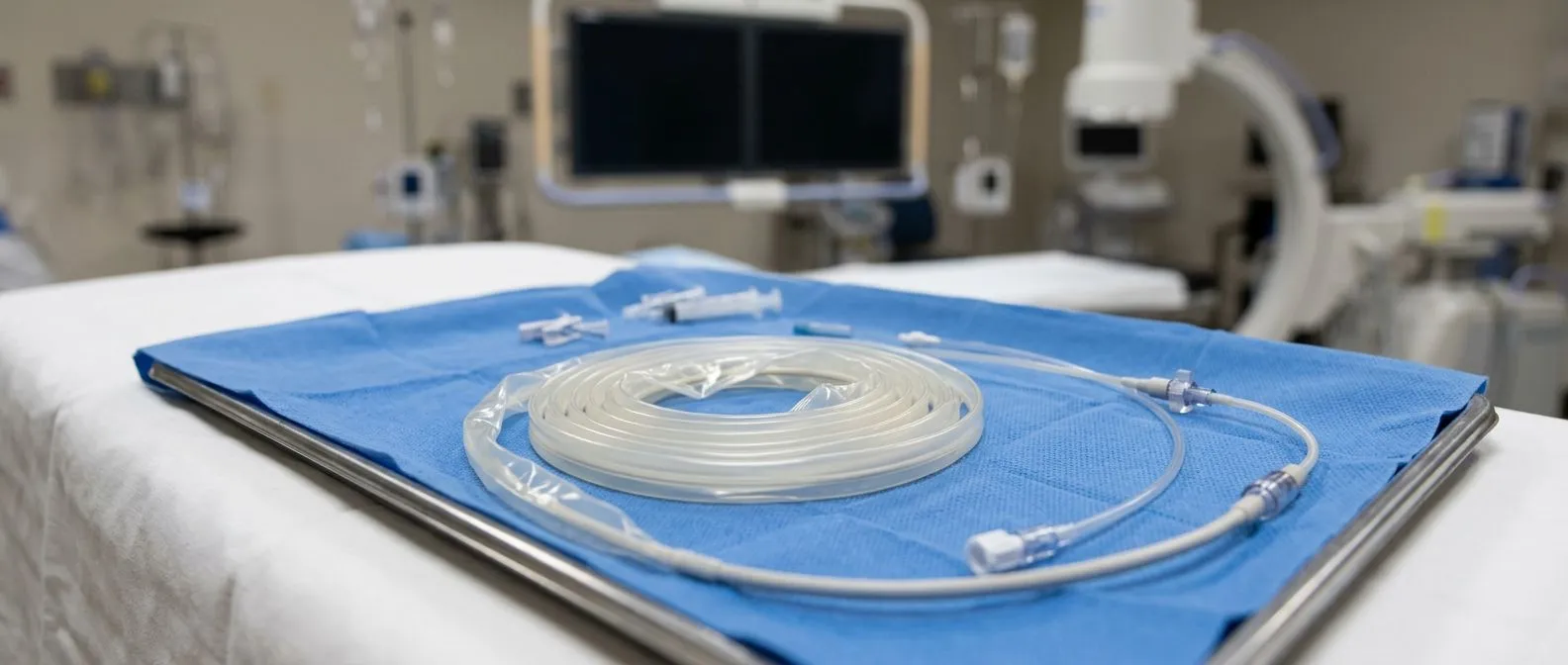
A complementary technique for pain management. It involves applying extreme cold to the intercostal nerves during surgery to reduce sensation for several weeks. It improves comfort after the operation and reduces the need for painkillers.
Outlook and life afterwards
Most patients see a significant improvement in the appearance of the chest and, in those with symptoms, in heart and lung function. Self-esteem and quality of life usually improve noticeably after treatment.
Although surgery corrects the deformity, medical follow-up is needed to monitor recovery and pick up any complications. In many cases the material implanted during the corrective surgery has to be removed; this is usually done years later.
Conclusion
Pectus excavatum is a treatable condition, and treatment options have evolved significantly, offering effective and less invasive solutions. If you or your child have this condition, I encourage you to see one of our specialists to assess the best treatment option for your particular case.