

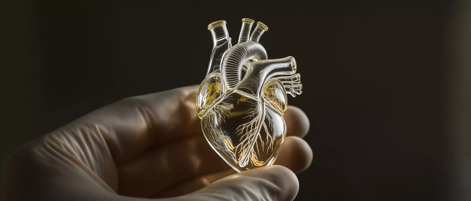
Sinuses of Valsalva
Explore the sinuses of Valsalva, their anatomy, function, and related pathologies, and learn how they affect cardiac health.
What Are the Sinuses of Valsalva?
The sinuses of Valsalva are three small, naturally occurring pouches or bulges located at the very base of the aorta, just above the heart. They sit directly behind the three leaflets of the aortic valve, which is the gateway that controls blood flow out of the left ventricle. When the heart muscle contracts and pushes blood into the body, these pouches expand slightly to accommodate the surge of circulating fluid.
Why this anatomy matters to cardiac patients: Understanding the aortic root structure helps patients visualize how the heart manages high-pressure blood flow. The sinuses are not merely passive containers; they are highly specialized anatomical features that protect the heart's valves and ensure that the coronary arteries receive the blood they need to keep the heart muscle itself alive.
For patients facing cardiac surgery, knowing about this specific region is essential because it is a common site for complex surgical intervention. Whether a surgeon is repairing a valve, replacing an ascending aorta, or addressing a structural defect, the sinuses of Valsalva are central to the surgical landscape. Their structural integrity is vital for long-term cardiac health and optimal circulatory function.
How Do Valsalva Sinuses Anatomy Support Blood Flow?
The mechanics of valve closure: The primary mechanical role of the sinuses is to create a zone of low pressure and gentle turbulence immediately behind the aortic valve leaflets. When the heart finishes squeezing blood into the aorta, the pressure drops, and the blood naturally wants to flow backward. The sinuses catch this backward-flowing blood, creating small eddies or swirls that push the valve leaflets closed smoothly, preventing blood from leaking back into the heart.
Without these anatomical pockets, the aortic valve leaflets would slam shut against the flat wall of the aorta, causing rapid wear and tear. The sinuses provide the necessary physical space for the leaflets to open fully without hitting the vessel wall, reducing mechanical stress. This elegant biological engineering ensures the valve remains competent over millions of cardiac cycles throughout a person's lifetime.
Furthermore, the unique shape of the ascending aorta sinuses helps to distribute the force of each heartbeat evenly. By absorbing and dissipating the immense pressure generated by the left ventricle, the sinuses protect the more delicate downstream vessels from sudden, damaging surges. This protective function is why surgeons take great care to preserve or recreate natural sinus geometry during cardiac operations.
Where Are the Coronary Artery Origins Located?
The coronary ostia, which are the small openings where the coronary arteries originate, are strategically positioned within two of the three sinuses of Valsalva. The left coronary artery typically arises from the left sinus, while the right coronary artery emerges from the right sinus.
Protecting the heart's oxygen supply: Because the coronary arteries originate directly within the sinuses, this anatomy allows the heart muscle to receive its oxygen-rich blood supply during diastole—the brief resting phase between heartbeats. The low-pressure environment inside the sinuses creates a suction effect that actively draws blood into the coronary arteries precisely when the heart muscle needs to relax and refuel.
If the origins of these arteries are malformed or compressed, the heart's blood supply is compromised, which can lead to severe symptoms. Surgeons must have a deep understanding of this coronary ostia anatomy to avoid damaging these critical openings during aortic valve or root surgeries. Reattaching the coronary arteries to a new aortic graft requires precise spatial awareness of the sinuses to ensure lifelong, unobstructed blood flow.
What Is the Aortic Sinus Function During the Cardiac Cycle?
During the cardiac cycle, the aortic sinus function is highly dynamic. As the left ventricle contracts in systole, the aortic valve opens, and blood rushes past the sinuses. The sinuses expand to accommodate the massive volume of blood being ejected, acting as a shock absorber that mitigates the sheer force of the cardiac output.
Transitioning to diastole: Once the ventricle stops contracting, the pressure inside the aorta forces blood backward. This backward flow enters the sinuses, creating the hydrodynamic forces that seal the aortic valve. The sinuses effectively convert the kinetic energy of the moving blood into the mechanical action required to snap the valve shut safely and efficiently.
This seamless transition is crucial for maintaining unidirectional blood flow throughout the body. When evaluating valsalva sinus echocardiography, cardiologists look closely at how blood moves through this region during both phases of the cardiac cycle. Abnormal flow patterns often indicate structural problems within the aortic root that may require surgical correction.
What Causes a Sinus of Valsalva Aneurysm?
A sinus of valsalva aneurysm occurs when the wall of one of these sinus pouches becomes abnormally thin and stretches outward like a small balloon. This weakening is most often congenital, meaning the patient is born with a structural deficiency in the aortic wall tissue, though it can also develop later in life due to infection, trauma, or degenerative connective tissue disorders.
Progression and risk factors: Over time, the continuous pounding of high-pressure blood causes the weakened sinus wall to gradually balloon outward. In many cases, patients are completely unaware of this condition until it grows large enough to compress nearby heart structures or shows up incidentally on a routine cardiac imaging scan.
- Congenital weakness: Most common in the right coronary sinus, where a developmental flaw leaves the tissue unusually thin.
- Connective tissue disorders: Conditions like Marfan syndrome can weaken the aortic root structure globally.
- Infections: Endocarditis or untreated bacterial infections can erode the sinus walls over time.
As the aneurysm grows, it poses a risk of rupture or can interfere with normal valve function, leading to aortic regurgitation. Cardiac surgeons monitor the size of these aneurysms closely, recommending surgical intervention before they reach a dangerous diameter that risks tearing or bursting under pressure.
How Is Aortic Sinus Dilation Diagnosed?
Aortic sinus dilation is typically discovered through advanced cardiac imaging, often before the patient ever experiences physical symptoms. Echocardiography is the frontline tool for assessing the sinuses of Valsalva, as it uses sound waves to create detailed, real-time images of the heart's structures in motion.
The role of advanced imaging: If a transthoracic echocardiogram reveals a suspicious enlargement, doctors will often order a cardiac MRI or a CT angiogram to get a highly precise, three-dimensional view of the aortic root. These detailed scans allow cardiologists to measure the exact diameter of the sinuses and monitor how quickly they are expanding over time.
Early diagnosis is highly beneficial because it allows for proactive monitoring rather than emergency intervention. Patients diagnosed with mild aortic sinus dilation can often be managed with medications and regular follow-up imaging, ensuring that surgery is only recommended when the anatomical changes truly warrant it.
What Happens During a Sinus of Valsalva Rupture?
A sinus of valsalva rupture is a rare but serious complication where the thin, stretched wall of an aneurysm suddenly tears open. This rupture creates an abnormal connection, or fistula, between the aorta and another chamber of the heart, most commonly the right ventricle or the right atrium, causing a massive and sudden shunting of blood.
Symptoms of acute rupture: When a rupture occurs, patients usually experience immediate and dramatic symptoms, including sudden, severe chest pain, breathlessness, and a feeling of a racing heart. A loud, continuous murmuring sound can often be heard through a stethoscope as blood rushes through the newly formed hole under high pressure.
This event is a surgical emergency that requires rapid intervention to repair the defect and restore normal blood flow. With prompt diagnosis and skilled surgical repair, many patients recover well and return to a good quality of life.
When Is Valsalva Sinus Surgery Necessary?
Valsalva sinus surgery becomes necessary when an aneurysm reaches a dangerous size, when the sinuses are actively leaking blood, or when the structural integrity of the aortic root is compromised by valsalva sinuses pathology. The decision to operate is based on the size of the dilation, the rate of growth, and the presence of symptoms like chest pain or shortness of breath.
Surgical approaches and goals: The primary goal of the surgical team is to replace the weakened sinus tissue with a durable, synthetic graft while preserving or repairing the aortic valve. In procedures like the David operation, the surgeon spares the patient's native valve, sewing it into a new artificial aortic root, which avoids the need for lifelong blood-thinning medications.
Cardiothoracic surgeons may utilize custom aortic root biological replacement conduits to perfectly match the patient's anatomy during these complex reconstructions. The objective is always to eliminate the risk of rupture while maintaining the heart's natural mechanical efficiency, ensuring the sinuses continue to function optimally for decades.
How Do Congenital Heart Defects Affect the Aortic Root?
Certain congenital heart defects fundamentally alter the normal structure of the aortic root, directly impacting the sinuses of Valsalva. Conditions such as bicuspid aortic valve disease or Marfan syndrome can cause the aortic root to stretch abnormally from a very young age, leading to early-onset sinus dilation.
Monitoring structural changes: Patients with these underlying conditions require lifelong, specialized cardiac surveillance. The altered hemodynamics of a malformed valve place uneven pressure on the sinus walls, accelerating tissue wear and increasing the likelihood of aneurysm formation much earlier than in the general population.
- Marfan syndrome: Causes a generalized weakness in connective tissue, leading to early root dilation.
- Bicuspid aortic valve: Alters blood flow turbulence, stressing the sinus walls over time.
- Unicuspid valve: A rare defect causing severe outflow obstruction that distorts sinus anatomy.
By understanding the patient's congenital background, cardiologists can predict how the sinuses will behave over time and plan preventive surgical strategies before a life-threatening rupture occurs. This proactive approach is a cornerstone of modern structural heart disease management.
Key Takeaways on Aortic Root Structure
Understanding the sinuses of Valsalva provides crucial insight into how the heart manages high-pressure blood flow while simultaneously feeding its own muscle. These small, pouch-like structures are anatomical marvels that protect the aortic valve from mechanical wear and ensure the coronary arteries receive a steady, reliable supply of oxygen.
Navigating pathology and treatment: Whether dealing with congenital weaknesses, progressive aortic sinus dilation, or the urgent need for valsalva sinus surgery, understanding this anatomy demystifies the surgical process. Patients can approach their cardiac care with greater confidence, knowing that modern surgical techniques are specifically designed to replicate and preserve these natural dynamics.
Ultimately, the health of the sinuses of Valsalva is closely tied to overall heart function. Through vigilant monitoring, early diagnosis via echocardiography, and precise surgical repair when necessary, patients can successfully manage conditions affecting the aortic root and maintain robust cardiovascular health for the long term.






