

Sinus of Valsalva Aneurysm: Diagnosis and Repair
Sinus of Valsalva aneurysm diagnosis and repair explained by Guadalajara heart surgeons.
If you or a family member has just been told about a sinus of Valsalva aneurysm, the name alone can be frightening. It refers to a small bulge in one of the three tiny pockets that sit just above the aortic valve, where blood leaves the heart. Many of these aneurysms grow quietly for years without a single symptom. Others can rupture suddenly and demand urgent surgery. This guide walks you through what the anatomy actually is, how the diagnosis is made, and what repair involves, so you can face the decisions ahead with clear, calm information.
What Are the Sinuses of Valsalva?
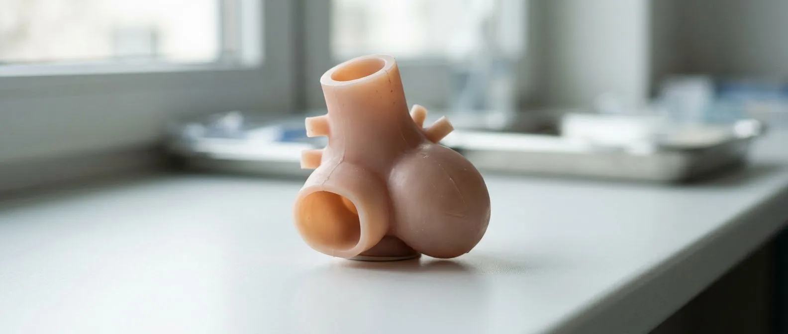
The sinuses of Valsalva are three small dilations of the aorta located immediately above the aortic valve. Think of them as three shallow pockets carved into the wall where the aorta begins. Each one sits behind one of the three leaflets of the aortic valve, and two of them give origin to the coronary arteries — the vessels that feed the heart muscle itself.
Because they are so close to the valve and to the origins of the coronary arteries, even a small abnormality here matters. Cardiologists and surgeons pay close attention to this region on every imaging study. It is also a region we examine carefully in every case we review, because what happens at the sinuses affects both the valve and the arteries that supply the heart.
The name comes from the Italian anatomist Antonio Valsalva, who described these structures in the eighteenth century. Despite the intimidating title, the anatomy is simple: three pockets, each a few millimetres to about a centimetre and a half wide in a typical adult.
What Is the Function of the Sinuses of Valsalva?
These pockets are not decorative. When the heart relaxes between beats, the aortic valve snaps shut, and blood swirls inside the sinuses. That swirl keeps the valve leaflets floating away from the aortic wall so they can close smoothly again. It also fills the two coronary arteries with blood while the heart rests — the coronary arteries are fed during relaxation, not during contraction.
Coronary perfusion: without the sinuses acting as small reservoirs, the heart muscle would receive far less blood with each heartbeat. This is why surgeons protect this region during any aortic valve operation.
In short, the sinuses of Valsalva keep the valve closing properly and keep the heart's own blood supply flowing. Any disease that stretches or distorts them can disturb both functions at once.
What Is a Sinus of Valsalva Aneurysm?
A sinus of Valsalva aneurysm is a weakness in the wall of one of these pockets that allows it to balloon outward. Most are congenital, meaning the person was born with a thin spot in the wall where the muscle and the aortic tissue do not join firmly. Less often, the wall is weakened by infection, inflammation, or long-standing high blood pressure.
Congenital sinus of Valsalva aneurysm: this is the most common form, particularly in younger adults. The wall between the aortic sinus and an adjacent heart chamber is missing its normal supporting layers, so pressure gradually pushes the pocket into a Windsock-like pouch.
Acquired causes: infections of the aortic root, connective tissue conditions that weaken artery walls, and long-standing hypertension can all produce similar dilation. Distinguishing between the two origins matters, because the underlying cause shapes the treatment plan.
For years the aneurysm may simply enlarge. The danger comes when it presses on neighbouring structures, distorts the aortic valve, or tears open into an adjacent chamber.
What Are the Three Sinuses of Valsalva?
Doctors name the three sinuses by the coronary artery that arises from them, or by their position. The usual naming is:
- Right coronary sinus: sits at the front-right of the aortic root and gives origin to the right coronary artery. This is the sinus most often involved in aneurysms that rupture into the right ventricle.
- Left coronary sinus: sits at the left and gives origin to the left main coronary artery, which then divides to supply most of the left side of the heart.
- Non-coronary sinus: sits at the back, gives origin to no artery, and lies close to the right atrium. Aneurysms here tend to rupture into that receiving chamber.
Knowing which sinus is involved tells the surgeon where the aneurysm will point and which chamber it could rupture into. That is why the exact location is spelled out in every report and every surgical plan.
Sinus of Valsalva Aneurysm Symptoms to Watch For
Unruptured aneurysms are frequently silent. Many are discovered by accident on an echocardiogram ordered for a heart murmur, an abnormal chest X-ray, or a family evaluation. When symptoms do appear, they usually come from pressure on nearby structures or from distortion of the aortic valve.
Common warning signs: breathlessness during exertion, an irregular heartbeat, palpitations, chest discomfort, or a new heart murmur your doctor hears through a stethoscope. A large aneurysm can also compress the vessel that carries deoxygenated blood to the lungs, producing a rare but distinctive picture of persistent shortness of breath.
Symptoms of valve leakage: if the aneurysm stretches the aortic valve, blood leaks backwards into the heart. Over time this produces fatigue, breathlessness on stairs, and swelling of the ankles. Any of these changes deserves a prompt cardiology visit.
It is worth saying plainly: none of these symptoms is unique to this condition. They are reasons to get checked, not reasons to panic. In our clinics we see that most patients who reach us with an image showing dilation arrive worried but stable, and the consultation exists to replace guesswork with a clear plan.
What Is a Normal Sinus of Valsalva Measurement?
On ultrasound, the aortic root is measured at the level of the sinuses. In adults, a typical diameter at that level is in the range of roughly 2.9 to 3.9 centimetres, varying with age, sex, and body size. Measurements are always compared to what is expected for the person's height and build, not to a single number.
A measurement above the expected range is called aortic root dilation, and it triggers closer follow-up. Guideline thresholds used across cardiology — such as operating on an enlarged aorta above about 5.5 centimetres, or earlier in certain genetic conditions — exist to prevent disaster rather than to alarm. Between normal and those thresholds lies a zone of surveillance, usually with imaging every six to twelve months.
One measurement is a snapshot. Trends over time matter more, which is why your cardiologist will want to compare each new study with the previous one rather than react to a single number.
Sinus of Valsalva Aneurysm Echocardiogram and Imaging Tests
The echocardiogram — an ultrasound of the heart — is almost always the first test. It shows the size of the sinuses, the function of the aortic valve, and any abnormal blood flow. A transoesophageal echocardiogram, in which the probe is passed down the throat for a closer view, gives much sharper images of the aortic root.
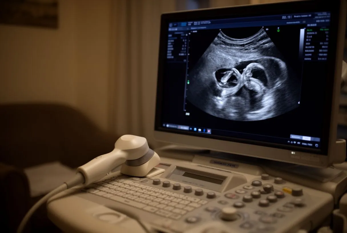
Sinus of Valsalva aneurysm cardiac MRI: magnetic resonance imaging measures the root precisely, shows the exact shape of the aneurysm, and reveals how blood flows through it. It is often the study of choice for planning and for follow-up, because it involves no radiation.
CT angiography: a contrast CT scan provides a rapid, detailed map of the aorta and coronary arteries. It is the standard test before surgery. Together, echo, MRI, and CT answer three questions: how big, where exactly, and what else is affected. In our team every case is reviewed against international practice guidelines with these studies in hand before a recommendation is made, and we take the time to explain in plain words what the images actually show.
Ruptured Sinus of Valsalva Aneurysm: Emergency Signs
A ruptured sinus of Valsalva aneurysm is a sudden event. The thin wall tears, and arterial blood pours from the aorta directly into a neighbouring heart chamber. This creates an abnormal shortcut that overloads the heart within hours to days.
The classic presentation is abrupt, sharp chest or upper abdominal pain followed rapidly by breathlessness. Many patients describe a racing or pounding heartbeat. A loud, continuous murmur appears, and the person can deteriorate quickly into heart failure — the heart simply cannot handle the extra volume being dumped into it.
This is a surgical emergency, and it is the reason a known, unruptured aneurysm is never ignored. Urgent repair of a rupture is possible and often successful, but the outcome is far better when the problem is found and repaired before it tears. If sudden severe chest pain with breathlessness occurs, the correct action is emergency services, not a scheduled appointment.
How Is a Sinus of Valsalva Aneurysm Repaired?
Repair means closing the weak pouch and restoring the normal wall between the aorta and the neighbouring chamber. This is done through open heart surgery, usually by patching the mouth of the aneurysm with a small piece of material or closing it directly with sutures. The heart-lung machine takes over circulation during the operation.
Repair with valve preservation: if the aortic valve leaflets are healthy, the valve is left in place and only the aneurysm is fixed. When the valve itself is damaged, repair or replacement of the valve is performed in the same operation, and the surgeon may combine this with a broader aortic root aneurysm treatment if the whole root is enlarged.
Choosing the approach: in some carefully selected situations the problem can be approached less invasively, for example closing a small rupture through a catheter. We recommend the less invasive route only where the evidence supports it for that specific patient, and our team — trained in conventional, minimally invasive, robotic and transcatheter techniques — reviews every case as a group before proposing anything. The patient then receives a clear proposal with the alternatives, and the final decision is always theirs.
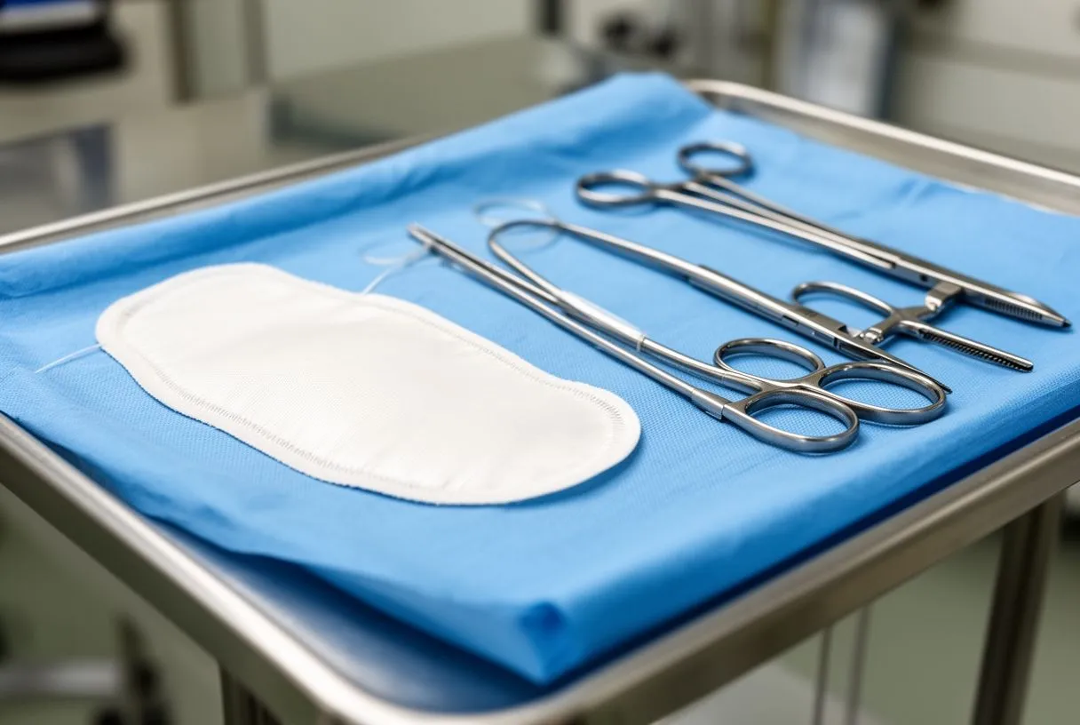
Because the aneurysm sits beside the coronary arteries, the surgeon also checks and protects their origins during the operation. This is standard, and it is one more reason repair belongs in experienced hands.
Sinus of Valsalva Aneurysm Surgery Recovery
Recovery after repair follows the same broad arc as other open heart operations. Most patients spend the first night or two in intensive care, then several days on the ward before going home. Breathing tube, drains, and monitoring lines are removed step by step over the first days.
The first weeks at home: expect tiredness, a healing breastbone, and gradual return to walking. Sternum precautions — no lifting, pushing, or pulling heavy objects — are followed for several weeks while the chest bone knits. Cardiac rehabilitation, a supervised programme of progressive exercise, helps rebuild confidence and fitness safely.
Follow-up imaging: echocardiograms are repeated after surgery and then periodically, to confirm the repair holds and the valve works well. Most patients who have an elective repair before rupture return to normal daily life, work, and moderate exercise. Recovery after an emergency rupture repair is longer, because the heart has already been strained by the event.

One further point families often ask about: seeking a second opinion is a normal, sensible part of deciding about heart surgery, and we encourage it openly. If you are weighing an elective repair or want clarity about what your images show, a free initial video consultation with our team is the natural first step, followed by a face-to-face visit in Guadalajara, Querétaro or Hermosillo when you decide to move forward.
A sinus of Valsalva aneurysm is uncommon, but the logic of its care is straightforward: know the anatomy, measure it accurately, watch it closely, and repair it on planned terms rather than emergency ones. A silent aneurysm found on imaging is an opportunity; a rupture is a race. If you have been told you have dilation at the aortic root, or if any of the symptoms above sound familiar, the next step is not self-diagnosis but a conversation with a qualified cardiac team that can read your studies and lay out your options. We invite you to book a consultation with Dr Alejandro Molina and our multidisciplinary group, bring your questions, and decide with every fact on the table.






