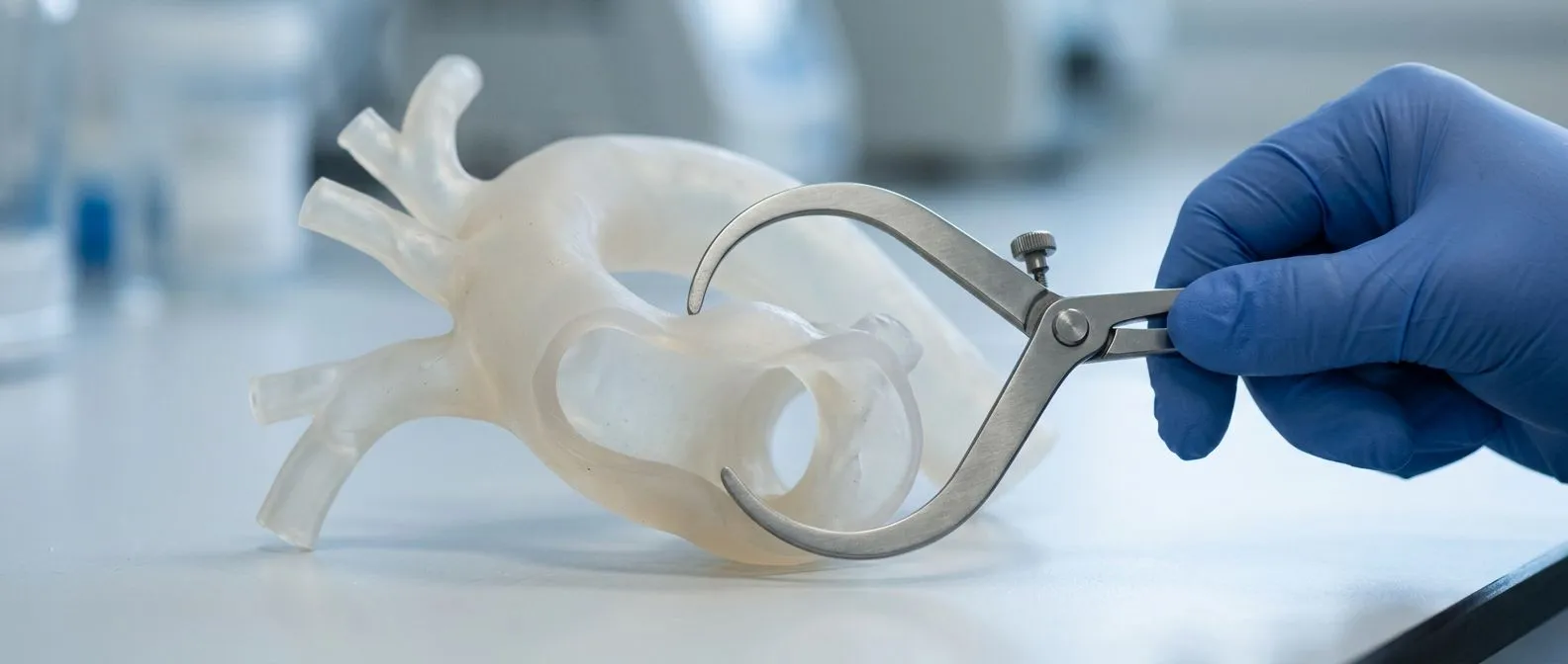
Acute Aortic Syndrome: Recognising the Emergency
Acute aortic syndrome emergency: learn the warning signs, how it differs from a heart attack, diagnosis and treatment.
An acute aortic syndrome emergency is one of the most time-critical events in all of medicine, yet many people have never heard the term. It describes a sudden, life-threatening problem inside the aorta — the large artery that carries blood from your heart to the rest of your body. The three conditions it covers share one frightening feature: the wall of the aorta begins to fail, and hours matter. This guide explains what happens inside the artery, what the symptoms feel like, how doctors confirm the diagnosis, and what treatment and recovery actually involve. Our aim is to replace panic with understanding, so that if you or someone close to you ever faces this, you know exactly what to do.
What Is Acute Aortic Syndrome?
The aorta under pressure. Every heartbeat pushes blood into the aorta at high pressure, and the artery wall is built in three layers to withstand that stress for decades. Acute aortic syndrome is the umbrella term for sudden conditions in which those layers separate, tear or ulcerate. It includes aortic dissection (a split between the layers), intramural hematoma (bleeding into the wall itself) and penetrating atherosclerotic ulcer (an ulcer that bores through the inner lining).
Why it is a true medical emergency. Once the wall layers separate, the aorta can rupture or cut off blood supply to vital organs. MedlinePlus classifies aortic dissection as a life-threatening condition requiring immediate care, and that urgency is exactly why emergency teams treat sudden, severe chest or back pain in a middle-aged or older adult as an aortic problem until imaging proves otherwise.
Who is most at risk. High blood pressure over many years is the single biggest driver, because it stiffens and stresses the wall. Connective tissue conditions such as Marfan Syndrome and Ehlers-Danlos Syndrome weaken the wall from birth, and a Thoracic Aortic Aneurysm — a balloon-like widening of the chest portion of the aorta — raises risk further. Every aneurysm patient should know the warning signs of acute aortic syndrome long before an emergency ever develops, because recognising it early is the whole game.
Acute Aortic Syndrome Symptoms to Watch For
The classic tearing pain. The hallmark symptom is severe pain that begins abruptly, usually in the chest or between the shoulder blades, and is often described as tearing, ripping or shearing — unlike the crushing pressure of a heart attack. The pain characteristically moves as the dissection extends, travelling from the chest toward the back, abdomen or legs. Sudden tearing chest pain that radiates to the back is the pattern every emergency doctor is taught to suspect first.
Symptoms in women and atypical presentations. Women, older adults and people with diabetes may present differently: the pain can be milder, located in the abdomen, or mistaken for indigestion or a muscle strain. Symptoms of an aortic aneurysm in females often overlap with these atypical patterns, which is one reason the diagnosis is delayed in women more often than in men. Fainting, weak or absent pulses in an arm or leg, sudden hoarseness, or a stroke-like weakness on one side of the body can all be the first sign.
Other warning signs that demand action. Shortness of breath, sweating, nausea, a blood pressure difference between the two arms, and a feeling of impending doom all appear in published case series. If any of these arrive together with abrupt chest or back pain, treat it as a Medical Emergency: call emergency services immediately and mention the possibility of an aortic problem, because saying those words can change how the ambulance team triages you.
Aortic Dissection vs Heart Attack: How They Differ
Why the confusion matters. Both conditions cause sudden chest pain, and both are deadly, but they are opposites in mechanism. An Acute Myocardial Infarction — a heart attack — happens when a blocked coronary artery starves the heart muscle of blood. An aortic dissection happens when the aorta's own wall splits. Giving a clot-busting drug meant for a heart attack to someone with a dissection can be catastrophic, so distinguishing the two quickly is critical.
Clues that point toward the aorta. Pain that is maximal at the very first second, tears through to the back, migrates downward, or comes with unequal pulses or blood pressures between arms favours the aorta. Pain that builds gradually, feels like pressure on the chest, and comes with cold sweat and shortness of effort favours a heart attack — though Takotsubo Syndrome, a stress-induced heart condition, can mimic both.
How doctors sort it out. An ECG is done within minutes, but a normal ECG does not exclude a dissection, so a CT angiogram remains the decisive test. Family members who raise the possibility of an aortic cause in the emergency department can help, because that single sentence often accelerates the imaging that settles the question.
Types of Aortic Emergencies: Dissection, Hematoma and Ulcer
Aortic dissection. This is the most common form. A small tear in the innermost layer lets blood surge between the wall layers, creating a false channel and splitting the artery in two. Everything downstream — brain, kidneys, spine, legs — depends on which branches the false channel steals.
Intramural hematoma. Here, the vessel's tiny feeding vessels bleed directly into the wall without an obvious tear, thickening it from the inside. It behaves like a dissection in waiting: some hematomas stabilise on medication, others progress, which is why close monitoring in an intensive care setting is standard.
Penetrating atherosclerotic ulcer. Typically in older patients with heavy cholesterol disease, an ulcer in the lining drills deeper until it reaches the middle layer, causing pain and sometimes localised bleeding. A Penetrating Aortic Ulcer can also be the starting point for a full dissection or rupture, so it is never dismissed as a simple ulcer.
- Dissection: a tear creates a false channel splitting the wall layers — the classic acute aortic syndrome emergency.
- Intramural hematoma: bleeding inside the wall, no visible tear, monitored closely because it can convert to dissection.
- Penetrating ulcer: a cholesterol ulcer erodes through the inner lining, usually in older, hypertensive patients.
How Acute Aortic Syndrome Is Diagnosed
CT angiography is the workhorse. A CT scan with contrast dye injected into a vein shows the aorta in fine detail within minutes: the tear, the false channel, the hematoma, or the ulcer, plus any leak or reduced organ blood flow. This is why the standard workup of sudden chest pain in higher-risk patients includes CT angiography of the chest, abdomen and pelvis as a first-line test.
Other imaging and tests. Transesophageal echocardiography — an ultrasound probe passed into the esophagus — is used when a patient is too unstable for a CT scan or when the aortic valve and heart function need assessing. MRI can map chronic dissection and is useful for younger patients with connective tissue disease who need repeated imaging over the years, and published guidance on acute aortic syndrome MRI supports it as a reliable alternative when CT is not appropriate. Blood tests, an ECG and a chest X-ray are done in parallel to exclude or confirm other causes.
What we explain to patients beforehand. When we review imaging with a patient and family, we take the time to show them exactly what the studies show — where the tear sits, which branches are affected, and what the options are — in plain words, and we answer every question before any decision is made. Understanding your own anatomy makes the treatment choices far less frightening.
How Doctors Fix a Tear in Your Aorta
Immediate medical stabilisation. Whatever the type, the first hour is about controlling blood pressure and heart rate with intravenous medication, so the tear stops propagating. Pain relief, oxygen, and admission to an intensive care unit follow. This medical phase is universal, whether or not surgery comes next.
Surgery for tears in the ascending aorta. When the tear is in the segment closest to the heart, open emergency surgery is required: the damaged segment is replaced with a synthetic tube, often with repair of the aortic valve, under deep hypothermic circulatory support. Patients sometimes ask how they fix a tear in your aorta without opening the chest — for tears further along the artery, a stent graft can be placed through an artery in the groin, a far less invasive option.
Treatment for descending aortic tears. Uncomplicated tears below the left arm branch are usually managed first with aggressive medication and close observation, escalating to stent-graft repair if the false channel grows, organs lose blood flow, or pain persists. In our multidisciplinary team, every case is reviewed together against international practice guidelines before a recommendation is made, and the patient receives a clear proposal with all the alternatives — the final decision is always theirs.
Type A vs Type B Aortic Dissection: Why the Location Matters
Type A: the ascending aorta. A Type A Aortic Dissection involves the ascending aorta, the segment that rises directly from the heart. This is the most dangerous form because the dissection can rupture into the sac around the heart, cause a heart attack, or stroke. Treatment is emergency surgery, almost always on the day of diagnosis. Aortic Dissection: a true cardiovascular emergency is not a figure of speech for this type — it is the standard description in the literature.
Type B: the descending aorta. A Type B Aortic Dissection begins beyond the left subclavian artery, further from the heart. If the vital organs are well supplied and the pain is controlled, treatment begins with medication in intensive care; stent grafting is reserved for complications. Many Type B patients do well for years under medical management with regular imaging.
Why the descending aorta deserves your attention. People often ask what is the descending aorta: it is the portion of the aorta running down through the chest and abdomen, supplying the spinal cord, kidneys and legs. Knowing this geography helps you understand why tears near the heart demand surgery today, while tears further down can often be watched — at least initially.
Recovery After Aortic Dissection Surgery: What to Expect
The hospital phase. Patients ask how long it takes to recover from aortic dissection surgery, and the honest answer is that recovery happens in stages. Expect several days in intensive care, then roughly one to two weeks in hospital overall for an uncomplicated repair, longer if complications such as kidney or spinal cord effects occur. Some patients need temporary heart support after the operation — devices such as an Intra-aortic Balloon Pump are sometimes used in high-risk situations to ease the heart's workload, a use documented in studies of high-risk cardiac interventions.
The first weeks at home. Walking is encouraged early; lifting anything heavy, driving, and straining are restricted for about six to twelve weeks as the breastbone heals (in open surgery) and the aorta settles. Blood pressure control becomes a lifelong discipline: most patients take two to three medications permanently, because uncontrolled pressure is the main trigger for the dissection to extend.
Follow-up imaging and the long view. Regular CT or MRI scans — often at intervals of months, then yearly for life — check that the repaired aorta is stable and that no new aneurysm is forming. Make sure this follow-up plan is scheduled before you leave, so you are not left guessing what comes next.
Survival After Aortic Dissection Surgery
What the numbers actually say. Questions about acute aortic syndrome survival deserve a careful answer. Emergency surgery for Type A dissection carries meaningful operative risk even in expert centres, but the alternative — leaving it untreated — is far more lethal, with a substantial proportion of patients not surviving the first day or two without repair. For stable Type B dissections treated with medication and monitoring, long-term outcomes are considerably better. The key point is that survival figures are averages across many different patients, and your own risk depends on age, organ function and how quickly you reach a hospital.
What improves the odds. Three factors dominate: how fast the diagnosis is made, whether blood pressure is controlled from the first hour, and whether the operation is done by an experienced aortic team. Coming to surgery in stable condition rather than in shock changes the picture dramatically.
Life after the emergency. Most survivors return to normal or near-normal lives. The essentials are simple and non-negotiable: take your medications daily, keep every imaging appointment, avoid smoking, and never stop blood pressure treatment because you feel well. Dr Molina trained in cardiothoracic surgery at the Universidad de Guadalajara and in cardiac, minimally invasive, robotic and structural surgery at the University of Montreal; the surgery is only the first chapter of the story, and these habits shape the rest.
When to Call a Cardiac Team About an Aortic Emergency
Red flags that mean act now. Sudden severe chest or back pain — especially tearing pain radiating to the back — a syncopal episode, unequal pulses, or new weakness on one side of the body all justify calling emergency services immediately. Do not drive yourself, and do not wait to see if it passes.
Second opinions in non-acute situations. For planned questions — an aneurysm found on a scan, a family history of aortic disease, or deciding between open repair and a stent graft — there is time to think, and a second opinion is a normal part of that process. We encourage second opinions and treat them as standard practice in heart surgery decisions; the first contact is a free video consultation, where we get to know you, discuss your condition, and tell you which studies are genuinely still needed so none are ordered unnecessarily. If surgery is the right path, a face-to-face visit at our consulting rooms follows.
Planning ahead if you are at risk. If you have high blood pressure, a known aneurysm, or a family history of Marfan Syndrome or Ehlers-Danlos Syndrome, talk to a qualified cardiac team before an emergency finds you. Learn your own baseline, keep your blood pressure at target, and know where your nearest centre with aortic surgery capability is. An acute aortic syndrome emergency rewards preparation more than almost any other cardiac crisis — and if you are ever unsure about your own situation, the right move is to discuss it with a specialist team rather than to self-diagnose. Our team also performs conventional, minimally invasive, robotic and transcatheter procedures, and we recommend the less invasive route only where the evidence supports it for that specific patient.

Questions People Ask About Aortic Emergencies
Could it be something less serious? Yes — severe chest pain has many causes, from muscle strain to reflux, and even Acute Respiratory Distress Syndrome or Metabolic Syndrome complications can produce frightening symptoms. That is precisely why self-diagnosis is dangerous in both directions. Only proper imaging can tell an aortic emergency from something benign.
What about the aortic root and valve? When a dissection or an Aortic Root Aneurysm involves the base of the aorta, the Aortic Annulus — the ring where the valve sits — may be affected, and valve repair or replacement can be part of the operation. Knowing your heart and its structures beforehand makes these conversations far easier to follow.
How do I get evaluated? Start with your cardiologist or a dedicated aortic centre. Bring any previous imaging on disc or digitally, your full medication list, and your family history. If anything in your story fits the patterns described above, insist on CT angiography rather than a plain chest X-ray alone — it is the test that finds the problem.

A final word. Acute aortic syndrome is frightening, but it is also survivable when recognised early and treated by an experienced team. Know the symptoms, control your blood pressure, and never let anyone tell you that severe sudden pain is nothing until it has been properly investigated. If you live with an aortic condition or a worrying family history, book a consultation with a qualified cardiac team — starting with a free video consultation costs you nothing and could change everything. Low Cardiac Output Syndrome, organ complications and prolonged recovery are real risks, but with modern surgery, careful follow-up and honest conversations between you and your surgeons, most patients get their lives back. Take the first step: ask your questions, get your studies, and put a specialist team on your side before the emergency ever arrives.