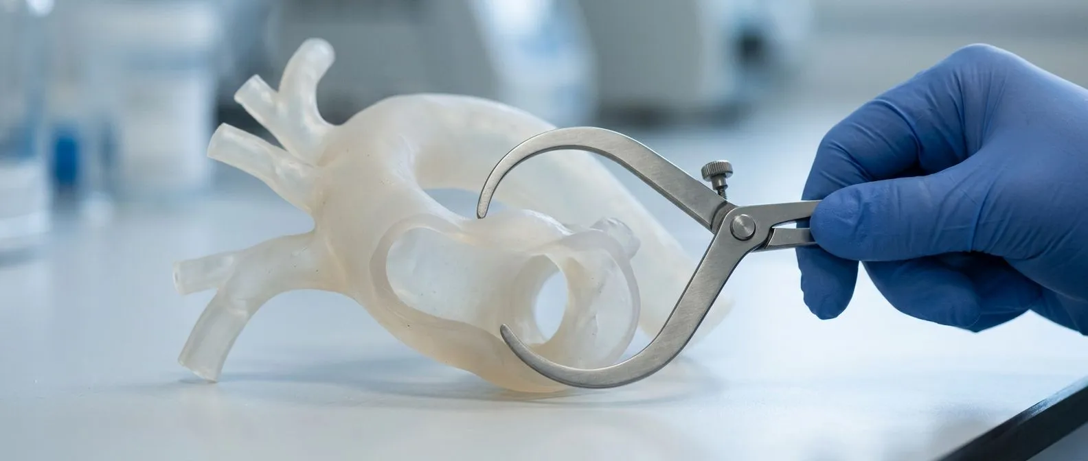
Penetrating Aortic Ulcer: Diagnosis and Treatment
Penetrating aortic ulcer treatment options explained by heart specialists in Guadalajara.
A penetrating aortic ulcer is a small sore that breaks through the inner lining of the aorta, the large artery that carries blood from the heart to the rest of the body. It sounds alarming, and it deserves serious attention, but it is also a condition that modern heart teams understand well. In this guide we explain penetrating aortic ulcer treatment in plain language: what the condition is, how doctors find it, when monitoring is enough, and when a procedure is the safer path. Our goal is that you leave this page able to ask better questions at your own consultation.
What Is a Penetrating Aortic Ulcer?
Definition in simple terms. The aorta has three layers, like a garden hose with an inner lining, a muscular middle layer, and an outer wall. Over many years, cholesterol plaque can form in that inner lining, especially in people with high blood pressure, smoking history, or high cholesterol. Sometimes the plaque erodes inward until it actually tunnels through the inner layer and into the middle layer of the vessel wall. That tunnel is called a penetrating aortic ulcer, and the term "penetrating" refers to how deep it goes, not to any injury from outside.
Doctors group this condition together with two related problems, aortic dissection and intramural hematoma, under the name Acute Aortic Syndrome. All three involve blood finding its way into or through the wall of the aorta. A penetrating atherosclerotic aortic ulcer is usually the starting point: blood enters through the ulcer crater and may then spread along the wall, creating one of the other two conditions.
Where it happens. Most ulcers form in the descending thoracic aorta, the section of the aorta that runs down through the chest behind the heart. Less often they appear in the abdominal aorta or in the arch. The location matters enormously for treatment, because an ulcer near the branches that supply the brain needs a different approach from one lower down. MedlinePlus offers reliable general background on aortic disease for patients who want to read further (MedlinePlus).
What Causes a Penetrating Aortic Ulcer?
Atherosclerosis comes first. Nearly every penetrating aortic ulcer begins as long-standing atherosclerosis, the same disease that causes heart attacks and strokes. Plaque builds up in the aortic wall over decades. As the plaque ages it can ulcerate, meaning its surface breaks down. When the erosion reaches the internal elastic lamina, the tough boundary between the inner and middle layers of the artery, blood under pressure can push through and create the ulcer crater.
Risk factors you can influence. The list is familiar, and that is good news, because these are factors patients can work on:
- Uncontrolled high blood pressure: constant pressure on the artery wall is the single strongest driver, which is why blood pressure control is the foundation of all thoracic aortic ulcer treatment.
- Smoking: damages the artery lining directly and accelerates plaque growth.
- High cholesterol and diabetes: both speed up the atherosclerosis that creates the plaque in the first place.
- Age: the condition is rare before 55 and becomes more common with each decade.
Cause versus trigger. It helps to separate the slow cause from the fast trigger. The plaque took years to form, but the ulcer itself can break through in a short time, sometimes during a spike in blood pressure. That is why a patient with known severe atherosclerosis who develops new, sudden chest or back pain should be evaluated urgently rather than waiting to see if it passes.
Penetrating Aortic Ulcer Symptoms and Warning Signs
The classic symptom is sudden pain. Most patients describe a sharp, tearing, or ripping pain in the chest, upper back, or between the shoulder blades. The pain often begins abruptly and is severe from the first minute, which is different from the gradual chest discomfort of a blocked coronary artery. Some patients also feel pain move downward as the ulcer extends along the aorta.
Symptoms that suggest something more serious. Aortic ulcer symptoms chest pain aside, there are signs that the ulcer may already be bleeding or threatening rupture, and these need emergency care, not a scheduled appointment:
- Pain that suddenly changes or worsens: often the first sign that an ulcer has progressed to dissection or Aortic Rupture.
- Faintness, sweating, or cold extremities: possible signs of significant blood loss or poor circulation.
- Weakness on one side, trouble speaking, or vision changes: warning signs that debris from the ulcer may be affecting arteries to the brain.
When there are no symptoms at all. Here is the part that surprises patients: many penetrating ulcers cause no pain at the time they form. They are discovered by chance on a CT scan done for another reason. A silent ulcer is not automatically a safe one. Every silent ulcer still needs a proper assessment before anyone can say monitoring is enough. If you have sudden severe chest or back pain, call emergency services; do not wait to book a consultation.

Penetrating Aortic Ulcer Diagnosis: Why the CT Scan Matters
CT angiography is the standard test. Penetrating aortic ulcer diagnosis rests on a CT angiogram, a scan in which contrast dye is injected into a vein and images are taken of the aorta in thin slices. This shows the ulcer crater, its depth, its location, and whether blood has already spread into the vessel wall. It also rules out or confirms dissection and intramural hematoma, the two conditions that travel alongside ulcers.
What we measure on the scan. A CT angiogram tells the team several things that decide treatment: how deep the ulcer penetrates, how wide its opening is, how far the affected segment is from the arteries to the brain and spinal cord, whether there is fluid around the aorta, and whether an Abdominal Aortic Aneurysm or Thoracic Aortic Aneurysm is present at the same time. In the consultation we take the time to explain what the studies actually show, in plain words, and to answer every question before any decision is made.
Other tests that play a supporting role. A transthoracic echocardiogram, an ultrasound of the heart done from the chest, does not show the ulcer itself but tells us how the heart is working and whether the Aortic Root and valve are involved. Blood tests, an electrocardiogram, and troponin levels help rule out a heart attack, which can mimic the pain. An MRI may be used when radiation exposure needs to be limited during follow-up, especially in younger patients.
Aortic Ulcer vs Dissection vs Intramural Hematoma
Three related but different problems. Patients often leave a scan appointment confused because their report mentions all three terms. Understanding aortic ulcer vs dissection makes the rest of the conversation much easier. All three belong to Acute Aortic Syndrome, but they describe different stages and different anatomy.
- Penetrating aortic ulcer: a localized crater where plaque has eroded through the inner lining into the wall of the aorta. Focal and deep.
- Aortic dissection: blood tears a channel between the layers of the aortic wall and travels along it, sometimes for long distances. Type A Aortic Dissection involves the ascending aorta and is a surgical emergency, while Type B Aortic Dissection involves the descending aorta and is often managed with medication first.
- Intramural hematoma: bleeding contained within the aortic wall without a visible tear; the usual causes of an intramural hematoma in the aorta are a penetrating ulcer breaking through or rupture of the tiny vessels that feed the wall itself.
Why the distinction changes treatment. A small, stable ulcer in the descending aorta may safely be watched with scans and medication. A dissection that involves the ascending aorta needs surgery within days or hours. An intramural hematoma sits in between, monitored closely in hospital at first. This is why the exact wording in the radiology report matters, and why we review the actual images rather than relying only on the written summary. Every case is reviewed by our multidisciplinary team against international practice guidelines before a recommendation is given, and the patient receives a clear proposal with the alternatives.
Penetrating Aortic Ulcer Treatment Options
Three broad paths. Penetrating aortic ulcer treatment falls into three categories: medication alone with careful imaging follow-up, minimally invasive stent repair, and in selected cases conventional surgery. Which path is right depends on the ulcer's depth, location, whether it is symptomatic, and whether it is growing. There is no single correct answer for everyone, which is exactly why a specialist assessment matters.
Medical management first. For a small, stable, asymptomatic ulcer, the first line is aggressive control of blood pressure and heart rate, using medication to reduce the force that blood exerts on the artery wall. This is the same medical approach used for uncomplicated Type B Aortic Dissection. Smoking cessation and cholesterol control run alongside it. Repeat CT scans at intervals, typically at one month, six months, and then yearly, confirm the ulcer is stable.
When repair is recommended. Intervention enters the picture when certain features appear, and each one is a genuine reason to act rather than wait:
- Ongoing pain despite medication: suggests the ulcer is actively progressing.
- Growth of the ulcer or the aorta on repeat scans: enlargement means the wall is weakening.
- Associated aneurysm or hematoma: an ulcer arising on an aneurysm or accompanied by intramural hematoma carries higher risk and usually tips the balance toward repair.
In every case the recommendation is made by the multidisciplinary team, and the final decision is always the patient's.
Endovascular Repair (TEVAR) for Aortic Ulcer
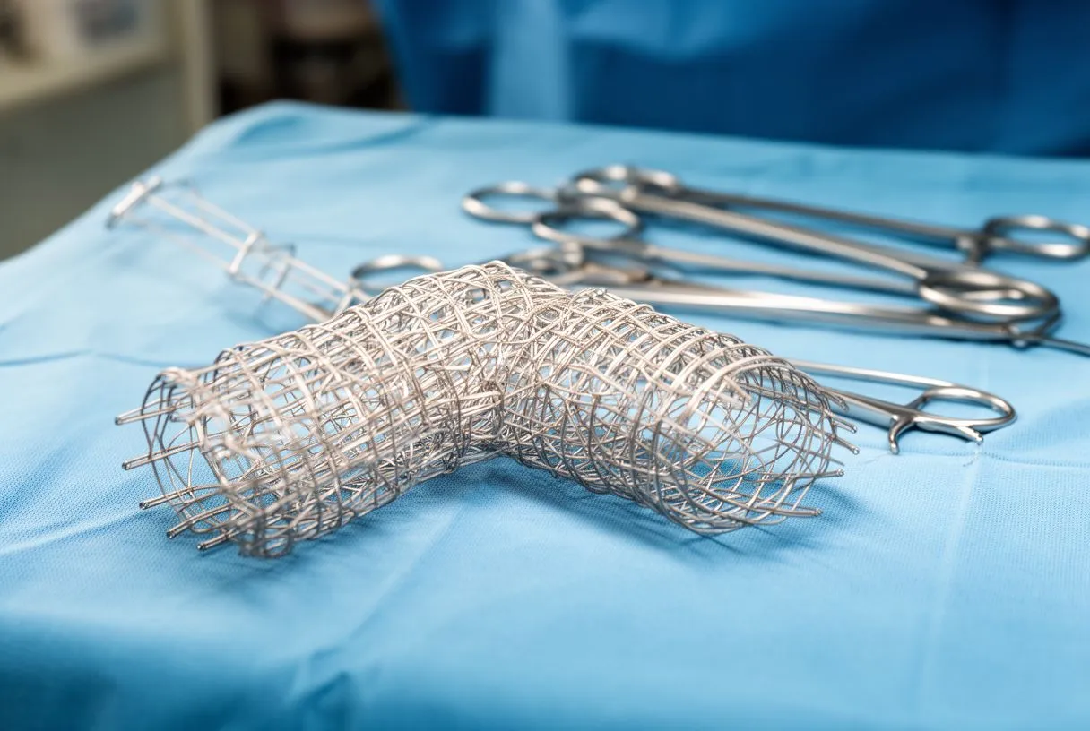
What the procedure involves. The most common repair for an ulcer in the descending aorta is thoracic endovascular aortic repair, usually called TEVAR. The surgeon makes a small incision, typically in the groin, and threads a catheter carrying a compressed fabric-covered stent, called an endograft, up to the diseased segment under X-ray guidance. The graft is released, expands against the healthy wall above and below the ulcer, and seals the crater off from blood pressure. No chest is opened, the heart is not stopped, and most patients go home within a few days.
Why minimally invasive repair is often chosen. Endovascular repair aortic ulcer treatment avoids the physiologic stress of open surgery: no Aortic Cross-clamp on the aorta, no heart-lung machine for straightforward descending cases, and a much shorter hospital stay. For elderly patients or those with other conditions, this difference in recovery is often decisive. In our practice we perform conventional, minimally invasive, robotic and transcatheter procedures, and we recommend the less invasive route only where the evidence supports it for that patient.
Where the technique reaches its limits. TEVAR works best for ulcers in the mid and lower descending aorta, where there is a stretch of healthy vessel for the stent to anchor on. Ulcers near the arch, close to the arteries that supply the brain and left arm, are more technically demanding, and options there include carefully planned hybrid approaches; recent reports describe physician-modified endograft techniques for symptomatic arch ulcers that preserve flow to critical branches (Journal of Vascular Surgery Cases and Innovative Techniques). An Aortic Arch Replacement or aortic root operation, including Aortic Root Replacement for ulcers with associated Aortic Root Aneurysm, remains the open-surgery route when the anatomy does not suit a stent.
Monitoring vs Surgery: When to Treat an Aortic Ulcer
The decision framework. Patients often ask when to treat aortic ulcer findings and when watching is genuinely safe. The honest answer is that both options can be correct, and the choice rests on specific features. A monitoring plan is reasonable when the ulcer is small and asymptomatic, located in the descending aorta away from the arch branches, and stable on repeat imaging with a normal aortic diameter. Repair is favored when pain persists, when the ulcer enlarges, when the aorta is dilated beyond guideline thresholds, or when the ulcer sits on an aneurysm.
How monitoring actually works. Being "under surveillance" is not a passive waiting game. It means strict blood pressure control, often with a target agreed in writing, repeat CT or MRI imaging on a schedule, and clear instructions about which symptoms should send the patient for urgent evaluation. We encourage patients to think of it as active management with a low threshold for re-assessment. Chronic penetrating aortic ulcer management fails only when the follow-up lapses or the blood pressure does.
Natural progression without treatment. Aortic ulcer natural progression varies widely. Some ulcers stay unchanged for years. Others deepen and provoke dissection, contained rupture, or free rupture into the chest. Because that progression is unpredictable, the safest posture is a documented plan: either a scheduled repair or a scheduled scan series with defined triggers that move the patient from watching to treating. In our consultations we write this plan down with the patient, so no one is left guessing which category they are in.
Recovery After Minimally Invasive Aortic Ulcer Surgery
The first days. After aortic ulcer surgery by the endovascular route, patients typically spend one night in an intensive care or step-down unit for monitoring, then one or two more on a regular ward. Walking usually starts the day after the procedure. The groin puncture site is checked for bleeding or swelling, and leg circulation is monitored. Most people are home within two to four days.
The first weeks. Recovery at home is faster than after open aortic surgery, but it is not instant. Expect two to four weeks before returning to most normal activities, with heavy lifting restricted during that period. Blood pressure medication usually continues permanently, and follow-up imaging is essential, because the stent needs periodic checks to confirm it has not shifted and that the aneurysm sac, if one existed, is not enlarging. Aortic ulcer surgery recovery from open procedures, such as an Aortic Aneurysm Repair through the chest or abdomen, is longer, often six to twelve weeks, because the operation itself is more invasive.
What we tell our own patients. Ask for the follow-up imaging schedule to be planned before you leave the hospital, so the first appointment is already on the calendar. Each surveillance scan should be compared with the last, with a plain explanation of what changed and what did not.
Risks, Complications and Choosing Your Team
Honest risks of repair. Aortic ulcer bleeding complications and other adverse events deserve a straightforward discussion. With endovascular repair, the main risks include leakage of blood around the stent, stroke, impairment of blood flow to the spinal cord in rare cases, groin complications, and kidney strain from the contrast dye used in imaging. With open surgery, risks add heart attack, bleeding requiring transfusion, infection, and a longer recovery. None of this means repair is wrong; it means the risks must be weighed against the risk of leaving a dangerous ulcer untreated.
Choosing where to have it done. Outcomes in aortic disease correlate strongly with the experience of the team and the volume of the center. Questions worth asking any team include how many TEVAR procedures they perform per year, what their imaging follow-up protocol is, and whether a multidisciplinary group reviews each case. If you want a second opinion, we treat that as a normal part of deciding about heart surgery and will happily review scans performed elsewhere.
How the process works with our team. Our cardiac surgeons, led by Dr Alejandro Molina, see patients in consulting rooms in Guadalajara, Querétaro and Hermosillo. The first contact is a free video consultation: we get to know the patient, talk through the condition, and say which studies are still needed so no unnecessary ones are ordered. Dr Molina trained in cardiothoracic surgery at the Universidad de Guadalajara, in cardiac, minimally invasive, robotic and structural surgery at the University of Montreal, and in structural cardiology and TAVR at CHU Bordeaux; the team also trained at Jena, Bad Berka and Buffalo. We also treat related valve and structural conditions, from Transcatheter Aortic Valve Implantation for aortic stenosis to Balloon Aortic Valvuloplasty and evaluation of Aortic Insufficiency and the Aortic Annulus, and we performed the first robotic mitral valve repair in the Ángeles Health System and the first robotic heart surgery in Guadalajara.
The takeaway. A penetrating aortic ulcer is serious, but it is also a condition with a clear decision path: confirm the diagnosis with CT angiography, control blood pressure immediately, and then choose between structured monitoring and repair based on defined features, not fear. The most damaging outcomes happen when an ulcer is found and then followed casually, without a plan. If you or a family member have been told you may have an aortic ulcer, or you carry a scan report with this finding, book a consultation with a qualified cardiac team and bring the images. A considered opinion from specialists who see these cases regularly is the best first step, and a free video consultation is a sensible place to start.