

TEVAR
Learn about TEVAR, the advanced minimally invasive aortic repair performed by heart surgery specialists in Guadalajara.
What is TEVAR
Thoracic endovascular aortic repair, commonly known as TEVAR, is a modern, minimally invasive surgical procedure used to treat diseases of the thoracic aorta. The aorta is the largest artery in the body, and the thoracic portion runs through the chest, supplying oxygen-rich blood to the lower body. When this critical vessel weakens or tears, patients require prompt and expert surgical intervention. For a frightened patient or family member, understanding the medical terminology is the first step toward feeling confident in the treatment plan. In the field of cardiac surgery, this technique has revolutionized how surgeons approach complex aortic pathologies.
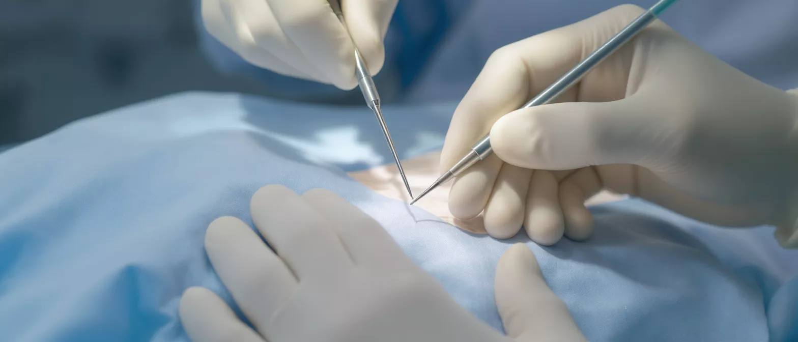
During a TEVAR procedure, a surgeon uses a specialized fabric-covered metal tube, called an endograft or stent graft, to reinforce the damaged section of the aorta. Instead of cutting open the chest to replace the vessel manually, the surgeon threads the device through small incisions in the groin, advancing it through the blood vessels directly to the site of the damage. Once positioned, the endograft expands against the inner walls of the aorta, creating a safe new pathway for blood flow while sealing off the weakened or torn area.
For patients facing heart or vascular surgery, the development of TEVAR represents a significant leap forward in medical technology. This technique achieves the critical goal of stabilizing the aorta without the intense physical trauma associated with traditional open surgeries, making it a vital option for patients who need effective, less invasive cardiac care. Choosing a minimally invasive aortic repair device represents a modern standard of vascular care.
How does the TEVAR procedure work
The mechanics of the TEVAR procedure rely on advanced imaging and precise catheter navigation. The patient is typically given regional or general anesthesia. The surgical team makes small incisions in the groin to access the femoral artery, which serves as the gateway to the rest of the vascular system. Using real-time X-ray guidance, known as fluoroscopy, the team carefully threads a delivery catheter containing the compressed stent graft up to the diseased portion of the thoracic aorta.
Device deployment: Once the catheter reaches the exact location of the aneurysm or dissection, the surgeon carefully withdraws the outer sheath, allowing the stent graft to expand and spring into shape. The device is designed to exert radial force against the healthy tissue above and below the damaged area, securely anchoring itself in place. This creates a durable, artificial lining that relieves pressure on the weakened aortic wall.
Because the procedure avoids large surgical incisions in the chest, the physiological impact on the body is drastically reduced. The entire deployment process usually takes a few hours, depending on the complexity of the patient's anatomy. To ensure the best structural support, surgeons rely on a high-quality thoracic aortic stent graft tailored to the patient's specific vessel dimensions. The precision of this technique allows for immediate stabilization of life-threatening aortic conditions.
What conditions require TEVAR aortic repair
TEVAR is primarily used to treat three major conditions affecting the descending thoracic aorta. The most common indication is a thoracic aortic aneurysm, which is an abnormal ballooning or widening of the aortic wall. When an aneurysm reaches a certain size, the risk of rupture increases dramatically, and a surgeon will recommend TEVAR to reinforce the vessel and prevent a life-threatening bleeding event. If a patient needs intervention for an aortic aneurysm endovascular stent, TEVAR is often the chosen approach.
Dissection repair: Another critical condition treated with TEVAR is an aortic dissection, specifically Type B dissections. An acute dissection occurs when the inner layer of the aorta tears, causing blood to surge through the false channel and separate the vessel walls. TEVAR is used to cover the tear, redirect blood flow into the true lumen, and promote healing of the separated aortic tissues.
Beyond aneurysms and dissections, surgeons may use TEVAR to manage traumatic aortic injuries resulting from severe accidents, or to treat penetrating aortic ulcers and complex pseudoaneurysms. In all these scenarios, the primary goal remains the same: to exclude the damaged segment of the aorta from high-pressure blood flow and prevent catastrophic rupture.
TEVAR vs open repair differences
Understanding the difference between TEVAR and traditional open surgical repair is essential for patients evaluating their treatment options. Open repair involves a large incision in the chest, temporarily stopping the heart and using a heart-lung bypass machine to replace the damaged aortic segment with a synthetic tube. While highly effective, this approach places immense stress on the body and requires extensive time in the intensive care unit.
Surgical impact comparison: TEVAR fundamentally changes the surgical experience. By accessing the aorta through small groin incisions, it eliminates the need for a massive chest opening and prolonged heart-lung bypass. This minimally invasive nature translates to significantly less blood loss, reduced pain, and a lower risk of severe respiratory complications following the operation.
For many patients, especially older individuals or those with underlying health conditions that make open surgery dangerous, TEVAR offers a safer, more tolerable alternative. The survival advantage in the immediate postoperative period is a major reason vascular surgeons prefer this method when the patient's anatomy permits. By utilizing advanced endovascular tools, surgeons achieve similar long-term durability with a fraction of the acute trauma.
TEVAR technique and stent graft placement
The success of TEVAR relies heavily on meticulous preoperative planning and precise stent graft placement. Before the procedure, patients undergo detailed CT scans or MRIs to map the exact dimensions of their aorta. Surgeons measure the length of the damaged area and the diameter of the healthy landing zones above and below it. These measurements determine the specific size and shape of the stent graft required for a custom fit.
Proper placement is critical because the endograft must create a seal without blocking important branching arteries. The surgeon must ensure the stent does not cover the blood vessels that supply the spinal cord or the arteries leading to the head and arms. Achieving a perfect seal requires selecting the correct endovascular stent graft system that accommodates the natural curves and tapering of the patient's aorta.
The technique involves balancing the need for aggressive anchoring against the risk of damaging the delicate aortic tissue. Surgeons may use overlapping stent grafts to cover long segments of the diseased aorta safely. The entire deployment is monitored in real-time, allowing the surgeon to make immediate adjustments if the graft migrates or if an endoleak, or persistent leakage around the graft, becomes visible during the operation.
What to expect during TEVAR recovery time
The recovery journey following TEVAR is generally much smoother and faster than open surgery. Immediately after the procedure, patients are taken to a specialized monitoring unit. Medical staff closely watch for any signs of complications, particularly monitoring kidney function and checking the strength and sensation in the legs to ensure spinal cord blood supply remains healthy. Most patients remain in the hospital for two to four days.
Post-operative milestones: Because there are no large chest incisions, postoperative pain is minimal and easily managed with standard medications. Patients are encouraged to sit up and walk within the first day. The groin incisions heal quickly, allowing patients to resume light daily activities within a week or two of returning home, though heavy lifting remains restricted for a short period.
Complete TEVAR recovery time varies depending on the individual's overall health, but most people return to their normal routines within a month. Follow-up appointments and imaging are crucial to ensure the stent remains securely in place and the aorta continues to heal. Many patients benefit from a supportive postoperative care plan to guide their rehabilitation safely.
Understanding potential TEVAR complications
While TEVAR is a highly advanced and generally safe procedure, it carries certain risks that patients should understand. One of the most specific risks is spinal cord ischemia, which occurs when the stent graft inadvertently covers the arteries supplying blood to the spine. Surgeons mitigate this risk by carefully planning the deployment and sometimes maintaining slightly elevated blood pressure postoperatively to ensure adequate spinal perfusion.
Device-related concerns: Another potential complication is an endoleak. This happens when blood continues to leak into the damaged aneurysm sac, bypassing the seal of the stent graft. While some endoleaks resolve on their own, others require a secondary procedure to place additional grafts or use specialized coils to seal the leak. Device migration or structural failure over time is rare but requires ongoing surveillance.
Other risks are standard for any vascular procedure and include bleeding or infection at the groin incision, kidney injury from contrast dye, and adverse reactions to anesthesia. It is important for patients to discuss these risks openly with their surgical team, understanding that the benefits of repairing a dangerous aortic aneurysm or dissection far outweigh the potential complications for most candidates.
TEVAR follow up and long-term monitoring
Successful TEVAR placement is only the first phase of a lifelong partnership with the cardiac care team. Because the aorta and the implanted stent graft are subject to constant blood pressure and dynamic movement, long-term monitoring is mandatory. Patients must return for regular follow-up appointments, typically scheduled at one month, six months, and annually thereafter.
Imaging and surveillance: At each follow-up visit, patients undergo imaging studies, usually CT scans or echocardiograms. These scans allow the surgeon to verify that the stent graft remains in the correct position, that the aneurysm or dissection is shrinking, and that there are no signs of new endoleaks or device fatigue. This ongoing vigilance ensures the repair remains durable over the years.
Alongside medical monitoring, patients must manage their cardiovascular risk factors. Strict blood pressure control, smoking cessation, and healthy lifestyle choices are vital to prevent the progression of aortic disease in other areas of the body. Engaging in consistent cardiovascular monitoring protects the surgical repair and ensures ongoing health.
TEVAR indications and patient selection
Not every patient with aortic disease is a candidate for TEVAR. The decision to proceed with this endovascular technique depends heavily on the patient's individual anatomy. The most critical factor is the presence of adequate landing zones, which are sections of healthy, straight aortic tissue above and below the damaged area. The stent graft needs at least two centimeters of healthy vessel wall to create a secure seal.
Anatomical constraints: If the aneurysm or dissection is too close to the heart or involves critical branching arteries like the carotid arteries, standard TEVAR may not be possible. In complex cases, surgeons might use modified techniques, such as branched stent grafts, or perform a hybrid operation that combines open and endovascular methods to achieve a safe repair.
Patient selection also considers the overall health profile. TEVAR is frequently recommended for elderly patients or those with severe comorbidities who cannot tolerate the stress of open surgery. However, younger, healthier patients might still be directed toward open repair to avoid the need for future reinterventions that can sometimes accompany endovascular devices.
- Adequate landing zones: Sufficient healthy aorta to secure the graft.
- Overall health status: Ideal for high-risk patients needing minimally invasive options.
- Specific pathology: Best suited for descending thoracic aneurysms and Type B dissections.
Conclusion
Navigating a diagnosis of an aortic aneurysm or dissection is daunting, but modern medicine offers powerful solutions. TEVAR provides a minimally invasive, highly effective method to reinforce the body's most vital artery. By understanding the procedure, recovery expectations, and necessary follow-up care, patients can approach their treatment with confidence rather than fear.
The journey does not end with the procedure; it requires a commitment to long-term monitoring and healthy lifestyle management. Through careful patient selection, precise technique, and ongoing vigilance, TEVAR allows patients to return to their daily lives safely.
Ultimately, open communication with an experienced cardiovascular surgeon is the best way to determine the right path forward. By asking the right questions and understanding the details of the TEVAR procedure, patients become active, empowered participants in their own cardiac health.






