

Penetrating Aortic Ulcer
Learn about penetrating aortic ulcer symptoms, diagnosis, and treatment at Cirugía de Corazón in Guadalajara.
What Is a Penetrating Aortic Ulcer?
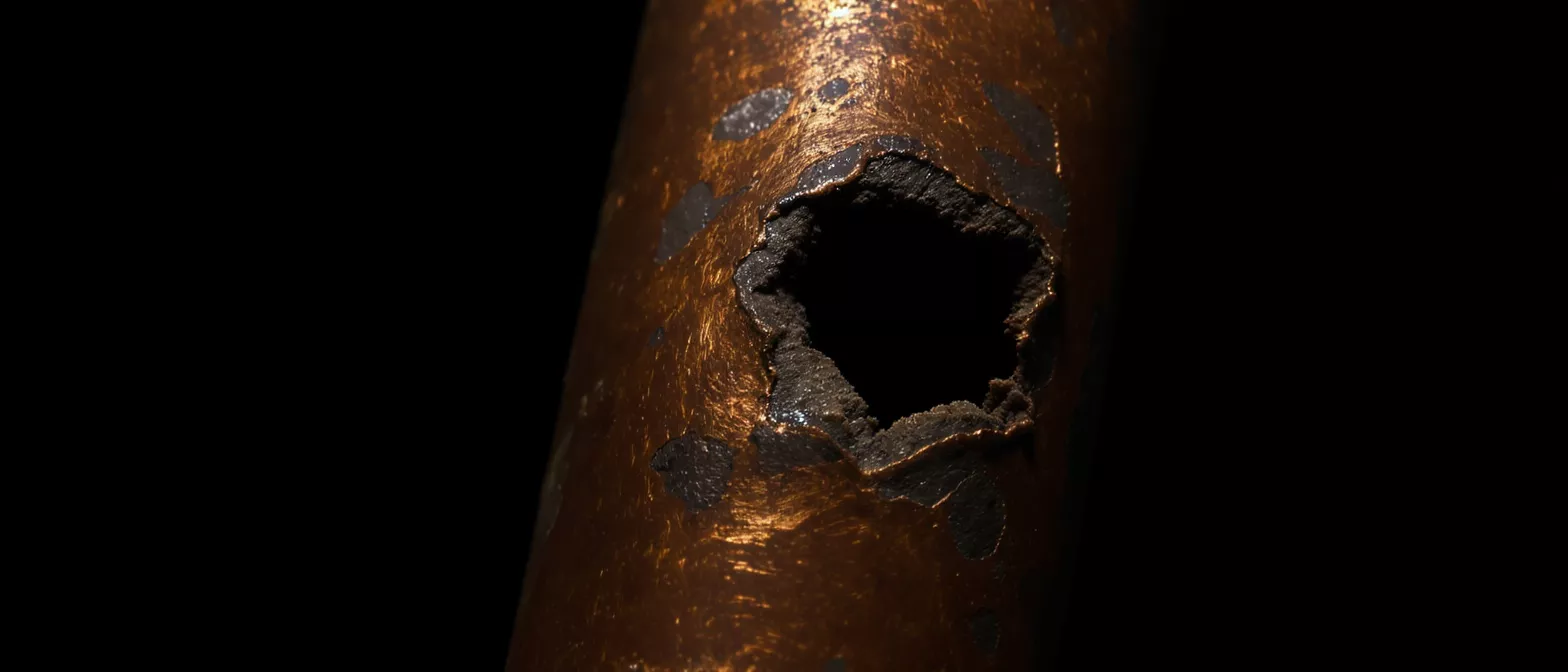
A penetrating aortic ulcer is a condition where a small sore, similar to a peptic ulcer, erodes through the inner lining of the aorta. The aorta is the largest blood vessel in the human body, responsible for carrying oxygen-rich blood away from the heart to the rest of the body. When the protective inner layer breaks down, the ulceration can push deeper into the artery wall. This creates a vulnerable point in one of the most critical structures in the circulatory system. Understanding this condition is vital for any patient facing complex cardiac care.
Unlike a tear that suddenly rips across the artery, this condition typically develops slowly over time. It often begins as a minor disruption in the lining but carries the risk of serious complications if left unmonitored. The erosion can allow blood to enter the wall of the aorta, potentially causing dangerous swelling or even a rupture. For patients evaluating their heart health, identifying these structural weaknesses early provides a crucial window for effective, life-saving intervention.
Medical specialists view this erosion as a specific type of acute aortic syndrome. The progression of the disease depends heavily on how deeply the ulcer penetrates and the overall health of the patient's blood vessels. Seeking expert evaluation is the safest approach when dealing with any abnormality in the aorta. A cardiovascular specialist provides the precise imaging and monitoring required to ensure the blood vessel remains stable.
Penetrating Aortic Ulcer Causes and Risk Factors
Understanding penetratingating aortic ulcer causes begins with looking at the wear and tear of the cardiovascular system. The primary driver is atherosclerosis, a condition where plaque builds up inside the arteries over many years. As this plaque accumulates, it damages the inner lining of the blood vessel. Eventually, the pressure of circulating blood forces the plaque into the artery wall, creating an ulcer. This is why the condition is most frequently seen in older adults who have a long history of vascular disease.
Several underlying factors accelerate this damage and increase a patient's risk profile. Uncontrolled high blood pressure forces blood against the arterial walls with excessive force, accelerating tissue breakdown. Smoking severely degrades vascular health by restricting blood flow and damaging the inner lining. Patients with high cholesterol or a history of coronary artery disease are also at a significantly higher risk of developing these arterial erosions.
- Hypertension: Chronic high blood pressure that physically stresses the walls of the aorta.
- Atherosclerosis: Plaque accumulation that damages and weakens the endothelial lining.
- Age and Vascular History: Most common in patients over 65 with chronic cardiovascular conditions.
Recognizing Penetrating Aortic Ulcer Symptoms
Identifying penetrating aortic ulcer symptoms can be challenging because they often mimic other cardiac events. The most common warning sign is sudden, severe chest or back pain. Patients frequently describe this discomfort as a tearing, ripping, or sharp stabbing sensation that does not relent. Because the aorta runs from the chest down into the abdomen, the exact location of the pain depends entirely on where the ulcer is situated along the vessel. Any sudden, unexplained chest pain requires immediate emergency medical attention.
Sudden localized pain: A piercing sensation in the upper back or chest that feels distinctly different from muscle aches. This occurs because the ulcer is actively eroding through sensitive tissue layers in the artery wall. The pain may feel similar to a heart attack but is often unrelated to blocked coronary arteries.
Neurological changes or limb discomfort: If the swelling from the ulcer disrupts blood flow to spinal or peripheral arteries, patients may experience numbness or weakness. This happens because the damaged aortic wall pinches off smaller branching vessels. Anyone experiencing sudden weakness alongside chest pain should seek immediate care from an aortic emergency treatment center.
How Penetrating Aortic Ulcer Diagnosis Works
Achieving an accurate penetrating aortic ulcer diagnosis relies heavily on advanced cardiovascular imaging. Physicians cannot diagnose this condition through a physical examination alone. When a patient presents with characteristic chest pain, doctors immediately turn to high-resolution scans to look inside the aorta. Computed tomography, commonly known as a CT scan, is the gold standard for viewing the structure of the blood vessel. This scan provides detailed cross-sectional images of the ulcer, showing its exact depth and location.
During the diagnostic process, the medical team looks for specific characteristics to determine the severity of the disease. They measure the width of the ulcer, the degree of blood vessel swelling, and whether blood has leaked into the outer layers. Magnetic resonance imaging, or MRI, is sometimes used as an alternative if the patient cannot tolerate contrast dye. These precise images allow the surgeon to map out the exact anatomy of the aorta before planning any intervention.
The diagnostic goal is to confirm the presence of the ulcer and rule out other life-threatening conditions. Distinguishing between an ulcer, a dissection, and a regular aneurysm is critical because the treatment pathways differ.
Penetrating Aortic Ulcer vs Aortic Dissection
Distinguishing between a penetrating aortic ulcer and aortic dissection is a critical step in emergency cardiology. While both conditions involve damage to the aortic wall, their mechanisms are distinctly different. An ulcer acts like a localized crater, boring deeply into a specific spot of the artery lining. In contrast, an aortic dissection is a sudden, lengthwise tear that causes the inner layer to peel away. Blood then surges into the newly formed false channel, splitting the wall of the aorta apart over a long distance.
Differentiating the pathophysiology: An ulcer is a focal, deep erosion, while a dissection is a linear tear. A dissection typically presents with tearing pain that migrates, whereas an ulcer's pain is often sharply localized. Understanding this difference helps thoracic specialists predict how the condition is likely to progress and what complications might arise.
Despite these differences, the two conditions can overlap. A deeply penetrating ulcer can eventually cause the lining to tear, leading directly into a dissection. This is why continuous monitoring is essential even if the ulcer initially appears stable. By comparing the two conditions, patients can better understand why their medical team chooses specific diagnostic protocols and surgical timelines.
Acute Aortic Syndrome and Aortic Intramural Hematoma
Physicians group penetratingating aortic ulcer, aortic dissection, and aortic intramural hematoma under a single umbrella term known as acute aortic syndrome. These three interconnected conditions all involve the deterioration of the aortic wall and carry a high risk of fatal rupture. An intramural hematoma occurs when blood bleeds into the wall of the aorta without a visible tear in the lining. This creates a bruise within the artery wall that can cause severe pain and restrict blood flow. Recognizing the symptoms of acute aortic syndrome early is the most critical factor in patient survival.
A penetrating ulcer can actually lead to the formation of an intramural hematoma. As the ulcer burrows through the inner lining, it allows blood to seep into the middle layer of the tissue. If the blood remains trapped within the wall, it forms the hematoma. If the pressure builds until it shears the inner layer, a dissection occurs. Understanding these related syndromes helps explain why doctors treat even small arterial ulcers with significant urgency.
Because of the overlapping nature of these conditions, specialized cardiovascular monitoring equipment is necessary to track changes in the aortic wall. A patient diagnosed with an intramural hematoma might be prescribed medication to lower their heart rate and blood pressure. This reduces the mechanical stress on the artery wall, giving the tissue a chance to heal naturally without surgical intervention.
Aortic Ulcer Treatment and Management Options
Determining the right aortic ulcer treatment depends heavily on the size of the ulcer and the patient's overall health. If the ulcer is small and not causing severe symptoms, doctors often recommend a conservative medical approach. The primary goal is to reduce the physical stress on the artery wall. This is achieved through medications that lower blood pressure and slow the heart rate. By keeping the force of the heartbeat gentle, the risk of the ulcer rupturing decreases significantly.
When the ulcer is large, growing rapidly, or causing intense pain, surgical intervention becomes necessary. The decision to operate is based on the clear danger of the ulcer turning into a rupture or dissection. Surgeons weigh the risks of an open procedure against less invasive techniques. An aortic disease specialist evaluates the imaging scans to determine if the ulcer is in a location that is safe to repair surgically. The patient's age, lung function, and previous surgical history also factor heavily into this decision.
- Medical Management: Beta-blockers and antihypertensives to reduce stress on the aortic wall.
- Surgical Intervention: Required for symptomatic, large, or rapidly expanding ulcers.
- Active Surveillance: Regular imaging to monitor stable but high-risk structural changes.
Endovascular Aortic Repair for Thoracic Aortic Ulcer
For a thoracic aortic ulcer, endovascular aortic repair has become the preferred surgical method for eligible patients. This minimally invasive procedure avoids opening the chest. Instead, the surgeon makes a small incision in the groin to access the femoral artery. A specialized delivery catheter is guided upward into the aorta using real-time x-rays. Once the catheter reaches the ulcer, a stent graft is carefully deployed to cover the damaged section of the artery wall.
The stent graft acts as a new, durable inner sleeve for the aorta. It creates a barrier that seals off the ulcer, preventing blood from entering the crater and reducing the pressure on the weakened wall. Because the chest remains unopened, patients typically experience less pain, lower blood loss, and a significantly shorter hospital stay compared to open surgery.
Endovascular repair is highly effective, but not every patient is a candidate. The anatomy of the aorta must have suitable healthy sections above and below the ulcer to anchor the stent graft securely. If the arteries are too tortuous or heavily calcified, passing the delivery catheter safely becomes too risky. The surgical team meticulously measures the blood vessel on CT scans beforehand to ensure a precise fit for the graft.
Penetrating Aortic Ulcer Surgery Recovery
Recovery after penetratingating aortic ulcer surgery varies greatly depending on whether the procedure was endovascular or an open repair. After an endovascular stent placement, patients often spend just a few days in the hospital. The incisions in the groin are small, and physical mobility returns quickly. Patients are encouraged to walk short distances the day after the procedure to promote healthy blood circulation. Most individuals return to their normal routines within two to four weeks.
Open surgical repair is a much more extensive operation. It involves replacing the damaged section of the aorta with a synthetic tube graft. Because the chest must be opened and the heart temporarily bypassed, the physical toll is significantly higher. Patients spend the first night in the intensive care unit and usually remain in the hospital for a week or longer. A full recovery from open aortic surgery requires several months of structured cardiac rehabilitation and careful, gradual physical exertion.
Regardless of the surgical method, strict blood pressure control remains the cornerstone of a successful recovery. The medical team will frequently adjust medications to ensure the heart pumps gently, protecting the new graft and the remaining native aorta. Patients must attend all follow-up appointments for scheduled CT scans, ensuring the stent is functioning properly and no new ulcers are forming elsewhere in the chest.
Long-Term Management and Heart Health
Surviving a penetratingating aortic ulcer requires a lifelong commitment to cardiovascular health. Even after a successful repair, the underlying vascular disease that caused the ulcer remains. Patients must adopt strict lifestyle modifications to prevent future complications. This includes quitting smoking, adopting a heart-healthy diet, and maintaining a regular exercise routine approved by their cardiologist. Managing cholesterol and keeping blood pressure in a strict target range are the most critical pillars of long-term survival.
Ongoing surveillance is mandatory for the rest of the patient's life. The aorta is a living structure that continues to age and endure stress. Surgeons check the stent graft annually using CT or MRI imaging to ensure it has not shifted or developed endoleaks, a condition where blood leaks around the edges of the stent. These routine scans also check the rest of the aorta for new aneurysms or ulcers. Consistent monitoring allows doctors to catch structural changes before they become emergencies.
Living with a repaired aorta can feel daunting, but modern cardiovascular medicine provides excellent tools for a long, healthy life. By partnering closely with a cardiovascular specialist, patients regain their confidence and return to their daily activities. The key is unwavering adherence to medication, healthy lifestyle choices, and scheduled imaging. Proactive management turns a frightening diagnosis into a manageable condition.






