

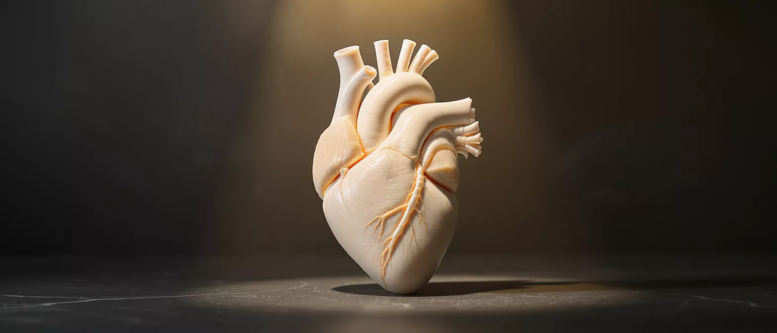
Aortic Root
Learn about aortic root anatomy, function, conditions like aneurysms and abscesses, and surgical treatments.
What is the aortic root?
The aortic root is the starting point of the aorta, the largest artery in the human body. Located just above the left ventricle of the heart, it serves as the critical bridge between the heart's main pumping chamber and the systemic circulation. When the heart beats, blood flows from the ventricle directly through this root to reach the rest of the body.
Why the aortic root matters to heart patients: This section of the heart is not just a simple pipe; it is a highly complex, dynamic structure. It houses the aortic valve, which ensures blood flows in one direction. Without a healthy aortic root, the valve cannot function properly, and the heart must work much harder to pump blood effectively.
For patients facing cardiac surgery, understanding this part of the heart helps demystify the procedure. Knowing how the heart's plumbing works reduces anxiety and helps families feel more confident discussing treatment options with their surgical team.
Aortic root anatomy and function
The aortic root anatomy is complex, consisting of several distinct components working in harmony. It includes the aortic annulus, the fibrous ring where the heart valve sits, and the sinuses of Valsalva, which are small bulges that help create the right blood flow dynamics to keep the valve leaflets moving smoothly.
How the aortic root function maintains blood flow: During systole, the heart contracts, and blood pushes the valve leaflets open. The unique shape of the root's sinuses creates small swirls of blood that wash behind the leaflets. This prevents them from sticking to the artery walls and slamming shut too forcefully, which protects the valve from physical damage over time.
Role of the aortic valve within the root: When the heart relaxes in diastole, the valve's three leaflets snap shut tightly to prevent blood from spilling backward into the ventricle. A structurally sound root is essential to keep this seal perfect. Any enlargement or distortion can cause the valve to leak, leading to a heart murmur and eventual heart failure if left untreated.
What causes aortic root dilation?
Aortic root dilation occurs when the walls of the root gradually stretch and enlarge. This stretching is often caused by chronic high blood pressure, which forces the aorta to handle higher pressure over years, weakening its elastic fibers. Other times, the cause is congenital, meaning the patient was born with tissue that is prone to stretching.
Some patients inherit genetic conditions, such as Marfan syndrome or bicuspid aortic valve disease, which directly affect the connective tissue of the arteries. In these cases, the arterial walls lack the structural strength to maintain their original shape, leading to progressive aortic root enlargement even if blood pressure is well controlled.
- Long-standing hypertension: Constant high pressure physically stretches the arterial walls over time.
- Genetic tissue disorders: Inherited conditions compromise the structural integrity of the aorta.
- Valve abnormalities: Conditions like a bicuspid aortic valve alter blood flow dynamics, stressing the root.
Understanding aortic root aneurysm
When the dilation becomes significant, it is classified as an aortic root aneurysm. An aneurysm is a localized bulging of the artery wall. Because the root is located inside the pericardium, an enlargement here is particularly dangerous and requires careful monitoring by a cardiologist or cardiac surgeon.
What are the risks of an aortic root aneurysm? The primary risk is dissection or rupture. As the wall stretches and thins out, it becomes fragile. If the inner layer tears, blood can split the artery wall apart, leading to a life-threatening emergency. Furthermore, a large aneurysm changes the shape of the root, pulling the valve leaflets apart and causing severe leakage.
Surgeons measure the diameter of the root to determine the risk. When the aortic root dimensions reach a critical size, or if the valve is leaking heavily, surgical intervention is recommended to prevent a rupture rather than waiting for symptoms to appear.
How is an aortic root abscess diagnosed?
An aortic root abscess is a localized infection that destroys tissue in the root, often as a complication of endocarditis. Endocarditis is an infection of the heart valves, and the bacteria can spread directly into the surrounding structures, creating a pocket of pus and inflammatory tissue that compromises the heart's foundation.
Recognizing the signs of aortic root pathology: Patients with an abscess may experience persistent fevers, chills, and abnormal heart rhythms. The infection can interfere with the heart's electrical pathways, causing heart block, which is a delayed or disrupted heartbeat that shows up clearly on an electrocardiogram.
Diagnosis requires advanced imaging. An echocardiogram, particularly a transesophageal echo, is the gold standard for visualizing the root. It allows the surgical team to see the exact extent of the infection and plan for aggressive surgical cleaning and reconstruction.
Aortic root dimensions and measurement
Measuring the aortic root is crucial for determining the severity of a patient's condition. Surgeons use echocardiography and CT scans to obtain precise measurements of the root's diameter. These dimensions tell the medical team exactly how much the artery has stretched compared to a normal baseline.
How is aortic root measurement used in treatment planning? The size of the root dictates the surgical timeline. If the root is only mildly enlarged, doctors may simply monitor it with annual imaging. However, if the diameter reaches 5.5 centimeters, or if it is growing rapidly, the risk of rupture outweighs the risk of surgery, and an operation is scheduled.
What is the normal aortic root definition? In a healthy adult, the aortic root diameter typically ranges between 2 and 3 centimeters. Understanding this baseline helps patients comprehend why their cardiologist is concerned when measurements exceed these limits. Size is only one factor; the rate of growth and family history are also heavily weighed.
When is aortic root surgery necessary?
Aortic root surgery is necessary when the risks of leaving the root untreated exceed the risks of an operation. This calculation depends on the size of the aneurysm, the presence of an abscess, and the function of the aortic valve. If the valve is leaking significantly due to the root's stretched shape, surgery is needed to restore normal blood flow.
For patients with genetic conditions like Marfan syndrome, surgical guidelines often recommend operating at smaller diameters, around 5.0 centimeters, because their tissues are inherently weaker and more prone to sudden tearing. Similarly, if a patient needs another heart surgery, such as a coronary bypass, the surgeon will often address a moderately dilated aortic root simultaneously.
Exploring aortic root treatment options: The surgical team will evaluate whether the valve can be spared or if it needs replacement. This decision determines the specific type of operation.
What is aortic root replacement surgery?
Aortic root replacement surgery is a highly specialized procedure where the enlarged or infected section of the aorta is removed and replaced with a synthetic tube. The surgeon also replaces the aortic valve, attaching a mechanical or biological valve directly into this new tube, creating a combined structure known as a valved conduit.
What happens during aortic root replacement surgery? The patient is placed on cardiopulmonary bypass, which takes over the work of the heart and lungs. The surgeon carefully excises the diseased root, protecting the coronary arteries, which must be reattached to the new synthetic conduit to ensure the heart muscle continues to receive oxygen-rich blood.
This surgery is complex and requires an experienced cardiac surgeon. The goal is to permanently remove the threat of rupture or ongoing infection while ensuring the heart pumps efficiently. Recovery is significant, but the long-term outcomes are excellent for patients who follow their post-operative care plans closely.
Aortic root repair vs replacement
The choice between aortic root repair and replacement depends heavily on the condition of the aortic valve. If the valve itself is healthy but simply leaks because the root is too wide, surgeons prefer a valve-sparing root replacement. This involves replacing the enlarged artery while delicately sewing the patient's own valve back into the new graft.
Benefits of aortic root repair: By keeping the native valve, patients avoid the need for lifelong blood thinners, which are mandatory with mechanical valves. The risk of blood clots and bleeding complications is significantly lower, and the natural hemodynamics of the heart are preserved. This approach is the gold standard for younger patients with otherwise healthy valve tissue.
When is total replacement unavoidable? If the valve is diseased, heavily calcified, or destroyed by an infection like endocarditis, it cannot be saved. In these cases, the entire root and valve are replaced together.
Recovery after aortic root treatment
Recovery after aortic root treatment is a gradual process that requires patience and careful monitoring. Patients usually spend a few days in the intensive care unit immediately after surgery to ensure their heart function and blood pressure are perfectly stable. They are then moved to a regular hospital room to begin walking and breathing exercises.
What to expect during cardiac rehabilitation: Once home, patients will feel fatigued for several weeks. Strict rules against heavy lifting or driving are enforced to allow the breastbone to heal. Cardiac rehabilitation, a structured exercise and education program, is critical for safely rebuilding cardiovascular strength under medical supervision.
Long-term follow-up includes regular imaging to ensure the new aortic graft remains secure and functioning perfectly. Patients must maintain healthy blood pressure and adopt heart-healthy lifestyle habits to protect their cardiovascular system. The surgical team remains a lifelong partner in the patient's cardiac care journey.






