

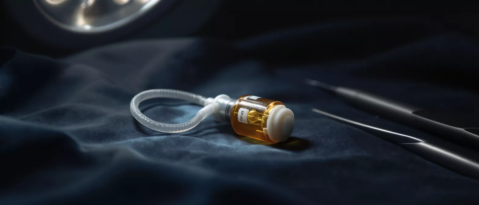
Valved Conduit
Learn how a valved conduit restores heart blood flow and valve function.
What is a valved conduit?
A valved conduit is a specialized tube-like device used in cardiac surgery to replace or bypass a damaged section of the heart or its major blood vessels. It combines a durable, flexible pipe, known as the conduit, with a functional heart valve built directly into it. This design allows the surgeon to restore both unobstructed blood flow and proper valve function in a single, integrated implant. For patients facing complex heart conditions, this medical device offers a reliable pathway for blood to travel from the heart to the lungs or the rest of the body.
Why a valved conduit matters to cardiac patients: When a heart valve and its connecting vessel are both diseased, simply repairing the valve may not be enough. The surrounding tissue might be enlarged, weakened, or scarred from congenital defects or infections. A valved conduit provides a complete structural replacement, ensuring the new valve has a stable, healthy landing zone. This approach is often necessary when treating severe aneurysms or complex congenital heart defects where the native anatomy is severely compromised.
Understanding this surgical tool can ease much of the anxiety surrounding heart operations. Surgeons rely on these devices to fix problems that standard valve repair cannot solve, making them an essential option in modern cardiac reconstruction and a vital component in adult and pediatric heart surgery.
How does a valved conduit work?
Restoring unidirectional blood flow: The valved conduit functions by acting as a new, clear highway for blood while simultaneously preventing backflow. The conduit portion replaces the damaged native vessel, providing a rigid yet flexible channel. The integrated valve opens to allow blood to flow forward, then closes tightly to prevent blood from leaking backward. This ensures that oxygen-depleted blood reaches the lungs efficiently, or that oxygen-rich blood is pumped out to the body.
The mechanics rely heavily on the pressure changes within the heart. When the heart beats, pressure forces the valve inside the conduit open, pushing blood through the tube. As the heart relaxes, the pressure drops, and the valve snaps shut. This precise timing mimics the natural function of a healthy heart, maintaining efficient circulation and reducing the workload on the cardiac muscle.
By replacing both the vessel and the valve simultaneously, the device addresses multiple points of failure at once, creating a more robust and long-lasting repair that helps the heart pump more effectively.
Types of valved conduits
Choosing between pulmonary and aortic placements: The two main categories are the pulmonary valved conduit and the aortic valved conduit. A pulmonary valved conduit connects the right ventricle to the pulmonary artery, carrying blood to the lungs. An aortic valved conduit connects the left ventricle to the aorta, carrying blood to the rest of the body. The choice depends entirely on which side of the heart requires surgical reconstruction.
Biological versus mechanical valve options: Within these categories, surgeons choose between biological and mechanical components. Biological valves are made from animal tissue, offering excellent natural blood flow without requiring long-term blood thinners. Mechanical valves are made from durable synthetic materials like carbon, lasting longer but requiring lifelong anticoagulant therapy. The decision balances the patient's age, lifestyle, and bleeding risk.
The specific type of conduit chosen is tailored to the patient's unique anatomy and the underlying disease. Your surgical team will evaluate imaging scans to determine the exact size, shape, and material composition that will provide the best long-term outcome for your heart.
Materials used in valved conduit implants
Biological and synthetic materials: Cardiac valved conduits are constructed from a variety of materials designed for durability and compatibility with the human body. Biological conduits often use treated bovine pericardium (cow tissue) or porcine valves (pig tissue), which are specially processed to prevent rejection. The tube itself might be made from woven polyester, a strong, synthetic material that integrates well with surrounding tissues.
The engineering of these materials ensures the device withstands the constant, high-pressure pumping of the heart. Woven polyester is highly resistant to stretching and degradation, making it an ideal material for the conduit portion. The biological valves are treated to remain flexible while eliminating the cells that could trigger an immune response, ensuring they function quietly and efficiently.
Some modern designs combine a mechanical valve with a polyester graft, known as a prosthetic valved conduit. Selecting the right material is crucial for a successful cardiac reconstruction implant, as it dictates the longevity and maintenance of the new valve over time.
When is valved conduit surgery necessary?
Addressing complex congenital defects: Surgeons frequently use a valved conduit in pediatric and adult congenital heart surgery. Conditions like Tetralogy of Fallot or pulmonary atresia often result in missing or severely underdeveloped pulmonary arteries. In these cases, the conduit provides a new pathway from the right ventricle to the lungs, correcting the anatomical deficit and restoring proper oxygenation to the blood.
Treating aneurysms and severe infections: In adults, a valved conduit replacement is often required when an aortic aneurysm stretches the vessel so widely that the valve can no longer close properly. It is also used to treat aortic dissections, where the layers of the aortic wall tear, or endocarditis, an infection that severely damages the heart valve and surrounding tissue.
By removing the diseased tissue and replacing it with a sturdy, functional device, surgeons can eliminate life-threatening complications and restore healthy cardiac function for patients with advanced structural heart disease.
Valved conduit surgical procedure
Cardiopulmonary bypass and exposure: The valved conduit procedure is an open-heart surgery performed under general anesthesia. The surgical team places the patient on a heart-lung machine, which takes over the job of pumping blood and oxygenating it while the heart is stopped. This allows the surgeon to open the heart and remove the diseased native vessel and valve with precision.
Suturing the new conduit into place: Once the damaged tissue is removed, the surgeon carefully sews the new valved conduit into position. They use fine, permanent sutures to create a secure, leak-proof seal between the heart and the prosthetic graft. The surgeon then checks the connections to ensure blood flows correctly and the valve opens and closes without obstruction.
After confirming proper function, the team restarts the heart, carefully removes air from the chambers, and separates the patient from the heart-lung machine. The chest is then closed, and the patient is transferred to the intensive care unit for close monitoring.
Choosing the right aortic aneurysm repair device is a critical step the surgeon takes before the operation begins, ensuring the specific dimensions match the patient's anatomy.
Valved conduit recovery and hospital stay
Intensive care and early mobilization: Valved conduit surgery recovery begins in the intensive care unit, where nurses monitor vital signs continuously. Patients typically have several tubes and drains to help clear fluids and support breathing. Within the first 24 hours, the team encourages gentle movement, such as sitting on the edge of the bed, to prevent blood clots and promote lung expansion.
Once transferred to a standard hospital room, the focus shifts to walking and regaining strength. Physical therapists guide patients through safe exercises. Pain management is carefully controlled, transitioning from intravenous medications to oral pain relievers as healing progresses. Most patients stay in the hospital for about a week, depending on their overall health.
Complete recovery takes several months.
Potential valved conduit complications
Risks of open-heart surgery: Like any major operation, cardiac conduit surgery carries risks. General complications include bleeding, infection at the incision site, or adverse reactions to anesthesia. Patients may also experience irregular heart rhythms, known as arrhythmias, which are usually temporary but may require medication or a pacemaker.
Device-specific complications over time: While rare in the short term, long-term valved conduit complications can occur. The biological valve inside the conduit may wear out over time, requiring a replacement procedure. The artificial tube itself can develop calcifications, narrowing the pathway and restricting blood flow. In mechanical conduits, blood clots remain a risk, necessitating strict adherence to anticoagulant therapy.
Regular follow-up appointments and echocardiograms are essential to monitor the device and catch these issues early. Patients should immediately report symptoms like shortness of breath, chest pain, or fever to their surgical team.
Long-term outlook with a valved conduit
Monitoring valve function and durability: The lifespan of a valved conduit depends on the materials used and the patient's age. Biological conduits generally last 10 to 15 years before the tissue degenerates, while mechanical valves can last a lifetime. However, the synthetic graft material usually remains durable. Patients will need lifelong cardiac checkups to ensure the device functions correctly.
Returning to daily activities and exercise: Most patients return to normal activities within three months, though heavy lifting and strenuous exercise must be cleared by a surgeon. Walking is highly encouraged right from the start of recovery. Over time, the heart adapts to the new valve, and many patients report a significant improvement in their energy levels and overall quality of life.
Living with a conduit means taking simple precautions, such as informing dentists about the artificial valve to prevent endocarditis. With proper care and regular monitoring, patients can enjoy a full, active life after their cardiac reconstruction.
Preparing for valved conduit surgery
Medical evaluations and clearances: Before surgery, patients undergo a series of tests to ensure they are healthy enough for the operation. These include blood work, electrocardiograms, and detailed imaging like CT scans or echocardiograms. Patients must also provide a complete medical history, including all medications and supplements, to avoid interactions during the procedure.
Lifestyle adjustments before admission: Doctors will instruct patients to stop smoking immediately, as tobacco use severely impacts healing and increases surgical risks. Certain medications, like blood thinners, may need to be paused or adjusted several days before admission. It is also essential to arrange for post-discharge support at home, including meal preparation and transportation.
Mental preparation is just as important. Understanding the surgical plan and asking the surgical team questions can significantly reduce anxiety. Many hospitals offer pre-surgical education classes that explain the procedure, recovery timeline, and what to expect in the intensive care unit.
Patients should pack a small bag with personal items, like compression socks for cardiac surgery, comfortable loose clothing, and toiletries, to make their hospital stay as comfortable as possible.
Frequently asked questions summary
Understanding the core concepts: The most common questions revolve around the durability of the implant and the need for blood thinners. If a mechanical valve is used, lifelong blood thinners are required. If a biological valve is chosen, blood thinners are usually only needed for a few months post-surgery.
Managing expectations for recovery: Patients often ask when they can return to driving, work, and exercise. Most people can drive in 4 to 6 weeks and return to non-physical jobs in 6 to 8 weeks. Always follow the specific timeline provided by your surgeon, as healing varies from person to person.
This surgical intervention is highly effective for complex heart conditions. By replacing both the damaged vessel and the faulty valve, the valved conduit restores healthy circulation and offers a second chance at an active, healthy life.
- Pulmonary vs. Aortic: The conduit is placed based on whether blood needs to go to the lungs (pulmonary) or the body (aortic).
- Material lifespan: Biological conduits last 10-15 years, while mechanical valves can last a lifetime but require blood thinners.
- Recovery timeline: Hospital stay is typically one week, with full recovery taking up to three months.






