

Valved Conduit Surgery: Replacing Valve and Vessel Together
Valved conduit surgery replaces a damaged heart valve and artery together in one operation.
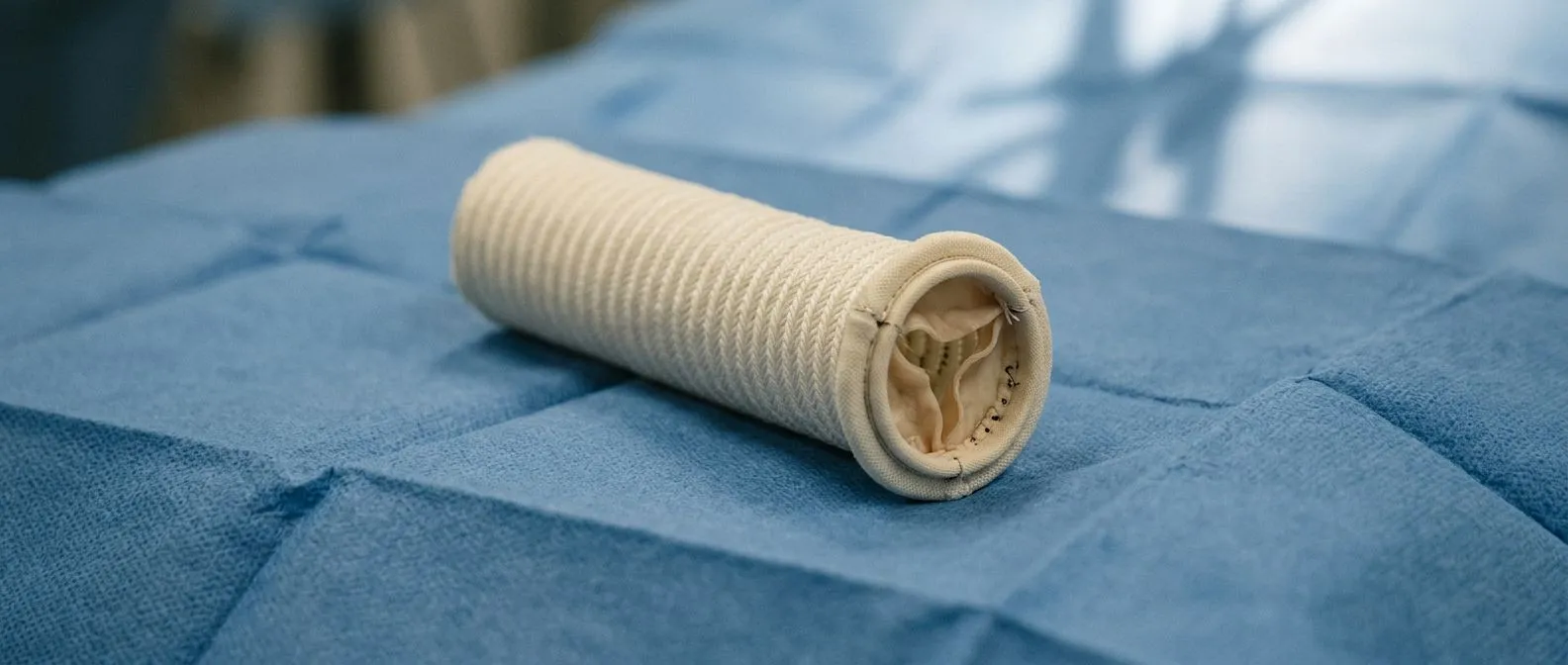
If your cardiac team has mentioned valved conduit surgery, you are likely facing a problem that involves more than the heart valve alone: the great vessel around it also needs rebuilding. A valved conduit is a synthetic tube — usually woven polyester — with a heart valve sewn inside it, and valved conduit surgery replaces a damaged valve and the vessel segment together in one operation. It sounds daunting, but it is a well-established operation performed around the world, and understanding what it involves is the first step toward feeling calmer about it. This guide walks through why it is done, how it is performed, what recovery really looks like, and how to choose the right surgical team.
What Is Valved Conduit Surgery?
Valved conduit surgery is an operation in which a surgeon removes a diseased heart valve and a segment of the adjoining great vessel — usually the aorta, the body's main artery, or the pulmonary artery, which carries blood to the lungs — and replaces both with a single prefabricated tube containing a valve. Think of it as replacing a stretch of worn pipe and its faulty one-way gate at the same time, rather than trying to patch the gate while leaving the pipe weak.
Why not just replace the valve? In some conditions, replacing the valve alone would leave diseased, stretched, or infected vessel wall behind, which can keep enlarging, leak, or tear. When the vessel itself is the problem — as in an aneurysm (a permanent bulge in the artery wall), certain infections, or complex congenital heart defects — the durable answer is to replace the whole segment. The conduit provides both a new valve and a new, strong vessel wall in one implant.
Where is it placed? Most often the conduit sits where the aortic valve and ascending aorta live, in a reconstruction often called a Bentall procedure. It can also be placed between the right ventricle — the lower pumping chamber that sends blood to the lungs — and the pulmonary artery, particularly in congenital heart surgery. The technique varies, but the principle is always the same: restore a secure channel for blood with a valve that opens and closes properly inside it.
What Does "Conduit Heart" Mean?
You may hear the phrase "conduit heart" or "conduit patient" in clinic. It does not describe a special type of heart; it simply describes a heart that now relies on an implanted tube-and-valve assembly for part of its circulation. Many adults with congenital heart disease have lived with a conduit since childhood and are described this way by their cardiology teams.
Living with a conduit. A conduit heart works the way any repaired heart works: blood flows through the artificial valve inside the tube, which opens to let blood forward and closes to prevent backflow. Most patients with a well-functioning conduit feel well, exercise, work, and live full lives. The conduit does, however, need lifelong follow-up with echocardiography — an ultrasound of the heart — because conduits can narrow or leak gradually over the years.
What follow-up involves. Expect regular imaging, sometimes every six to twelve months depending on the conduit's age and type, plus dental hygiene vigilance, since bacteria from the mouth can seed artificial valves. Your team will also discuss medication needs, such as anticoagulants (blood thinners) if the valve inside the conduit is mechanical.
When Does a Heart Need Valve and Artery Replacement?
The most common adult scenario is a diseased aortic valve combined with an enlarged ascending aorta, or an aortic root — the segment of aorta just above the valve — that has stretched beyond safe limits. Guidelines generally consider surgery when the aortic root or ascending aorta reaches about 5.5 cm in diameter in most adults, or earlier in connective tissue disorders such as Marfan syndrome, where thresholds are lower. Combining the valve replacement with vessel replacement in a single operation avoids a second sternotomy (opening of the chest) later.
Infection is another major reason. Prosthetic Valve Endocarditis — infection of an artificial valve — can destroy both the valve and the surrounding tissue, leaving abscess cavities and weakened structures. In these cases, radical removal of all infected tissue and reconstruction with a valved conduit is often the safest path to cure, because leaving infected tissue behind invites relapse.
Congenital and reoperative situations. Patients born with conditions such as tetralogy of Fallot or truncus arteriosus often need their pulmonary conduit replaced when it wears out — and modern designs, including bioprosthetic models like the KONECT RESILIA aortic valved conduit, aim to extend the time between operations. In our practice, every case is reviewed by the multidisciplinary team against international guidelines before a recommendation is made, and the patient receives a clear proposal with the alternatives laid out side by side.
Valved Conduit vs Valve Replacement: Which Is Right?
For many patients, a straightforward Mitral Valve Replacement or aortic valve replacement — without vessel disease — is all that is needed, and adding a conduit would be unnecessary surgery. The decision comes down to whether the vessel around the valve is healthy. If the aorta is normal in size and tissue, an isolated valve replacement through a standard Surgical Aortic Valve Replacement approach is less extensive.
When the conduit wins. If imaging shows an aneurysm, severe calcification, dissection (a tear in the artery layers), or infection extending beyond the valve, valve-only replacement is incomplete. In that setting, valved conduit heart surgery is not "more surgery for its own sake" — it is the operation that addresses the whole problem at once. Our team recommends the less invasive route only where the evidence supports it for that specific patient, and we say so plainly when a conduit is genuinely required.
Older patients and TAVR vs SAVR. Families often ask whether a 78-year-old should have transcatheter aortic valve replacement (TAVR) or conventional surgery. TAVR — delivering a valve on a catheter through the groin — is an excellent option for many older or higher-risk patients, but it does not replace a diseased ascending aorta. When the vessel needs rebuilding, a surgical approach with a conduit usually remains the right answer, and age alone does not decide it; overall condition, anatomy, and frailty do.
- Healthy vessel + diseased valve: valve replacement alone, surgical or transcatheter, is usually sufficient.
- Diseased vessel + diseased valve: a valved conduit addresses both in one operation.
- Congenital conduit worn out: pulmonary valved conduit surgery restores flow from the right ventricle to the lungs.
Types of Valved Conduits
Conduits are not all alike, and the choice inside the operating room shapes the years that follow. Broadly, the valve within the tube is either mechanical (made of durable synthetic material, requiring lifelong blood thinners) or a Tissue Valve (made from animal or human tissue, usually sparing patients from anticoagulation but with a finite lifespan).
Commercial and handmade options. Manufacturers produce prefabricated conduits, including bioprosthetic conduits and newer designs that use a treated tissue valve inside a tube. Surgeons also construct handmade valved woven polyester conduits, sewing a valve into a tube themselves — a technique that can be tailored to unusual anatomy. Research even explores novel restorative and custom-designed pulmonary conduits, including work applying machine learning to validate handmade trileaflet valve designs for pulmonary valved conduit reconstruction (IEEE Xplore).
Biological alternatives. Some centers use homografts — donor human valves and vessels — or decellularized grafts, where donor tissue is processed to remove cells and reduce immune reaction; a decade of clinical experience with decellularized fresh homografts for pulmonary valve replacement has been reported in the literature (IBN). Meanwhile, stentless and Stentless Valve configurations, Sutureless Valve technologies, and Self-expanding Valve designs continue to expand the toolbox, particularly for minimally invasive and transcatheter-era practice.
How the Valved Conduit Procedure Works
Valved conduit replacement is performed under general anesthesia with the heart-lung machine, which takes over circulation while the heart is stopped and opened. The surgeon removes the diseased valve and vessel segment, then sews the conduit in place — the valve end at the valve annulus (the ring where the valve sits) and the tube end joined to the remaining healthy vessel. Coronary arteries, which feed the heart muscle itself, are reattached to the conduit wall through small openings called buttons.
How long does it take? Expect the operation itself to take several hours, typically three to six depending on complexity, whether it is a first operation or a redo, and whether other procedures are combined. You will wake up in an intensive care unit with a breathing tube briefly in place, then step down to a cardiac ward within a day or two in an uncomplicated course. Before any of this, we take the time in consultation to explain what the studies actually show, in plain words, and to answer every question — including the ones families hesitate to ask.
What the incision looks like. Most valved conduit operations use a full midline breastbone incision, though selected cases can be done through smaller incisions. Dr Molina's team performs conventional, minimally invasive, robotic, and transcatheter procedures across our centers, so the approach is chosen to fit the anatomy — not to fit a habit.
Valved Conduit Surgery Recovery: What to Expect
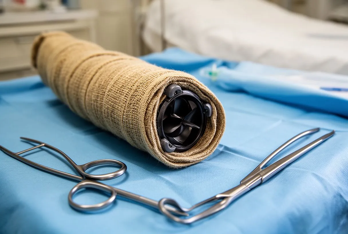
Patients usually ask first: how long does it take to fully recover from an aortic valve replacement or a conduit operation? The honest answer is staged. Most people are up walking within days, home within one to two weeks, and driving again around four to six weeks if healing is on track and they are not taking strong pain medication. Full recovery — feeling like yourself, back to work and normal exercise — commonly takes about two to three months, because the breastbone needs that long to knit solidly.
The first six weeks. Plan on cardiac rehabilitation: a supervised program of graded exercise that rebuilds confidence and fitness safely. Follow sternal precautions — no lifting, pushing, or pulling more than a few pounds with your arms — while the breastbone heals. Expect tiredness that surprises you; major surgery demands energy, and fatigue lifting week by week is normal, not a setback.
Life afterward. If your conduit contains a mechanical valve, warfarin (a blood thinner) becomes a permanent companion with regular blood tests. Tissue valves generally avoid this but may eventually need re-replacement. Many patients ask about life expectancy after aortic valve surgery: published experience shows that most patients return to a near-normal life expectancy when surgery is done for the right reasons at the right time, though exact figures depend on age, other conditions, and valve type. Long-term follow-up with regular imaging of the conduit helps catch small problems while they are still small.
Valved Conduit Surgery Complications and Success Rate
Every open-heart operation carries risks, and you deserve them stated plainly. The main valved conduit surgery complications include bleeding requiring reoperation, stroke, Prosthetic Valve Endocarditis, kidney strain, arrhythmias such as atrial fibrillation, and — specific to conduits — leakage at the coronary reattachment sites or progressive narrowing of the conduit over years. Your surgical team estimates your personal risk with tools such as EuroSCORE II, a scoring system that combines age and health factors to estimate operative risk.
Interpreting "success rate." For standard valved conduit operations in experienced centers, operative success is high and long-term function is good, but no honest team quotes a single universal number — outcomes depend on your specific disease, urgency, and overall health. What matters more for you is the specific discussion of your own risk profile, not a statistic from a registry.
Reducing your personal risk. The strongest levers are preparation and team experience: controlling diabetes and blood pressure before surgery, stopping smoking, meticulous dental care beforehand, and choosing a team that performs this operation regularly. We encourage second opinions and treat seeking one as a normal, sensible part of deciding about heart surgery — a good team welcomes it.
Pediatric and Adult Valved Conduit Surgery
Pediatric valved conduit surgery differs from the adult operation in one crucial way: children grow, and conduits do not. A conduit placed in a child will eventually become relatively small, and tissue conduits in the pulmonary position calcify and stiffen with time. Families should understand this is expected biology, not a surgical failure — the plan from day one anticipates future catheter or surgical revisions as the child grows.
Right ventricle valved conduit considerations. When the conduit connects the right ventricle to the pulmonary artery, problems show up as exercise intolerance, breathlessness, or rhythm issues as the conduit degrades. Monitoring focuses on the pressure gradient across the conduit and on leaking, and many modern programs can now place a valve inside a failing conduit by catheter — a valve-in-conduit approach with a balloon-expandable transcatheter valve — postponing open surgery in selected patients.
Adult valved conduit surgery. Adults usually face the aortic version: root aneurysm, dissection, or infection. Adults with repaired congenital heart disease form a bridge group — they are adult-sized now, but their anatomy and scar tissue demand congenital expertise. In both groups, the goal is the same: the longest possible interval of good conduit function between operations, with the least invasive reasonable intervention each time.
Ask specifically about: the surgeon's volume with this exact operation, whether a multidisciplinary heart team reviews the plan, which conduit brands and valve types the hospital stocks, intensive care capability, and what the all-inclusive package covers (implants, hospital stay, anesthesia, follow-up imaging). Be wary of any quote given before anyone has actually reviewed your echocardiogram and CT scan.
What a proper evaluation looks like. A trustworthy program starts with your imaging and history, not a price list. Our first contact is a free video consultation: we get to know the patient, talk through the condition, and identify exactly which studies are still needed so no unnecessary ones are ordered — a face-to-face visit at our consulting rooms in Guadalajara, Querétaro, or Hermosillo follows only when the patient decides to go ahead. Dr Molina trained in cardiothoracic surgery at the Universidad de Guadalajara, in cardiac, minimally invasive, robotic and structural surgery at the University of Montreal, and in structural cardiology and TAVR at CHU Bordeaux; the team also trained at Jena, Bad Berka and Buffalo. He performed the first robotic mitral valve repair in the Ángeles Health System and the first robotic heart surgery in Guadalajara.
Travel and planning questions. International patients should build in time: preoperative studies on arrival, the hospital stay, and enough recovery locally before flying — most teams advise waiting until the chest is stable and the surgeon clears you for air travel. Ask in writing about emergency aftercare, who to contact with concerns, and how follow-up imaging will be shared with your cardiologist back home. Harvard Health notes that realistic recovery expectations and clear communication with your surgical team are central to a good outcome (Harvard Health Publishing).
Valved conduit surgery replaces a failing valve and a diseased vessel together, and for the right patient it is the operation that solves the whole problem in one sitting. The key takeaways: the conduit is a tube-and-valve implant used when vessel disease, infection, or congenital anatomy makes valve replacement alone incomplete; recovery follows a predictable arc from ICU to walking to full activity over roughly two to three months; risks are real but manageable with an experienced team and honest risk assessment; and conduit choice — mechanical versus tissue — shapes your follow-up for years. None of this replaces a personal evaluation. If you or a loved one has been told a conduit may be needed, bring your questions and your imaging to a qualified cardiac team, ask about every alternative, and make the decision with people who explain, not just operate. Booking a consultation with a multidisciplinary heart team is the right next step — never a self-diagnosis.
Valved Conduit Surgery Cost in Mexico and Choosing a Team
For international patients, valved conduit surgery in Mexico can offer a significant cost difference compared with the United States, without compromising standards — but you should evaluate programs with the same rigor you would anywhere. Ask specifically about: the surgeon's volume with this exact operation, whether a multidisciplinary heart team reviews the plan, which conduit brands and valve types the hospital stocks, intensive care capability, and what the all-inclusive package covers (implants, hospital stay, anesthesia, follow-up imaging). Be wary of any quote given before anyone has actually reviewed your echocardiogram and CT scan.
What a proper evaluation looks like. A trustworthy program starts with your imaging and history, not a price list. Our first contact is a free video consultation: we get to know the patient, talk through the condition, and identify exactly which studies are still needed so no unnecessary ones are ordered — a face-to-face visit at our consulting rooms in Guadalajara, Querétaro, or Hermosillo follows only when the patient decides to go ahead. Dr Molina trained in cardiothoracic surgery at the Universidad de Guadalajara, in cardiac, minimally invasive, robotic and structural surgery at the University of Montreal, and in structural cardiology and TAVR at CHU Bordeaux; the team also trained at Jena, Bad Berka and Buffalo. He performed the first robotic mitral valve repair in the Ángeles Health System and the first robotic heart surgery in Guadalajara.
Travel and planning questions. International patients should build in time: preoperative studies on arrival, the hospital stay, and enough recovery locally before flying — most teams advise waiting until the chest is stable and the surgeon clears you for air travel. Ask in writing about emergency aftercare, who to contact with concerns, and how follow-up imaging will be shared with your cardiologist back home. Harvard Health notes that realistic recovery expectations and clear communication with your surgical team are central to a good outcome (Harvard Health Publishing).
Valved conduit surgery replaces a failing valve and a diseased vessel together, and for the right patient it is the operation that solves the whole problem in one sitting. The key takeaways: the conduit is a tube-and-valve implant used when vessel disease, infection, or congenital anatomy makes valve replacement alone incomplete; recovery follows a predictable arc from ICU to walking to full activity over roughly two to three months; risks are real but manageable with an experienced team and honest risk assessment; and conduit choice — mechanical versus tissue — shapes your follow-up for years. None of this replaces a personal evaluation. If you or a loved one has been told a conduit may be needed, bring your questions and your imaging to a qualified cardiac team, ask about every alternative, and make the decision with people who explain, not just operate. Booking a consultation with a multidisciplinary heart team is the right next step — never a self-diagnosis.






