

Valve Annulus
Learn valve annulus anatomy and its role in heart blood flow.
What is the valve annulus in the heart?
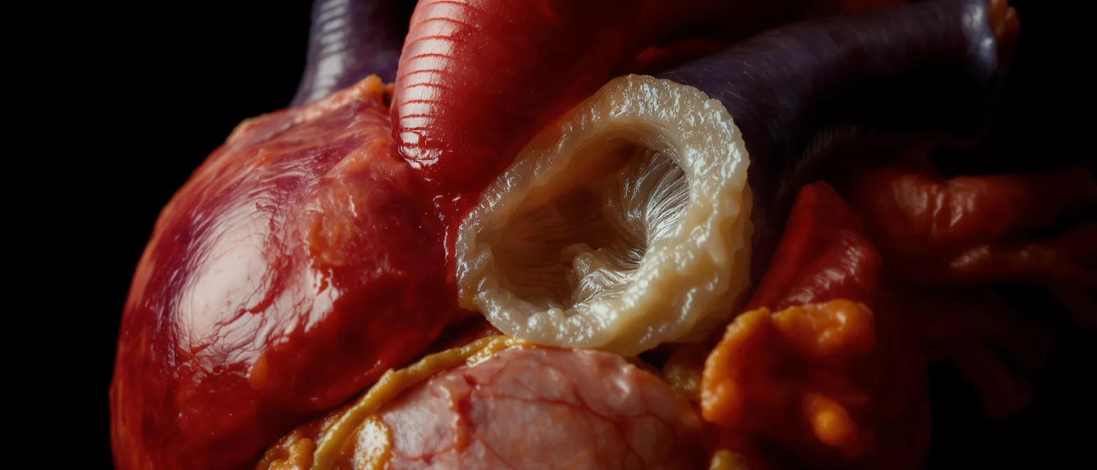
The valve annulus is a ring-shaped band of tough, fibrous tissue that serves as the anchor for each of the heart's four valves. Think of it as the sturdy frame surrounding a door, holding the leaflets firmly in place so they can open and close efficiently. In cardiac anatomy, this structure is also referred to as the annulus fibrosus, providing the necessary structural support to withstand the high pressures of blood pumping through the chambers.
Valve annulus definition and basic anatomy: To understand the valve annulus definition, picture a gasket or a washer that seals the edge of a pipe. In the heart, this fibrous ring ensures that when the valve leaflets close, they form a tight, leak-proof seal. Without a properly functioning annulus, the delicate leaflets would not be able to maintain their shape, leading to backward blood flow and inefficient circulation.
When you hear surgeons discuss heart valve disease, the annulus is frequently a central part of the conversation. It is not just a static ring; it is a dynamic, living structure that subtly changes shape with every single heartbeat, expanding and contracting to facilitate smooth, unidirectional blood flow throughout the body.
How does valve annulus anatomy support blood flow?
The valve annulus anatomy plays a critical role in maintaining unidirectional blood flow through the heart. Because the fibrous ring provides a rigid yet flexible base, the valve leaflets can open fully to allow blood to pass forward, and then snap shut securely to prevent any backward leakage, a condition doctors call regurgitation.
Mechanics of the cardiac annulus structure: The cardiac annulus structure acts like a flexible hinge. During the heart's pumping phase, or systole, the annulus contracts and slightly reduces its diameter. This dynamic narrowing helps bring the leaflets together perfectly, ensuring a tight coaptation zone that seals the valve completely against the high pressure of the bloodstream.
If the annulus loses its natural flexibility or becomes stretched out, the leaflets cannot meet properly in the center. This is why understanding the mechanics of this structure is so vital; the proper function of the entire cardiovascular system relies heavily on the mechanical integrity of these tiny, pressurized rings of connective tissue.
Types of heart valve annulus: mitral, aortic, and tricuspid
There are distinct types of heart valve annulus, each uniquely shaped to handle different pressures and blood volumes. The mitral valve annulus and the tricuspid valve annulus are saddle-shaped and highly dynamic, changing shape dramatically throughout the cardiac cycle to support the inflow of blood into the lower chambers. The aortic valve annulus, by contrast, is much more rigid and tubular, designed to withstand the intense, high-pressure ejection of blood from the left ventricle to the rest of the body.
Comparing the left and right side annulus fibrosus heart structures: The annulus fibrosus heart structures vary depending on their location. The left side of the heart handles much higher pressures, so the mitral and aortic annuli are heavily reinforced with dense collagen fibers. The right side, featuring the tricuspid and pulmonary annuli, operates under lower pressure and is slightly more compliant, accommodating the venous return of blood to the lungs.
- Mitral: Saddle-shaped, highly dynamic, prevents blood from leaking back into the left atrium.
- Aortic: Rigid, crown-shaped structure designed to withstand high systemic blood pressure.
- Tricuspid: Oval-shaped and flexible, manages lower-pressure blood flow returning from the body.
What happens during annulus dilation heart disease?
Annular dilation heart disease occurs when the fibrous ring stretches beyond its normal size. When the annulus becomes dilated, the valve leaflets are pulled apart and can no longer meet in the center to form a seal. This results in a leaky valve, allowing blood to flow backward, which forces the heart muscle to work significantly harder to pump the same amount of blood forward.
Causes and consequences of a stretched valve annulus: A stretched valve annulus is often caused by untreated high blood pressure, genetic connective tissue disorders, or age-related tissue degeneration. As the ring expands, the physical gap between the valve leaflets widens. Over time, this backward leakage increases the volume of blood in the heart chambers, leading to dangerous stretching of the heart muscle and eventual heart failure if left untreated.
Patients experiencing this condition may feel shortness of breath, fatigue, or irregular heartbeats. A surgeon evaluating this condition will measure the diameter of the ring to determine if it has pathologically expanded, which is the first step in planning an effective surgical repair strategy.
Why valve annulus calcification affects heart function
Valve annulus calcification happens when calcium deposits build up on the fibrous ring, turning the normally flexible tissue into a hard, unyielding shell. This hardening prevents the annulus from dynamically contracting during the heartbeat, severely restricting the valve's ability to open fully, a condition known as stenosis. The rigidity also prevents the leaflets from closing properly, which can cause a combination of blocked forward blood flow and backward leakage.
The impact of severe calcium buildup on heart valves: Severe calcium buildup makes surgical interventions much more complex. A heavily calcified ring can crack or fracture during traditional surgical procedures, making it incredibly difficult to securely implant a replacement valve. Surgeons must carefully assess the density and depth of calcium using advanced imaging before deciding whether to perform a repair, a mechanical replacement, or a tissue replacement.
For patients, understanding this process is essential because calcification often progresses silently over many years without noticeable symptoms. Regular monitoring and early intervention can prevent the heart muscle from thickening and weakening due to the constant struggle of pumping blood through a stiff, narrowed valve.
How surgeons perform annulus repair surgery
During annulus repair surgery, the primary goal is to reduce the size of the stretched fibrous ring back to its normal dimensions so the valve leaflets can close properly again. The surgeon accomplishes this by implanting a specialized prosthetic ring, known as an annuloplasty ring, around the base of the valve. This artificial ring acts as a new, sturdy foundation, pulling the leaflets closer together and restoring a tight, leak-proof seal.
Choosing the right heart valve repair ring for patients: Choosing the right heart valve repair ring depends on the specific valve being treated and the patient's unique pathology. Surgeons may select a rigid ring to completely stabilize a heavily calcified base, or a flexible, partial band that preserves the heart's natural geometric movement. The choice of implant is tailored to ensure long-term durability while maintaining optimal blood flow dynamics.
This surgical approach is highly effective because it addresses the root cause of the leakage rather than just treating the symptoms. By securing the base of the valve, surgeons can often preserve the patient's native leaflets, avoiding the need for a total valve replacement and reducing the reliance on long-term blood thinners.
Valve annulus measurement in echocardiography
Accurate valve annulus measurement is the cornerstone of both diagnosis and surgical planning. Using echocardiography, which is a painless ultrasound of the heart, doctors can precisely visualize the size, shape, and movement of the fibrous ring. These measurements are taken during different phases of the heartbeat to fully understand the dynamic changes in the valve's opening and closing.
How imaging guides surgical decisions and sizing: How imaging guides surgical decisions is a matter of millimeters; a slight miscalculation in measurement can result in a poorly fitting implant. Advanced imaging techniques like 3D echocardiography and cardiac MRI allow surgeons to view the annulus from multiple angles, ensuring they select an implant size that perfectly matches the patient's anatomy. This preoperative planning is critical for minimizing complications and ensuring a successful outcome.
During your pre-surgical evaluation, you will likely undergo several of these imaging tests. Finding a reliable cardiac echocardiography imaging center is vital, as the data collected from these scans directly dictates the surgical approach, the type of incision used, and the specific prosthetic devices prepared for the operating room.
Are annular ring heart implants safe long-term?
Annular ring heart implants have an excellent safety profile and are widely considered a gold-standard treatment for repairing leaking valves. These prosthetic devices are typically constructed from medical-grade titanium, silicone, or advanced polymer composites, which are highly biocompatible. This means the body readily accepts the material without triggering a severe immune reaction, allowing the surrounding tissue to heal naturally around the implant.
Durability and biocompatibility of repair devices: The durability and biocompatibility of these repair devices mean that patients can often enjoy a lifetime of normal heart function without needing another surgery. Unlike mechanical valves, which require lifelong blood-thinning medications, structural repair rings carry a much lower risk of blood clots. However, patients still need regular check-ups to ensure the implant remains secure and functional.
Long-term studies show that the vast majority of these implants continue to function perfectly decades after the initial surgery. Patients looking into their options might explore different types of cardiac valve repair devices alongside their surgeon to understand the materials and designs best suited for their specific heart anatomy and lifestyle needs.
Cardiac annulus structure and surgical implications
The cardiac annulus structure has profound implications for how a surgeon approaches a valve repair or replacement. Because the annulus is deeply integrated with the heart's electrical conduction system—especially in the tricuspid and mitral positions—surgeons must be incredibly precise when placing sutures. A misplaced stitch in the fibrous ring can inadvertently damage the heart's wiring, leading to a condition known as a heart block, which would require a pacemaker.
Anatomical landmarks and surgical risks: Understanding these anatomical landmarks helps surgeons navigate around critical structures like the coronary arteries and the aortic root. The proximity of the aortic valve annulus to the heart's primary electrical pathway means that operations in this area require specialized techniques to avoid permanent damage. Surgeons use decades of anatomical research to map out safe suture zones around the perimeter of the ring.
This is why heart valve surgery is such a delicate, highly specialized field. The surgeon must not only fix the mechanical problem of the leaking or narrowed valve but also protect the complex, intertwined architecture of nerves, muscle, and connective tissue that surrounds the annulus.
Preparing for heart valve annulus surgery
Preparing for heart valve annulus surgery involves both physical and mental readiness. Your surgical team will conduct a comprehensive evaluation, including blood tests, lung function tests, and detailed heart imaging, to ensure you are a strong candidate for the procedure. It is completely normal to feel anxious, but knowing exactly what to expect can significantly reduce preoperative stress and improve your overall recovery.
What to expect during recovery and rehabilitation: What to expect during recovery includes spending a few days in the intensive care unit for close monitoring, followed by a few weeks of gradual, guided rehabilitation at home. Your breastbone will need time to heal, so you will be given specific instructions on how to safely move, lift, and exercise without straining your chest. Most patients begin a cardiac rehabilitation program to safely rebuild their strength and stamina under professional supervision.
It is important to have a strong support system of family or friends to help with daily activities during the first few weeks of recovery. Attending all follow-up appointments is critical, as your surgeon will use echocardiography to verify that the repaired annulus and valve are functioning perfectly, ensuring a safe and full return to your normal daily life.






