

STEMI
Learn STEMI symptoms, ECG diagnosis, and emergency treatment.
What is a STEMI heart attack?
A STEMI is the most severe type of heart attack a person can experience. The term stands for ST-Elevation Myocardial Infarction, a highly technical phrase that essentially describes a specific pattern seen on an electrocardiogram (ECG) when a major artery supplying the heart has become completely blocked. When this critical blood flow is suddenly cut off, the heart muscle begins to starve, making immediate medical intervention absolutely essential to save the patient's life.
Why this distinction matters: Medical professionals use the term STEMI to rapidly differentiate a full-thickness heart attack from less severe cardiac events. A non-STEMI heart attack implies a partial blockage, whereas a STEMI means the blockage is total and the risk of permanent heart damage is imminent. This distinction triggers a massive, coordinated emergency response from the hospital staff.
Understanding the gravity of this condition helps patients and families recognize why emergency rooms act so quickly when a STEMI is suspected. The primary goal is not just to relieve chest pain, but to physically open the blocked artery before irreversible damage occurs. Every minute matters, making rapid diagnosis the first critical link in the chain of survival.
How does STEMI affect the heart muscle?
The heart is a powerful, tireless muscle that requires a constant, uninterrupted supply of oxygen-rich blood, delivered by the coronary arteries. In a STEMI, a blood clot suddenly forms on top of a ruptured cholesterol plaque, completely occluding one of these vital arteries. Without oxygen, the heart tissue downstream from the blockage begins to starve and die off rapidly.
Transmural damage explained: The damage in a STEMI is termed transmural, meaning it extends through the entire thickness of the heart muscle wall. This distinguishes it from less severe attacks where the damage is localized only to the inner surface. Full-thickness damage severely compromises the heart's ability to pump blood effectively to the rest of the body's organs.
Because the heart muscle lacks the ability to regenerate well, the tissue destroyed during a STEMI is typically replaced by stiff scar tissue as the patient heals. While scar tissue holds the heart together structurally, it does not contract or pump effectively. This is exactly why preserving muscle function by opening the blocked artery as fast as possible is the central pillar of acute cardiac emergency care.
What are the symptoms of STEMI?
Recognizing the symptoms of a STEMI early can be the difference between life and death. The most common and classic sign is sudden, severe chest pain that often feels like a crushing weight or intense pressure sitting directly in the center of the chest. This discomfort may radiate outward to the left arm, jaw, neck, or back, and it rarely goes away with rest or changing positions.
Atypical warning signs: Not everyone experiences the classic chest-clutching pain seen in movies. Many patients, particularly women, older adults, and individuals with long-standing diabetes, present with atypical symptoms such as profound shortness of breath, sudden nausea, cold sweats, or extreme unexplainable fatigue. These silent or subtle warning signs often delay treatment, which is why any sudden cardiac symptom warrants an emergency evaluation.
If you or a loved one experience these symptoms, calling emergency services immediately is critical. Paramedics can begin life-saving interventions in the ambulance and alert the hospital to prepare for a cardiac emergency before you even arrive. Attempting to drive yourself or a family member to the hospital wastes precious time and can be fatal if the heart's rhythm suddenly becomes unstable on the road.
How is STEMI diagnosed on an ECG?
The fastest and most reliable way to diagnose a STEMI is through an electrocardiogram, commonly known as an ECG or EKG. This completely non-invasive test measures the electrical activity of the heart through small patches placed on the skin of the chest, arms, and legs. Within just a few minutes, the machine can show doctors exactly how the heart's electrical pathways are behaving.
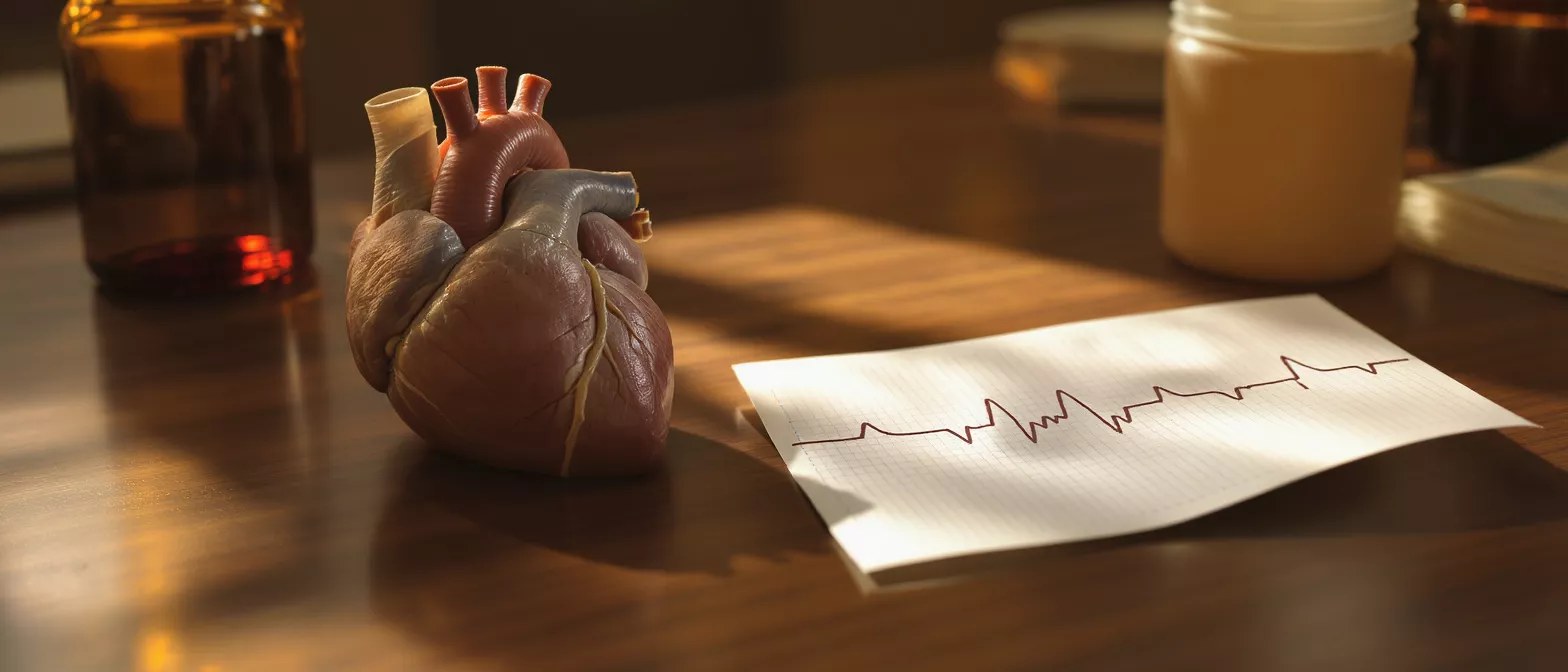
Recognizing ST elevation: In a healthy heartbeat, the ECG shows a very specific wave pattern. When a major coronary artery is fully blocked, a specific segment of the wave, called the ST segment, rises abnormally high above the baseline. This elevation is the absolute hallmark of a STEMI and instantly tells the medical team that a severe, full-thickness heart attack is actively in progress.
- Rapid ECG assessment: Paramedics and triage nurses use portable ECGs in the field to identify ST elevation quickly, transmitting the results to the waiting hospital.
- Reciprocal changes: Doctors also look for reciprocal depressions on the opposite side of the heart, which helps pinpoint the exact blocked artery.
- Blood tests for confirmation: Troponin blood levels are drawn to confirm heart muscle damage, though the visual ECG findings dictate immediate emergency treatment.
Doctors rely so heavily on the ECG because waiting for blood test results to confirm heart damage wastes critical time. The visual elevation of the ST segment is enough evidence to activate the cardiac catheterization laboratory, ensuring the patient is rushed to the appropriate treatment suite the moment they enter the hospital doors.
Types of heart attack and STEMI
Cardiologists broadly categorize acute heart attacks into two main types: STEMI and Non-STEMI. Both are incredibly serious medical emergencies, but they require distinctly different treatment strategies because of how they affect the heart's blood supply. Understanding the difference helps patients comprehend the rapid medical decisions made during a crisis.
Comparing STEMI and NSTEMI: A STEMI involves a complete blockage of a coronary artery, causing total interruption of blood flow to a specific section of the heart. A Non-STEMI involves a partial blockage, meaning some blood still gets through, but the patient is still at high risk of the blockage worsening. This distinction is vital because a STEMI requires immediate physical intervention to mechanically clear the artery.
Both conditions are usually caused by the sudden rupture of a cholesterol plaque inside the artery wall, which triggers the body's clotting system to form a clot. In a STEMI, the clot grows so rapidly and large that it completely seals the pipe. Doctors use this classification system to instantly communicate the severity of the patient's condition to the entire medical response team.
Emergency treatment for STEMI
The instant a STEMI is recognized, the entire focus of treatment shifts to reperfusion, which simply means restoring blood flow to the starving heart muscle. There is absolutely no time to waste, as the muscle continues to die minute by minute. Emergency departments have specific, highly rehearsed protocols to move patients to treatment as rapidly as possible.
Primary percutaneous coronary intervention: The gold standard for STEMI treatment is primary angioplasty. In this procedure, an interventional cardiologist threads a thin tube through a small incision in the wrist or groin, navigating directly to the blocked heart artery. A tiny balloon is inflated to push the plaque aside, and a coronary stent placement is performed to keep the artery permanently open, restoring immediate blood flow.
When a cardiac catheterization lab is not immediately available within the critical time window, doctors may use thrombolytics, commonly known as clot-busting drugs. These powerful medications are administered intravenously to dissolve the blockage from within the bloodstream. Rapid-acting thrombolytic therapy can buy vital time, but physical stenting remains the definitive long-term solution to keep the artery open.
STEMI benefits of immediate reperfusion
The primary and most urgent benefit of immediate reperfusion in a STEMI is the preservation of viable, functioning heart muscle. When blood flow is restored quickly, the heart tissue is saved before it undergoes irreversible cell death. This preserves the heart's pumping function and drastically reduces the long-term risk of debilitating heart failure.
Time is muscle: Cardiologists frequently use this phrase to emphasize that the sooner the artery is opened, the better the patient's outcome. The ideal timeframe from entering the hospital doors to opening the artery, known as the door-to-balloon time, is under ninety minutes. Achieving this target significantly lowers mortality rates and vastly improves the patient's future quality of life.
- Preserved ejection fraction: Quick treatment maintains the heart's percentage of blood pumped with each beat, preventing long-term cardiac failure.
- Reduced scar tissue: Less muscle death means smaller areas of stiff scar tissue, keeping the heart structurally sound and flexible.
- Long-term survival: Patients who undergo rapid reperfusion have significantly higher survival rates in the critical months and years following the attack.
Ultimately, the rapid restoration of blood flow prevents the heart from remodeling into a weaker, enlarged shape. By maintaining its natural geometry and strength, patients are able to return to their daily lives with a much lower risk of subsequent complications. Advanced cardiac monitoring equipment is often used to carefully track this recovery over time and ensure the heart is healing properly.
Are STEMI treatments safe and effective?
Modern STEMI treatments are incredibly safe and highly effective, representing some of the most successful emergency interventions in the history of modern medicine. Primary angioplasty has a very high success rate at opening the blocked artery immediately, resolving the severe chest pain almost instantly in the vast majority of cases.
Risks versus rewards: Like any invasive medical procedure, angioplasty carries some risks, such as bleeding at the insertion site, allergic reactions to contrast dye, or the rare possibility of the artery narrowing again over time. However, the risk of doing nothing during a STEMI is near-certain death or severe disability. The overwhelming rewards of restoring blood flow far outweigh the procedural risks.
Following the acute emergency intervention, patients are prescribed a strict regimen of medications to prevent new clots from forming inside the stent. Dual antiplatelet therapy, usually consisting of daily aspirin and a specific prescription clot-prevention drug, keeps the newly placed stent clear. Compliance with these medications is absolutely crucial to ensure the long-term safety and effectiveness of the emergency treatment.
Recovery and life after a STEMI
Recovering from a STEMI is a gradual, step-by-step process that involves both physical healing and significant emotional adjustment. Most patients spend a few days in the hospital for close observation, where doctors monitor their heart rhythm and ensure the newly placed stent is functioning perfectly. The physical damage heals over several weeks as the heart forms stable scar tissue.
Cardiac rehabilitation: A cornerstone of recovery is enrolling in a supervised cardiac rehabilitation program. This structured exercise and education plan helps patients safely rebuild their cardiovascular fitness under direct medical supervision. It also provides essential emotional support, helping patients overcome the very natural anxiety and fear that follow a severe heart attack.
Life after a STEMI involves taking medications exactly as prescribed by your cardiologist and attending all follow-up appointments without fail. Many patients go on to live full, active lives, returning to work, hobbies, and family activities. The key is understanding that a STEMI is a massive warning sign, and adopting a heart-healthy lifestyle is necessary to prevent future blockages from occurring.
Preventing a second STEMI
Preventing a second STEMI requires a lifelong, daily commitment to modifying cardiovascular risk factors. The underlying plaque that caused the initial blockage exists throughout the coronary arteries, meaning a future event is a very real risk if aggressive lifestyle changes are not made. Secondary prevention is just as important as the emergency treatment itself.
Key lifestyle modifications: Patients must adopt a heart-healthy diet low in saturated fats, sodium, and refined sugars. Quitting smoking is absolutely essential, as tobacco severely damages blood vessel linings and promotes clot formation. Regular physical activity, as approved and guided by a cardiologist, helps strengthen the heart muscle and control weight, blood pressure, and cholesterol levels.
Medical management is equally critical for long-term prevention. Patients will likely need to take medications to lower cholesterol, control blood pressure, and prevent blood clots for the rest of their lives. By combining daily medication with sustained, healthy lifestyle changes, patients significantly reduce their risk of experiencing another STEMI and protect their long-term heart health for years to come.






