STEMI Heart Attack: Emergency Treatment and Recovery
STEMI treatment requires emergency care within 90 minutes to save heart muscle.
What Is STEMI and Why Emergency Treatment Matters
A STEMI heart attack is the most severe form of acute myocardial infarction, where a coronary artery becomes completely blocked and heart muscle begins to die within minutes. The term STEMI stands for ST-segment elevation myocardial infarction, a pattern seen on an electrocardiogram (ECG) that tells the cardiac team a major artery is fully occluded. Understanding what STEMI treatment involves helps patients and families make sense of the urgency, the procedures, and the long road of recovery that follows. Every minute that passes without blood flow reduces the amount of heart muscle that can be saved, which is why emergency teams move so quickly once the diagnosis is confirmed.
Why time is muscle: Cardiologists often repeat this phrase because it captures the single most important principle in STEMI care. Heart cells deprived of oxygen begin to suffer irreversible damage after roughly twenty minutes, and after two hours the affected territory may be permanently lost. The goal of STEMI treatment is to open the blocked artery as fast as possible — ideally within ninety minutes of first medical contact. This window, known as door-to-balloon time, is a recognised benchmark in international guidelines because faster reperfusion translates into better survival and less long-term disability.
For families watching a loved one rushed into the catheterisation laboratory, the speed can feel frightening rather than reassuring. It helps to know that the urgency is not a sign of chaos but of a well-rehearsed protocol. The team is working against a clock measured in heart muscle cells, not hours. When that protocol works as designed, many patients leave the hospital with their heart function largely preserved and a clear plan for preventing another event. The rest of this guide explains each stage of that journey, from the first symptom to life at home.
How STEMI Differs From Other Heart Attacks
Not every heart attack is the same, and the distinction between STEMI and other types matters because it changes the urgency and the treatment strategy. A STEMI involves complete blockage of a coronary artery with full-thickness (transmural) damage to the heart wall, producing the characteristic ST elevation on the ECG. A non-STEMI, by contrast, involves a partial or temporary blockage — enough to injure muscle but not enough to produce that classic electrical signature. Unstable angina sits alongside these as chest pain at rest caused by reduced flow but without measurable muscle damage yet. These three conditions together are called acute coronary syndromes, and they share many causes but require different responses.
Partial blockage versus complete occlusion: In a non-STEMI, a fragment of an atheromatous plaque — a cholesterol-rich deposit inside the artery wall — ruptures and partially narrows the vessel, causing symptoms but leaving some flow intact. In a STEMI, that rupture triggers a clot that seals the artery completely. This is why STEMI patients are rushed to the catheterisation laboratory for immediate mechanical reopening, whereas non-STEMI patients may be stabilised with medication and treated within twenty-four to seventy-two hours depending on risk stratification. Research compiled through registries such as PubMed continues to refine these thresholds and help clinicians decide who needs immediate intervention versus careful observation.
Understanding the difference helps families grasp why one patient is wheeled to a procedure within minutes while another is monitored on a ward with medications first. It is not that one patient is less important — it is that the anatomy and the electrical findings dictate the safest, most effective strategy for that particular artery at that particular moment. Both pathways aim for the same destination: a stable patient, a reopened or protected artery, and a plan to prevent the next event.
Signs and Symptoms That Demand Immediate STEMI Treatment
Recognising a STEMI early is the first and most critical step in STEMI treatment, because nothing else matters if the ambulance is not called. The classic symptom is crushing central chest pressure, often described as a weight or squeezing sensation rather than sharp pain, lasting more than a few minutes and not eased by rest. This pressure may radiate to the left arm, both arms, the neck, jaw, or upper back. Some patients sweat profusely, feel nauseated, or experience a sense of impending doom that is hard to articulate but unmistakable once you have felt it.
Atypical presentations: Not every STEMI follows the textbook. Women, older adults, and people with diabetes frequently report shortness of breath, profound fatigue, indigestion-like discomfort, or arm pain without classic chest pressure. This variability leads to dangerous delays, as patients and bystanders wait to see if symptoms pass. An atypical infarction can be just as lethal as a textbook one, and the rule is simple: any new, persistent chest or upper-body discomfort lasting beyond a few minutes warrants an emergency call, not a wait-and-see approach.
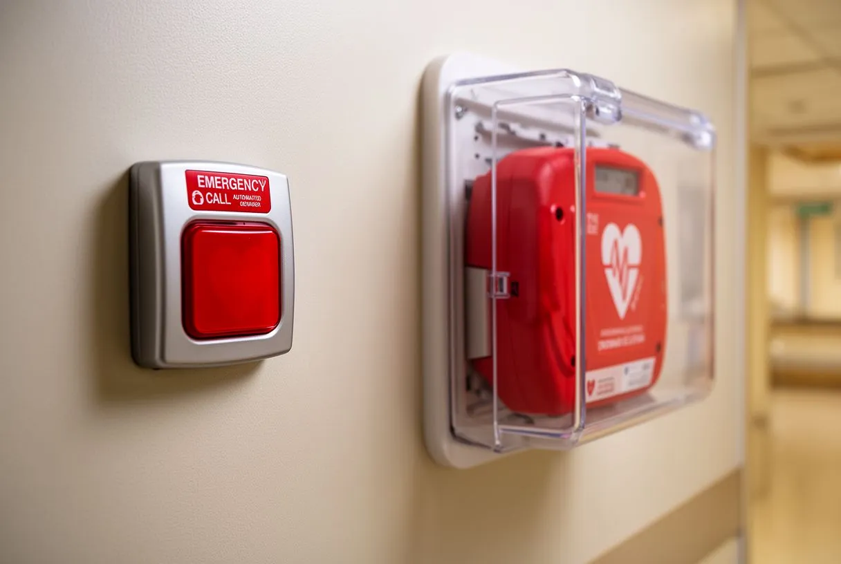
If someone collapses and is unresponsive, an Automated External Defibrillator (AED) should be used immediately if one is available, alongside CPR. STEMI can trigger lethal arrhythmias such as ventricular fibrillation within seconds, and defibrillation within the first few minutes can be the difference between life and death. The same applies in the hospital: Auscultation with a stethoscope and a rapid ECG are performed the moment the patient arrives, because the electrical tracing — not the symptoms alone — confirms whether this is a STEMI requiring immediate catheterisation.
What Happens During Emergency STEMI Treatment
Once a STEMI is confirmed on the ECG, the hospital activates a pathway often called a code STEMI or cath lab alert. The goal is to get the patient from the emergency department to the catheterisation laboratory and to reopen the artery as quickly as possible. Intravenous access is established, blood samples are drawn for cardiac enzymes and clotting studies, and medications such as Aspirin and a second antiplatelet drug are given immediately to limit clot growth. Oxygen is provided if blood oxygen levels are low, and pain relief is offered, though comfort measures never delay the procedure itself.
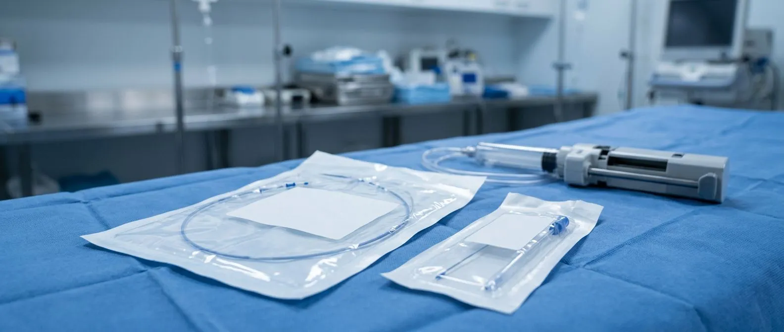
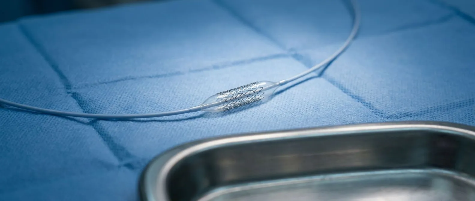
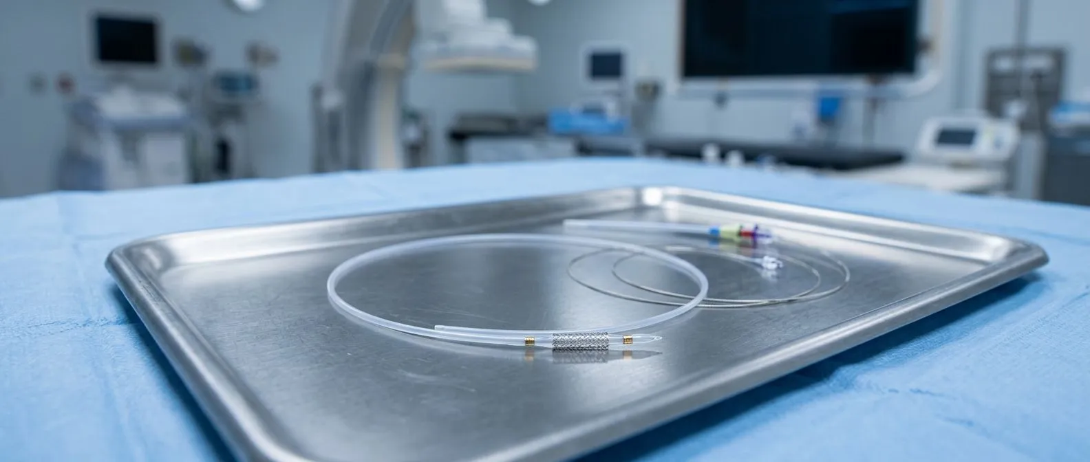
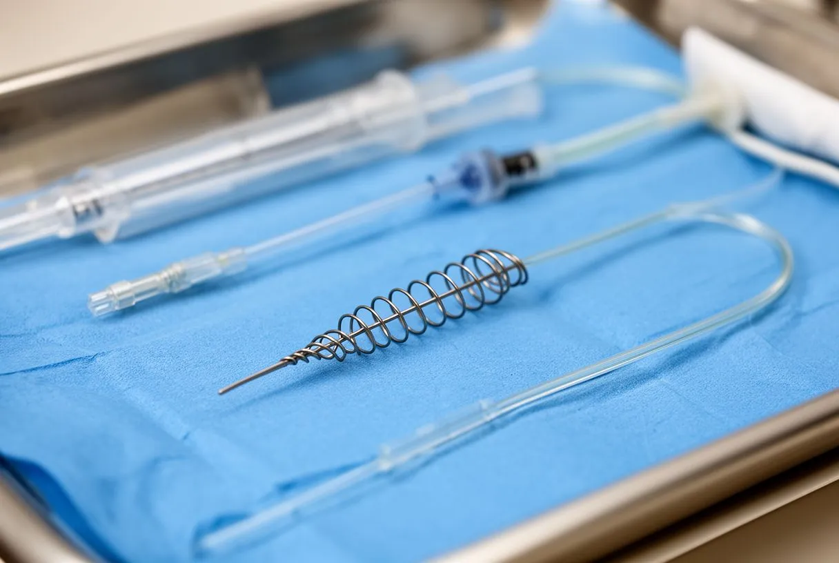
Inside the catheterisation laboratory: The interventional cardiologist threads a thin catheter from the wrist (radial artery) or the groin (femoral artery) up to the heart. Contrast dye is injected to map the coronary arteries and pinpoint the blockage. A guidewire is passed across the clot, and a small balloon is inflated to compress the plaque and restore flow — a technique known as balloon angioplasty. In nearly all STEMI cases a stent, a tiny mesh tube, is placed to keep the artery open. The entire reperfusion sequence may take less than thirty minutes once the artery is reached, though preparation and imaging add time.
Thrombotic complications are a real concern during and after this process. Studies of clotting behaviour in critically ill patients, including work such as the Venous and arterial thromboembolic complications in COVID-19 patients admitted to an academic hospital in Milan, Italy, illustrate how systemic inflammation can amplify clot risk and complicate recovery. The same principle applies in STEMI: the rupture that triggered the blockage signals a broader clotting tendency that must be managed with medication for weeks and months afterwards, not just in the catheterisation laboratory.
Types of Procedures Used in STEMI Treatment
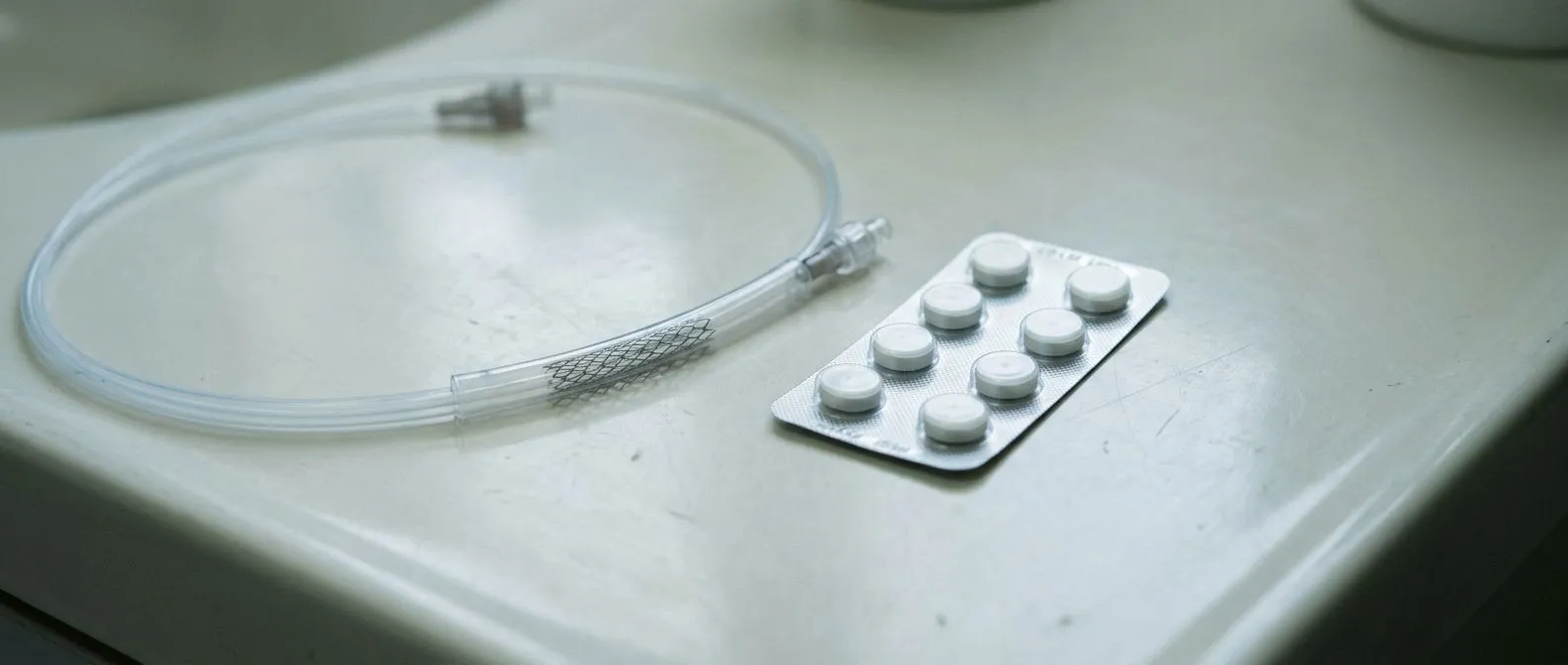
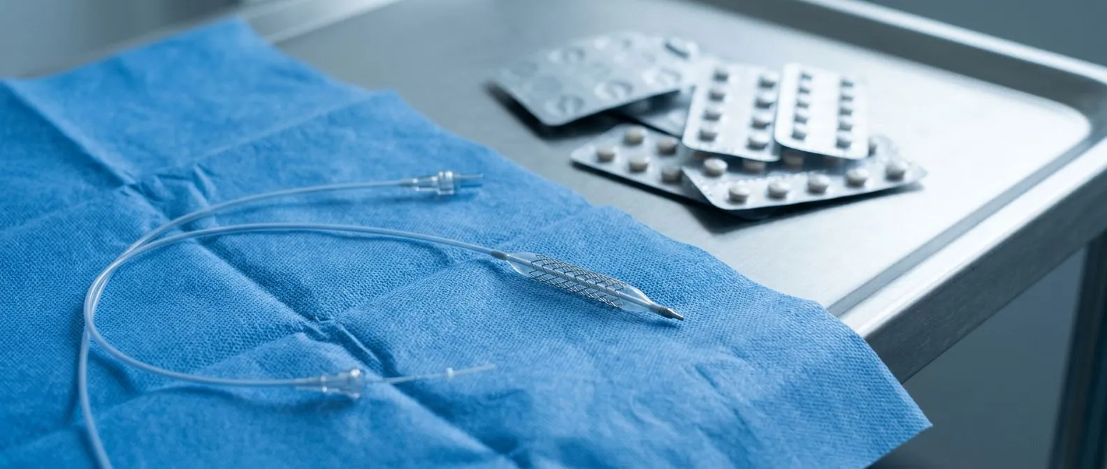
Primary percutaneous coronary intervention (PCI) is the preferred STEMI treatment when it can be performed within the recommended time window. This means mechanical reopening of the artery with a stent, as described above, and it consistently produces better outcomes than clot-busting drugs when a catheterisation laboratory is available. The stent may be bare metal or drug-eluting, with drug-eluting stents releasing medication slowly to reduce the chance of scar tissue re-narrowing the artery over the following months.
When fibrinolysis is used instead: In hospitals without catheterisation facilities, or when transfer would take too long, intravenous fibrinolytic therapy — clot-busting medication — may be given to dissolve the clot chemically. This is effective but carries a higher risk of bleeding and a slightly higher risk of re-blockage compared with PCI, which is why patients who receive fibrinolysis are often transferred to a PCI-capable centre afterward for evaluation. In rare cases of severe multi-vessel disease or failed PCI, emergency coronary artery bypass grafting is performed, using a healthy Arterial Graft or vein graft to bypass the blocked segment. Both approaches aim for complete revascularisation of the threatened muscle.
The choice between these options is not a matter of preference but of logistics, anatomy, and timing. A patient arriving at a rural clinic thirty minutes from a PCI centre will have a different pathway than one arriving at a major hospital with a catheterisation laboratory on site. What matters is that the team follows the fastest route to reperfusion for that patient's situation, and that the family understands why one approach was chosen.
Medications That Support STEMI Treatment and Recovery
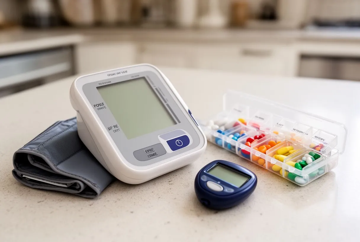
Medications after STEMI are not optional extras — they are a continuation of the treatment itself. Dual antiplatelet therapy, usually Aspirin plus a second drug such as clopidogrel or ticagrelor, is essential to keep the stent open and prevent new clots. This combination is typically continued for at least twelve months, though the exact duration depends on bleeding risk and the type of stent placed. Stopping these medications early, especially in the first month, can trigger a catastrophic re-blockage.
Beyond antiplatelets: A beta-blocker reduces the heart's workload and helps prevent dangerous rhythm disturbances such as atrial flutter. An ACE inhibitor or angiotensin receptor blocker protects the Atrium and ventricle from remodelling — the stretching and weakening that follows a large infarction. A high-intensity statin lowers cholesterol aggressively; elevated Apolipoprotein B levels are a key driver of plaque growth, and reducing this particle is central to preventing recurrence. Cardiometabolic research, including work such as the analysis titled Prevalence and impact of cardiovascular metabolic diseases on COVID-19 in China, reinforces how tightly cardiovascular and metabolic conditions intertwine, which is why STEMI care addresses the whole risk profile, not only the blocked artery.
Patients sometimes feel overwhelmed by the number of pills, especially when each comes with its own instructions and side effects. It helps to understand that each medication targets a specific mechanism: one prevents clots, one reduces heart strain, one controls blood pressure, one lowers cholesterol. Together they form a safety net that dramatically reduces the chance of a second heart attack. Skipping doses or stopping because you feel well undermines the entire strategy.
STEMI Recovery Timeline in the Hospital
Most patients stay in the hospital for two to five days after a STEMI, depending on the extent of damage and whether complications arise. The first twenty-four hours are spent in a coronary care unit with continuous monitoring, because the injured heart is electrically unstable in the early phase and dangerous rhythms can appear without warning. Nurses watch the ECG, blood pressure, and oxygen saturation constantly, and the access site — wrist or groin — is checked for bleeding.
Day by day: On the second day, many patients begin sitting up and walking short distances with assistance. Blood tests track the fall of cardiac enzymes, which confirms the infarction has stabilised. An echocardiogram assesses how much pumping function was preserved, which guides the long-term medication plan. By the third or fourth day, if all is stable, the team discusses discharge planning, cardiac rehabilitation referral, and follow-up appointments. This is the moment to ask questions about driving, returning to work, and physical limits at home.
Recovery in the hospital is not only physical but educational. The team will review modifiable risk factors — smoking, blood pressure, cholesterol, diabetes, weight, and physical activity — and help the patient build a realistic plan for change. Family members are encouraged to attend these conversations, because the home environment plays a decisive role in whether the patient sustains those changes after discharge.
Long-Term Recovery After STEMI Treatment
Recovery after a STEMI is measured in months, not days. The heart muscle that was deprived of blood forms a scar over several weeks, and during this time patients commonly feel fatigue, palpitations, and emotional fragility. These are expected responses, not signs of failure. Cardiac rehabilitation — a structured programme of supervised exercise, education, and psychological support — is one of the most effective interventions available, reducing mortality and improving quality of life significantly.
Building back strength: Supervised exercise typically begins two to six weeks after discharge, depending on the patient's condition. Sessions start gently, with walking and light resistance, and progress as confidence and capacity return. Most patients can return to non-manual work within four to six weeks and to moderate exercise within three months, though heavy lifting and intense sport require individualised clearance. Sexual activity, travel, and driving all have specific timelines that the cardiac team will outline based on the patient's ejection fraction and symptoms.
Emotional recovery is just as important as physical healing. Depression and anxiety are common after a heart attack and are associated with worse outcomes, partly because they reduce adherence to medication and exercise. Recognising this and seeking support — whether through the rehabilitation programme, a counsellor, or a support group — is part of the treatment, not a separate concern. Patients who address both body and mind recover more fully than those who focus only on the physical side.
Risks and Complications of STEMI Treatment
STEMI treatment is life-saving, but like any urgent intervention it carries risks. Bleeding from the access site is the most common complication, usually minor but occasionally requiring transfusion or intervention. More serious risks include stroke during the catheterisation, kidney injury from contrast dye (particularly in patients with pre-existing kidney disease), and allergic reactions to contrast or medications. These risks are weighed against the near-certainty of progressive muscle death without treatment, which is why emergency PCI remains the standard despite them.
Complications of the infarction itself: The damage from the blocked artery can cause its own problems independent of the procedure. Heart failure may develop if a large territory was affected, presenting as breathlessness and fluid retention. Arrhythmias, including atrial flutter and ventricular tachycardia, can persist after the artery is opened. In rare cases the damaged wall thins and bulges, forming an aneurysm of the left ventricle, or the muscular septum ruptures, creating a shunt that demands urgent surgical repair. These are the reasons patients remain monitored in the coronary care unit rather than being sent home the same day.
Being honest about these risks is not meant to frighten but to prepare. Families who understand that a complication is possible — and that the team is watching for it — are better able to cope if one arises, and better able to ask informed questions. The overall benefit of prompt STEMI treatment far outweighs the risks for the vast majority of patients, but each person's situation is discussed individually with the cardiac team before and after the procedure.
Life After STEMI: Preventing a Second Heart Attack
Surviving a STEMI is a second chance, and protecting it requires sustained change. The medications prescribed at discharge are the foundation: dual antiplatelet therapy, beta-blocker, ACE inhibitor or ARB, and statin. Taking them consistently, attending follow-up appointments, and reporting new symptoms early are the everyday habits that keep the repaired artery open and the rest of the coronary tree stable.
Lifestyle changes that change outcomes: Stopping smoking is the single most powerful change a patient can make, reducing recurrence risk dramatically within months. The other pillars are regular physical activity, a Mediterranean-style diet rich in vegetables, fish, and olive oil, blood pressure and diabetes control, and maintaining a healthy weight. These are not vague suggestions but evidence-based interventions that, combined with medication, substantially lower the risk of a second event. For patients with remaining blockages that were not treated during the emergency procedure, the cardiologist may plan a second, elective intervention once the patient is stable.
Finally, a STEMI is not the end of an active life — it is the beginning of a different, more intentional one. With the right treatment, medication, and habits, most patients return to the people and activities they love. The most important step is the first one: discuss your own situation with a qualified cardiac team, share your symptoms and concerns openly, and let the team build a plan tailored to your heart, your history, and your goals. If you or a loved one has questions about STEMI treatment or recovery, booking a consultation is the right next step — not self-diagnosis, not delay.