

Temporary Pacing Wires
Learn how temporary pacing wires support heart rhythm after cardiac surgery.
What Are Temporary Pacing Wires in Cardiac Surgery?
Temporary pacing wires are thin, insulated electrical leads gently attached to the outer surface of the heart during cardiac surgery. Their primary purpose is to deliver controlled electrical impulses to the heart muscle, ensuring it maintains a safe and steady rhythm while the patient recovers from the immediate stress of an operation. They serve as a critical backup system for the heart's natural electrical pathway.
When a patient undergoes heart surgery, the heart is often stopped or put on a bypass machine, which can temporarily stun the cardiac muscle and disrupt its normal signaling. The temporary cardiac pacing leads act as an external bridge, taking over the role of the heart's natural pacemaker if it fails to maintain an adequate heartbeat on its own. This ensures continuous, healthy blood circulation to the body's vital organs during the vulnerable early recovery phase.
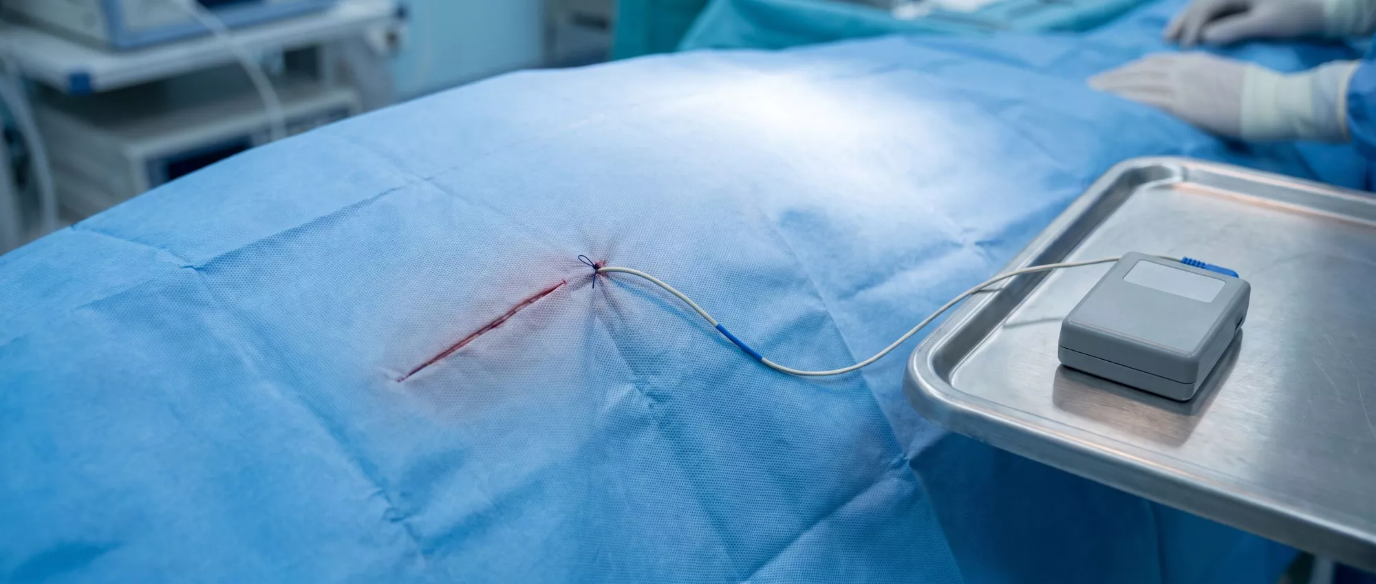
What do temporary pacing wires look like? They are very fine, flexible wires that exit the chest through a small puncture site in the skin, typically just below the surgical incision. The external portion of the wire is carefully secured to the skin with a small stitch and connected to an external pacing device, allowing the medical team to adjust the heart's rate and rhythm seamlessly without requiring a second operation.
How Do Temporary Epicardial Pacing Wires Work?
How does the electrical signaling function? The wires are placed on the epicardium, which is the outer protective layer of the heart. When the heart's natural electrical node fails to fire or fires too slowly, the external pacemaker generator sends a low-voltage electrical pulse down the wires, stimulating the heart muscle to contract and pump blood effectively.
This process is highly controlled by the cardiac surgery team in the intensive care unit. The medical staff can program the external generator to maintain a specific minimum heart rate, ensuring the patient's cardiac output remains stable. If the heart beats perfectly on its own, the pacemaker simply senses the rhythm and waits in standby mode, ready to intervene only if the heart rate drops below the programmed safety threshold.
The temporary epicardial pacing wires can function in different modes depending on the patient's needs. They can be set to simply pace the lower chambers of the heart, or they can be used to sense and track the heart's intrinsic electrical activity. This versatility allows doctors to fine-tune the cardiac support as the patient's heart muscle gradually wakes up and regains its natural, independent rhythm following the trauma of surgery.
Why Are Pacing Wires Used After Open Heart Surgery?
What rhythm issues do pacing wires after open heart surgery address? They are primarily used to manage transient arrhythmias, which are temporary irregular heartbeats caused by the physical manipulation of the heart, the use of the cardiopulmonary bypass machine, and the protective cooling of the heart muscle. It is very common for the heart to be slow or irritable in the first few days postoperatively.
When the heart tissue is healing, swelling can interfere with the heart's electrical pathways, leading to a dangerously slow heart rate known as bradycardia or intermittent heart blocks. By having the pacing wires already in place, the medical team can instantly correct these slow rhythms, preventing drops in blood pressure and maintaining vital organ perfusion without needing emergency medications or procedures.
They also provide a safety net when medical interventions are adjusted. For example, if a patient requires medications to control a fast heart rate, those same drugs might inadvertently slow the heart down too much. With temporary pacing wires active, doctors can safely administer necessary medications, knowing the pacemaker will prevent the heart rate from dropping to dangerous levels.
Temporary Pacing Wire Placement During Heart Surgery
During the cardiac procedure, while the chest is open and the heart is exposed, the surgeon meticulously attaches the pacing leads to the surface of the heart. This is a routine part of the operation, performed with great care to ensure the wires make solid electrical contact with the heart muscle without being placed too deeply, which could cause bleeding or muscle damage.
Typically, the surgeon places one or two wires on the right ventricle, and occasionally an additional wire on the right atrium, depending on the specific surgical approach and the anticipated needs of the patient. Once the wires are secured to the epicardium, they are tunneled safely out through the skin of the upper abdomen or lower chest.
How are the wires secured outside the body? The external ends of the wires are carefully looped and taped to the patient's skin to prevent accidental pulling or dislodgement. The exit site is covered with a sterile dressing to protect the area from infection. Patients and family members should never touch the wires or the pacing box, as accidental tugging could dislodge the leads from the heart surface.
Temporary Pacemaker Wires and Postoperative Care
In the intensive care unit, the external pacing generator is kept at the bedside. The nursing staff continuously monitors the patient's cardiac rhythm, adjusting the pacemaker settings only under the direct orders of the cardiac surgeon or attending cardiologist. This ensures the patient receives the precise level of electrical support needed as their heart begins to recover.
As the heart heals, the medical team will gradually lower the pacemaker's minimum rate setting to encourage the heart's natural pacemaker to take over. They will closely observe the monitoring screens to see if the heart maintains a safe, independent rhythm. This process of testing the heart's native function is crucial before deciding if the temporary cardiac pacing can be completely turned off.
Proper pacing wire management post-op requires strict hygiene and careful patient handling. The nursing team will inspect the wire exit site daily for any signs of redness, swelling, or drainage. Patients are encouraged to move and sit up in bed, but they must be mindful not to tug on the wires or accidentally pull the external generator off the bed.
What Are the Risks and Epicardial Pacing Wires Complications?
While temporary pacing wires are a standard and highly beneficial tool, they do carry some inherent risks. The most significant concern is infection at the wire exit site, as the wires create a direct pathway from the outside environment into the chest cavity. Strict sterile techniques and daily site care are the primary defenses against this complication.
Another potential risk involves the physical removal of the wires. When the wires are pulled out, there is a small chance of bleeding from the heart surface where the wire was attached. To manage this risk, doctors ensure the patient's blood clotting levels are normal before removal, and the procedure is always done in a monitored setting where immediate medical intervention is available.
Can pacing wire removal cause cardiac tamponade? In very rare cases, pulling the wire can cause a small tear in the heart muscle that leads to bleeding around the heart, a condition known as cardiac tamponade. This is why removal is performed in a clinical setting with echocardiogram capability, allowing doctors to quickly detect and treat any fluid accumulation around the heart.
Temporary Pacing Wire Removal Process and Recovery
How is temporary pacing wire removal performed? The removal procedure is typically done at the bedside a few days after surgery, once the heart has demonstrated a stable, independent rhythm. The doctor will carefully cut the small retaining stitch that holds the wire to the skin, apply gentle traction, and smoothly pull the wire out through the chest wall. The procedure usually takes only a few seconds and may cause a brief pulling sensation.
After the wire is removed, the nurse will apply firm pressure to the exit site for a few minutes to ensure there is no bleeding. A small sterile dressing is applied, and the patient's heart rate and blood pressure are monitored closely for the next few hours to ensure the heart continues to function perfectly without the electrical backup. Most patients feel very little discomfort during the actual removal.
Once the wires are out, the small puncture site heals quickly over a few days, similar to a minor scratch. The removal signifies a major milestone in the patient's recovery, indicating that the surgical team is confident in the heart's ability to maintain a healthy rhythm on its own.
Living With Temporary Cardiac Pacing at Home
In some cases, patients may be discharged from the hospital with the pacing wires still in place, connected to a small, portable pacing generator. This is done when the heart needs a bit more time to recover or if the medical team wants to ensure rhythm stability over a longer period. Living with these wires at home requires specific safety education and careful hygiene practices.
Patients must keep the pacing box dry at all times, which requires modifying how they bathe by using sponge baths or carefully shielding the area. They are advised to avoid vigorous physical activities, heavy lifting, or sudden stretching movements that could put tension on the wires or dislodge the pacing box. A loose garment or a specialized pouch is usually provided to keep the generator secure and protected.
What should you do if a pacing wire comes out at home? If a wire accidentally pulls out or the box disconnects, the patient should not panic. The heart will often continue to beat on its own, but they should immediately lie down flat to ensure optimal blood flow to the brain and call their surgical team or emergency services for immediate guidance. It is helpful to keep portable medical alert cards for heart surgery patients accessible at all times.
How Long Are Pacing Wires Left in After Surgery?
The duration the temporary pacing wires remain in place varies depending on the patient's specific recovery trajectory. For the vast majority of patients, the wires are removed before they leave the hospital, usually between three to seven days postoperatively, once the heart's rhythm is stable without pacing support.
If a patient has a slower-than-expected recovery of their natural electrical pathways, the wires might be left in for up to two weeks. The medical team will conduct daily evaluations of the heart's rhythm, gradually turning down the pacemaker to test the heart's independent function before making the final decision to remove the leads.
What factors influence the timing of temporary pacemaker after cardiac surgery removal? The presence of pre-existing conduction disorders, the specific type of valve surgery performed, and the degree of postoperative heart swelling all play a role in how long the team chooses to keep the safety net in place.
Key Takeaways on Heart Surgery Pacing Wires
Understanding the role of temporary pacing wires helps demystify a critical aspect of cardiac recovery. These leads are a temporary, highly effective safety net that ensures the heart maintains a steady, life-sustaining rhythm while the muscle heals from the physical stress of surgery. They are a standard precaution used to manage transient rhythm disturbances smoothly and safely.
- Immediate Support: They provide instant electrical backup if the heart's natural rhythm drops too low after surgery.
- Safety Net: The wires allow doctors to safely administer necessary heart medications without the risk of causing a dangerously slow heart rate.
- Safe Removal: Once the heart beats independently, removal is a quick, low-discomfort bedside procedure.
Patients and families should feel reassured that the use of temporary pacing wires is a routine, proactive measure taken by the surgical team to ensure a safe and stable recovery. If you have concerns about your postoperative rhythm or the management of these leads, always direct your questions to your cardiac care team, who can provide personalized guidance based on your specific surgical journey.






