

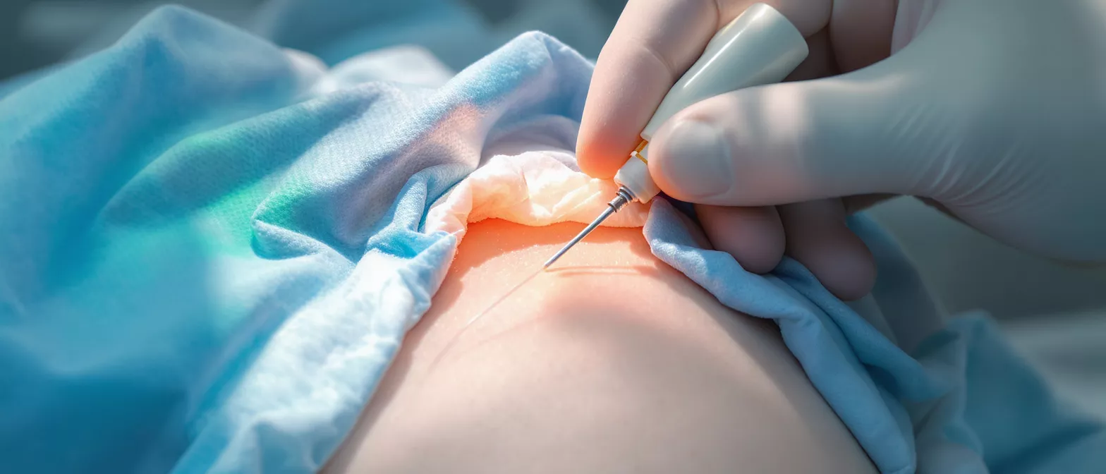
Transfemoral Access
Learn how transfemoral access enables minimally invasive heart surgery, including its technique, risks, benefits, and recovery time.
What is transfemoral access
Transfemoral access is a medical technique used to reach the heart by inserting a thin, flexible tube called a catheter through the femoral artery, which is located in the groin area. This method serves as a minimally invasive gateway, allowing cardiac surgeons and interventional cardiologists to perform complex heart procedures without making large incisions in the chest wall. For patients facing heart surgery, understanding this entry point helps remove much of the fear surrounding operating rooms and recovery. The approach is a cornerstone of modern cardiology because it provides a direct, navigable pathway from the leg directly to the heart.
Transfemoral access definition for patients means your surgeon can treat blocked arteries or replace faulty valves by threading instruments through your blood vessels rather than opening your breastbone. This is a highly planned procedure. The femoral artery is chosen because it is a large, strong blood vessel that offers a direct route upward to the aorta and the heart. By using this natural pathway, surgeons avoid the trauma of traditional open surgery, which inherently carries greater physical tolls and longer hospital stays.
When you first hear the term in a consultation, it may sound highly technical, but the concept is straightforward. Think of your vascular system as a highway system. The groin access site is the on-ramp. Once the medical team places the catheter, they can guide cameras, balloons, stents, or artificial valves directly to the affected area of the heart. This targeted precision is what makes minimally invasive closure devices and related techniques so effective for patients with severe cardiac conditions.
How transfemoral access works in cardiac procedures
The mechanics of this technique rely heavily on real-time imaging and precise vascular navigation. After the patient receives local anesthesia and a mild sedative, the physician locates the femoral pulse in the groin. A small needle is inserted into the artery, followed by a guidewire. Over this wire, the doctor slides a sheath, which acts as a secure tunnel holding the vessel open. Through this sheath, various catheters can be exchanged safely while maintaining continuous access to the cardiovascular system.
Transfemoral arterial access steps involve continuous X-ray guidance, known as fluoroscopy, to track the progress of the catheter. The doctor watches a monitor as the catheter moves through the femoral artery, up through the abdominal aorta, and into the heart chambers or coronary arteries. Contrast dye is often injected to make the blood vessels highly visible on the screen. This ensures the surgical team can see exactly where the blockages are or precisely where a new valve needs to be placed.
Patients remain comfortable throughout the process, often awake but relaxed. The entire setup focuses on minimizing trauma to the surrounding tissues. Because the femoral artery is naturally wide, it can accommodate the specialized tools required for complex interventions, including femoral artery compression straps used post-procedure. This direct vascular route explains why the transfemoral approach has become the default technique for many cardiac interventions in modern hospitals.
Transfemoral access vs transapical approach
When planning a structural heart intervention, surgeons must choose the safest and most effective route to the heart. The main comparison is between the transfemoral approach and the transapical approach. Transfemoral access goes through the groin, while the transapical approach involves making a small incision in the chest to enter the heart through its apex, or tip. The decision dictates the entire surgical strategy and heavily influences the recovery timeline.
- Entry Point: Transfemoral uses the groin artery, while transapical requires a surgical incision between the ribs.
- Recovery Speed: Transfemoral patients usually sit up and walk within hours, whereas transapical recovery takes days.
- Anesthesia Type: Transfemoral often uses moderate sedation, but transapical requires general anesthesia.
Transfemoral access vs transapical comparison ultimately favors the femoral route when patient anatomy allows it. The transapical route is reserved for patients whose blood vessels are too narrow, heavily calcified, or tortuous to safely pass a catheter from the groin. By evaluating CT scans, the surgical team determines which path offers the lowest risk of vascular injury and the highest chance of procedural success.
Transfemoral access TAVR and heart surgery
The most common application of this technique is in transcatheter aortic valve replacement. Transfemoral access TAVR allows surgeons to deliver a replacement aortic valve directly into the heart through the femoral artery. For patients suffering from severe aortic stenosis, where the heart's main valve becomes stiff and restricts blood flow, this procedure can be life-saving. It provides a viable treatment option for individuals who are too frail to undergo traditional open-heart surgery.
Transfemoral aortic valve replacement access represents a major shift in cardiac care. Instead of stopping the heart and using a heart-lung bypass machine, the medical team threads a collapsed artificial valve on a catheter. Once the catheter reaches the diseased aortic valve, the new valve is expanded, pushing the old valve out of the way. The newly placed valve immediately begins regulating blood flow, restoring normal cardiac function without the extreme trauma of a chest opening.
This method has revolutionized how cardiologists treat valvular disease. By avoiding the sternotomy, or cutting of the breastbone, patients experience significantly less pain and a drastically reduced risk of major infection. It is a testament to how vascular engineering can improve outcomes for vulnerable patient populations, turning a high-risk open surgery into a manageable, minimally invasive procedure.
Femoral artery catheterization cardiac process
Understanding the femoral artery catheterization cardiac process helps patients mentally prepare for their procedure. The femoral artery is a massive blood vessel that supplies blood to the lower extremities. Because it sits just beneath the skin in the groin, it is easily accessible. The puncture is typically just a few millimeters, yet it opens a pathway for instruments that can reach deep into the chest cavity.
Transfemoral access site preparation begins with sterilizing the groin area thoroughly to prevent infection. The medical team maps the vascular tree beforehand using advanced imaging. They measure the diameter of the femoral artery to ensure it can accommodate the sheath required for the procedure. If the right femoral artery is too small, the left side can be used, ensuring the patient still benefits from a minimally invasive route.
During the process, the catheter serves as a delivery system. It must be rigid enough to advance against the pressure of arterial blood flow but flexible enough to navigate the natural curves of the aorta without damaging the vessel walls. This balance is critical. The design of these hemostatic wound management dressings applied after the sheath is removed plays a crucial role in helping the puncture site heal safely.
Transfemoral access risks and benefits
Evaluating the transfemoral access risks and benefits is a standard part of preoperative planning. The benefits are substantial. Patients experience smaller scars, significantly less postoperative pain, and a quicker return to daily activities. There is also a lower risk of respiratory complications because the chest cavity is never opened. Avoiding large surgical wounds means the immune system is spared from fighting a massive recovery effort.
Transfemoral approach cardiology advantages also include lower blood loss and reduced need for blood transfusions. Because the procedure is minimally invasive, the inflammatory response in the body is much lower compared to traditional surgery. This makes it highly suitable for elderly patients or those with multiple comorbidities. The ability to avoid general anesthesia in some cases further reduces the risk of cognitive decline post-surgery.
However, the technique is not without risks. The primary concern is vascular damage to the femoral artery. If the artery is punctured incorrectly, it can lead to bleeding, pseudoaneurysms, or nerve injury. Careful patient screening and the use of ultrasound guidance during the puncture have drastically reduced these complications over the years.
Transfemoral access complications to watch for
While the procedure is generally safe, understanding transfemoral access complications helps patients recognize warning signs early. The most common issue is minor bleeding or a bruise at the groin puncture site. This is normal and usually resolves on its own within a few days. However, in rare cases, the bleeding can continue internally, forming a hematoma. This requires careful monitoring and sometimes additional intervention.
Transfemoral access complications signs include severe pain in the groin or leg, numbness, or a pulsating mass at the puncture site. If the hematoma grows, it can compress the nearby femoral nerve, causing weakness in the leg. Patients are taught to check the site regularly and report any sudden swelling immediately. Rapid response prevents minor issues from becoming serious threats.
Another rare but known risk is the formation of a pseudoaneurysm, where blood leaks outside the artery wall but is contained by surrounding tissue. This creates a false aneurysm that requires a specialized repair. By maintaining strict immobility immediately after the procedure, patients can significantly reduce the likelihood of these vascular complications occurring.
Transfemoral access closure techniques
Once the cardiac intervention is complete, removing the sheath safely is the final critical step. Transfemoral access closure techniques have evolved significantly. Manual compression, where a clinician applies pressure to the groin until the bleeding stops, was once the standard. While still used, it requires extended periods of strict bed rest, which can be uncomfortable and increase the risk of blood clots in the legs.
Transfemoral access closure devices now offer mechanical solutions that seal the artery immediately. These devices use small sutures, collagen plugs, or specialized clips to close the hole in the blood vessel from the inside. This immediate sealing allows patients to sit up and walk much sooner after the procedure. The quicker mobility reduces muscle stiffness and speeds up overall recovery.
The choice of closure method depends on the size of the catheter used and the patient's vascular health. By securing the puncture site efficiently, the surgical team ensures the artery heals properly without leaving a weak spot in the vessel wall. This meticulous attention to closing the entry point is what makes the entire minimally invasive approach viable.
Transfemoral access recovery time
One of the most appealing aspects of this approach is the rapid recovery timeline. Transfemoral access recovery time is typically measured in hours or days, rather than weeks or months. Patients who undergo traditional open-heart surgery face a grueling recovery process that can last up to three months. In contrast, those treated via the femoral artery often leave the hospital within a few days.
Transfemoral access recovery time expectations include a short stay in the recovery room for monitoring. Nurses frequently check the pulse in the foot to ensure blood is flowing properly to the leg. They also monitor the groin for any signs of swelling. Once the patient is stable, they are moved to a standard room and encouraged to sit on the edge of the bed.
Full recovery takes a bit longer as the artery heals completely, but the functional recovery is nearly immediate. Most patients return to normal daily activities within a week or two. The small puncture site heals rapidly, leaving a tiny mark that is barely visible. This rapid return to normalcy is a primary reason why the transfemoral approach is heavily favored by both patients and surgeons.
What to expect after groin access for heart procedures
After the procedure, patients play an active role in their own healing. Knowing what to expect after groin access for heart procedures empowers individuals to care for themselves properly. The first instruction is to avoid heavy lifting or strenuous exercise for a specified period. This prevents pressure from building up in the abdomen, which could dislodge the clot forming at the puncture site.
Groin access heart procedure aftercare involves keeping the area clean and dry. Patients should monitor the site for any signs of infection, such as redness, warmth, or discharge. It is normal to experience some mild soreness or bruising, which will fade over time. Walking is encouraged as it promotes healthy blood flow, but the intensity should be kept low initially.
Ultimately, the success of a transfemoral procedure relies on the partnership between the surgical team and the patient. By adhering to discharge instructions and attending follow-up appointments, patients ensure their new heart valves or stents function perfectly. This minimally invasive approach has transformed cardiac care, proving that major heart interventions do not always require major surgery.






