

Valvular Heart Disease
Discover valvular heart disease causes, symptoms, and treatments.
What is valvular heart disease?
Understanding the core definition: Valvular heart disease is a condition affecting one or more of the four valves in the heart. These valves are essentially one-way doors that keep blood flowing in the correct direction. When a valve is damaged or diseased, it can disrupt the normal, efficient flow of blood, forcing the heart muscle to work harder than it should to compensate.
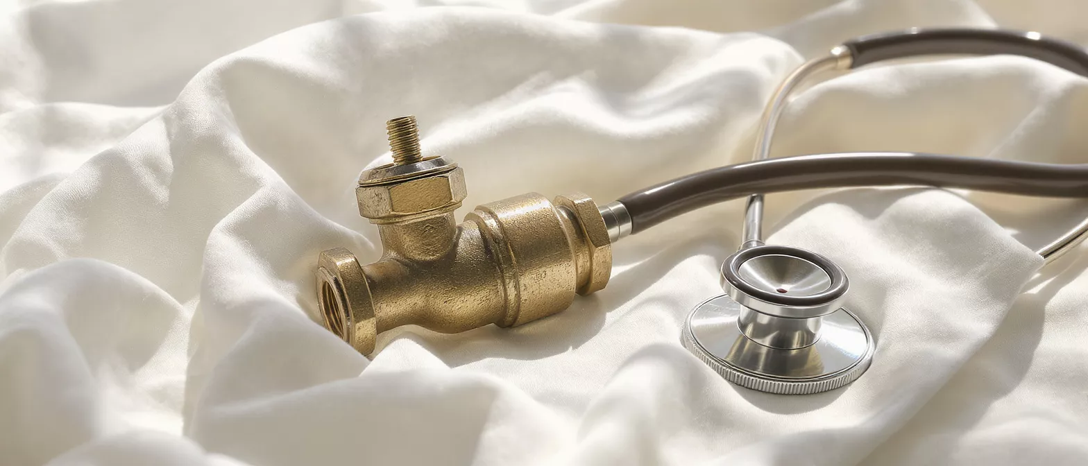
For a patient preparing for cardiac surgery, understanding this diagnosis is vital. The term itself simply means that a mechanical problem exists within the heart's plumbing system. It does not mean the heart muscle is irreparably broken; rather, it means a specific component needs medical attention to restore normal circulation and relieve strain on the heart.
Receiving this diagnosis can feel overwhelming, but modern medicine offers highly effective solutions. By addressing the structural issue directly, surgeons can dramatically improve a patient's quality of life, relieve uncomfortable symptoms, and restore the heart's natural rhythm and efficiency.
How do heart valves work?
The mechanics of blood flow: The heart consists of four chambers, and between them sit four crucial valves: the mitral, aortic, tricuspid, and pulmonary. Every time the heart beats, these valves open to let blood pass through to the next chamber or artery, and then close tightly to prevent any backward leakage. This synchronized opening and closing is what maintains a steady, forward circulation of oxygen-rich blood throughout the body.
Each valve has delicate flaps, known as leaflets or cusps. In a healthy heart, these flaps open fully and close completely. When disease strikes, it typically affects the leaflets' ability to open or close properly. This malfunction leads to either restricted blood flow or problematic leakage, both of which alter the heart's internal pressures over time.
Understanding this mechanical function helps patients grasp why their symptoms occur. If the valves cannot open or close correctly, the heart must generate extra force to push blood forward, leading to the fatigue and breathlessness characteristic of heart valve disease.
Types of valvular heart disease?
Valvular stenosis (narrowed valves): This occurs when the valve flaps become thick, stiff, or fuse together. As a result, the valve opening becomes narrowed, restricting the amount of blood that can flow from the heart to the body. The heart must pump with much greater force to push blood through the tight opening, eventually weakening the heart muscle.
Valve regurgitation (leaky heart valve): Also known as insufficiency, this happens when the valve flaps do not close tightly. Blood leaks backward into the previous chamber instead of moving forward. This forces the heart to pump the same volume of blood twice, increasing the volume of blood in the chamber and leading to thickening or enlargement of the heart muscle over time.
Both conditions can affect any of the four valves, but the aortic and mitral valves are most frequently involved. Understanding whether the problem is a blocked opening or a leaky seal helps the surgical team determine the most appropriate approach to repair or replace the malfunctioning structure.
What causes valvular heart disease?
Congenital and age-related factors: Some individuals are born with abnormal valve structures, such as a bicuspid aortic valve, which has only two flaps instead of three. This congenital anomaly can lead to early valve wear and tear. Conversely, age-related degenerative changes are a leading cause, where the valve tissue simply thickens and calcifies over decades of constant use.
Infections and secondary conditions: Rheumatic fever, resulting from untreated strep throat, can cause lasting damage to the heart valves. Additionally, endocarditis, an infection of the heart's inner lining, can destroy valve tissue. Other systemic issues, such as high blood pressure, connective tissue disorders, or a previous heart attack, can also alter the heart's structure and damage the valves.
- Calcification: Calcium deposits build up on the valve flaps, making them stiff and immobile.
- Infection: Bacterial infections like endocarditis directly attack and destroy valve tissue.
- Congenital defects: Abnormal valve structures present at birth wear down faster than normal ones.
Valvular heart disease symptoms to watch for?
Early warning signs: In its early stages, valve disease might not cause any noticeable symptoms. Often, a doctor first detects the condition by hearing a heart murmur through a stethoscope. As the disease progresses and the heart struggles to maintain adequate blood flow, patients may begin to experience shortness of breath, particularly during physical exertion or when lying flat.
Progressive physical changes: Patients often report unusual fatigue, dizziness, or even fainting spells. Swelling in the ankles, feet, or abdomen can also occur as the heart fails to circulate blood efficiently, causing fluid to back up in the body's tissues. Some individuals experience a fluttering sensation or irregular heartbeats as the heart muscle stretches to accommodate the abnormal blood flow.
Recognizing these symptoms is crucial for timely intervention. If you or a loved one experience these signs, a prompt evaluation by a cardiologist is necessary to determine the underlying cause and prevent further deterioration of the heart muscle.
How is valvular heart disease diagnosed?
Non-invasive imaging techniques: The primary tool for diagnosing this condition is an echocardiogram. This ultrasound test uses sound waves to create detailed, moving images of the heart. It allows the physician to see the valves in action, measure the size of the valve openings, and observe the direction and speed of blood flow, easily identifying any leakage or stenosis.
Additional diagnostic tools: Beyond the echocardiogram, an electrocardiogram (ECG) is used to record the heart's electrical activity and detect irregular rhythms. A chest X-ray can show if the heart is enlarged or if fluid is accumulating in the lungs. In some cases, a cardiac MRI provides highly detailed cross-sectional images to assess the structure and function of the heart valves more thoroughly.
Accurate diagnosis is the foundation of a successful surgical plan. By precisely mapping the extent and location of the valve damage, the surgical team can tailor the intervention to the patient's specific anatomy and condition severity.
Valvular heart disease treatment options?
Monitoring and medical management: For mild cases, treatment may simply involve active surveillance and regular check-ups. Medications cannot cure the structural problem, but they can manage symptoms and reduce the workload on the heart. Drugs like beta-blockers or diuretics might be prescribed to control blood pressure and eliminate excess fluid while the condition is monitored.
Surgical interventions: When symptoms become severe or the heart function begins to decline, surgery is required. The goal is to restore normal blood flow by fixing the mechanical problem. Depending on the specific valve and the nature of the damage, the surgeon may choose to repair the existing valve or replace it entirely with a mechanical or biological prosthesis.
Choosing the right treatment path requires a careful assessment of the patient's overall health, the severity of the valve defect, and the expected durability of the chosen surgical approach. The decision is always made collaboratively between the patient, cardiologist, and surgical team.
Heart valve repair vs replacement?
The advantages of valve repair: Whenever possible, surgeons prefer to repair the natural valve rather than replace it. Repairing preserves the patient's own tissue, which maintains better heart function and eliminates the need for lifelong blood-thinning medications. Repair techniques often involve separating fused leaflets, patching holes, or tightening the valve ring to prevent leakage.
When replacement is necessary: If the native valve is too badly damaged by calcification or infection, replacement is the best option. The surgeon will remove the diseased valve and substitute it with a mechanical valve, which is highly durable, or a biological tissue valve made from animal or human tissue.
Both repair and replacement have excellent long-term outcomes. The choice depends heavily on the patient's age, lifestyle, and specific valve pathology. Understanding these options empowers patients to have informed discussions with their surgeon about the best strategy for their long-term health.
Are valvular heart disease treatments safe?
Modern surgical safety: Advancements in surgical techniques, anesthesia, and post-operative care have made heart valve surgery remarkably safe and effective. Minimally invasive approaches allow surgeons to access the heart through smaller incisions, which can lead to less postoperative pain, shorter hospital stays, and quicker recoveries compared to traditional open-heart surgery.
Managing potential risks: As with any major medical procedure, risks exist. These can include infection, bleeding, or irregular heart rhythms. However, surgical teams take extensive precautions to minimize these risks. Patients are closely monitored in the intensive care unit immediately following surgery to ensure their heart is recovering stably and efficiently.
The risk of leaving severe valvular heart disease untreated often far outweighs the risks of the surgery itself. Without intervention, a severely damaged valve will eventually lead to heart failure. By proactively addressing the issue, patients can return to a normal, active lifestyle with a fully functional heart.
Living with valvular heart disease?
Post-surgical recovery and lifestyle: After successful valve surgery, most patients experience a dramatic improvement in their energy levels and breathing. Cardiac rehabilitation is a crucial part of recovery, involving supervised exercise and education on heart-healthy living. Adopting a balanced diet, managing blood pressure, and engaging in regular, moderate physical activity are essential for protecting the repaired or replaced valve.
Long-term monitoring: Even after a successful procedure, lifelong follow-up with a cardiologist is necessary. Regular echocardiograms ensure the repaired or replaced valve continues to function correctly. If a mechanical valve was used, patients must take blood thinners for life and require routine blood tests to monitor their clotting levels, ensuring they remain safe and healthy.
- Routine check-ups: Annual visits to the cardiologist are essential to track valve function over time.
- Heart-healthy habits: A low-sodium diet and regular exercise protect the structural integrity of the repair.
- Medication adherence: Strictly following prescribed blood thinners or rhythm control medications prevents complications.
With proper care and medical supervision, individuals with treated valvular heart disease can enjoy a full, vibrant life. The mechanical problem is fixed, and the heart is restored to its proper working order, allowing patients to return to the activities and people they love with renewed vitality.






