

Central Venous Catheter
Discover how a central venous catheter works in heart surgery, from insertion to removal.
What is a central venous catheter?
A central venous catheter definition describes a flexible, sterile tube placed into a large vein leading directly to the heart. When patients or families hear the term for the first time, it often sounds intimidating. In reality, this specialized central venous access device is a routine and vital tool used by cardiac surgery teams to provide safe, reliable intravenous access during critical care. It allows doctors to administer powerful medications, draw blood, and monitor the heart without repeatedly sticking the arms.
Understanding what is a central venous catheter helps reduce anxiety before a cardiac operation. Unlike a standard IV in the hand or arm, this line sits deep within the chest, neck, or groin, reaching a larger blood vessel that can handle strong medications and rapid fluid delivery. The placement is performed under strict sterile conditions by an experienced anesthesiologist or intensivist, ensuring the line functions optimally throughout the surgical stay.
For a frightened family watching a loved one prepare for heart surgery, seeing this line can be a source of comfort. It means the medical team has direct access to stabilize the cardiovascular system instantly. They can measure blood volume status and administer essential treatments without causing unnecessary pain from repeated needle sticks.
How does central line placement work?
What happens during central line placement? The procedure is typically performed in the operating room or intensive care unit just before your heart surgery begins. The doctor uses ultrasound guidance to identify a large, safe vein—usually the internal jugular vein in the neck or the subclavian vein beneath the collarbone. Local anesthesia is used to numb the skin before a small needle is gently inserted into the vein.
Once blood flow is confirmed, the physician passes a thin guidewire into the vein, followed by the catheter tube itself, which is advanced until its tip rests near the top of the heart. The entire central venous catheter insertion process takes only a few minutes, though the team takes great care to ensure absolute precision. An X-ray is often taken immediately afterward to verify the exact position of the line before any fluids are given.
Patients are usually sedated or already under general anesthesia for their operation, so they feel little to no discomfort during the process. The medical team covers the site with a sterile, transparent dressing to keep it clean and secure. This sterile surgical vascular line remains securely in place for the duration of the intensive care stay.
Central venous catheter uses in heart surgery
Why is central venous catheter heart surgery access necessary? During complex cardiac operations, the anesthesiology team must rapidly deliver medications that could damage smaller, fragile peripheral veins. A central line safely carries strong medications—such as vasopressors to support blood pressure or anti-arrhythmics to steady the heartbeat—straight to the heart. It serves as a lifeline for precise intraoperative management.
Beyond drug delivery, central venous catheter uses include the ability to swiftly give large volumes of blood or fluids if needed during a complicated operation. This access route is robust enough to handle rapid transfusions, making it indispensable for managing sudden hemodynamic changes. It gives the surgical team the immediate ability to expand blood volume without struggling to find smaller peripheral veins.
Additionally, having a CVC catheter in place means nurses can easily draw blood samples for essential laboratory tests during and after surgery. This reduces the need for multiple separate needle punctures, keeping the patient more comfortable while providing continuous, real-time data to the surgical team. It is a cornerstone of modern perioperative cardiac care.
Types of central venous catheters
What are the different types of central venous catheters used in cardiac care? The clinical team selects the appropriate line based on the specific needs of the operation and expected recovery time. The most common for heart surgery is the multi-lumen catheter, which features multiple separate channels inside a single tube, allowing the team to deliver different medications simultaneously without mixing them.
For longer-term needs or specific surgical requirements, variations like the sheath introducer—a larger catheter that allows devices like pacemakers to be passed through—might be used. When choosing the right CVC catheter for hemodynamic monitoring, physicians consider the patient's anatomy, clotting risk, and anticipated duration of intensive care. Each variation serves the same fundamental purpose of providing safe, direct venous access.
While some patients requiring long-term outpatient therapy use tunneled catheters or implanted ports, cardiac surgery patients almost always use non-tunneled, temporary central lines placed in the neck or upper chest. These are designed for easy insertion and removal once the critical postoperative period has passed and the patient stabilizes in the ward.
Central venous pressure monitoring explained
Central venous pressure monitoring is a critical function of the CVC during heart surgery and intensive care. The central venous pressure (CVP) reflects the amount of blood returning to the right side of the heart and the heart's ability to pump that blood forward. By attaching a pressure sensor to the catheter, the team can see continuous, real-time waveforms on the bedside monitor.
How does central venous pressure help cardiac patients? It helps the surgical team fine-tune fluid management. Giving too much fluid can strain a recovering heart, while giving too little can lead to dangerous drops in blood pressure. The CVC provides a continuous read of the patient's volume status, allowing doctors to maintain perfect balance during the delicate postoperative period.
This monitoring is particularly crucial for patients with compromised heart function following a major operation. By using the CVC to track central venous pressure, the intensive care team ensures the heart is neither overloaded nor starved for blood, optimizing conditions for recovery.
CVC insertion technique and process
The modern CVC insertion technique heavily relies on real-time ultrasound guidance, a standard of care that has vastly improved safety. The ultrasound allows the physician to clearly see the vein and surrounding structures, avoiding arteries and nerves entirely. This visual approach minimizes complications and makes the procedure much faster and safer for the patient.
During the central venous catheter placement, the Seldinger technique is standard. This involves sliding a thin wire through the needle, removing the needle, and passing the catheter over the wire into the vein. The wire acts as a secure track, guiding the catheter gently into the correct position.
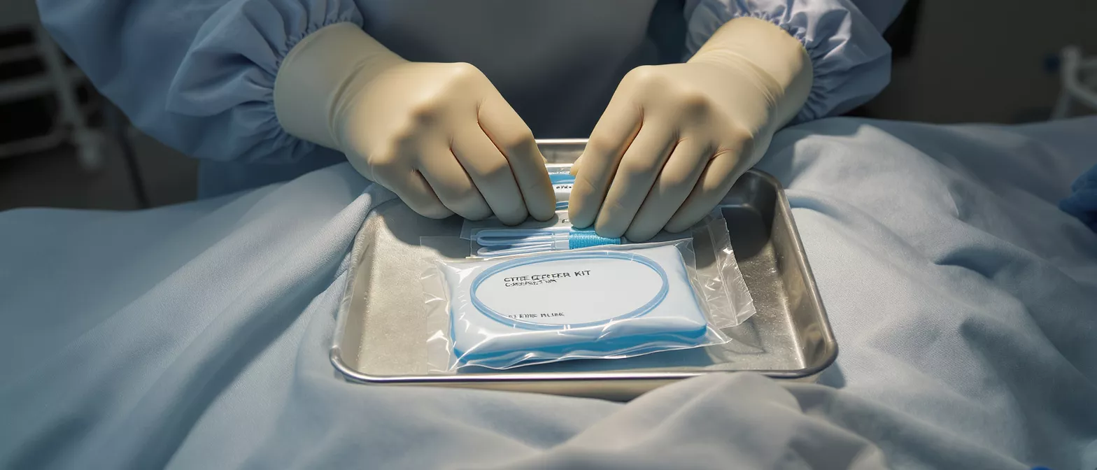
Strict sterile precautions are taken throughout the entire process. The medical team wears sterile gowns and gloves, uses large sterile drapes, and prepares the skin with chlorhexidine to drastically reduce infection risk. This meticulous attention to detail protects the immunocompromised cardiac patient from harm.
Central venous catheter complications and management
While central line placement is remarkably safe, understanding central venous catheter complications and management helps families feel informed. Potential risks include minor bleeding at the insertion site, a small risk of pneumothorax (a collapsed lung) when placing lines near the collarbone, or the rare possibility of arterial puncture. Ultrasound use has dramatically reduced these mechanical risks.
What is central line associated bloodstream infection? Often referred to as CLABSI, it is a serious but preventable complication where bacteria enter the bloodstream through the line. The cardiac intensive care team follows strict bundles of care—sterile dressings, daily reviews of necessity, and scrubbing the injection caps—to prevent this. If infection is suspected, the line is removed immediately and cultures are drawn.
- Sterile Technique: Maximum barrier precautions during insertion prevent bacteria from entering the bloodstream.
- Daily Review: The medical team evaluates the line every day, removing it the moment it is no longer essential to reduce infection risk.
- Cap Care: Nurses scrub the injection caps with alcohol before any medication is given, ensuring external bacteria cannot travel inward.
Effective management of these lines means weighing the immense benefits against these known, well-mitigated risks. The surgical team is highly trained to recognize early signs of any issue and act immediately.
Central venous catheter care guidelines
Proper central venous catheter care is essential while the line remains in place during the intensive care stay. Nurses handle all maintenance of the line, ensuring the dressing stays clean, dry, and intact. If the clear plastic dressing becomes wet or peels back, it is changed immediately using sterile technique to protect the entry point.
Family members and visitors should never touch the line or the surrounding tubing. It is vital to keep hands washed and to avoid catching clothing or blankets on the multiple IV lines connected to the patient. This helps prevent accidental pulling or dislodgement of the delicate equipment.
Patients should not feel nervous about the tubing while resting in bed. The nursing staff secures the lines safely to the skin and the bed linens, allowing gentle movement. Following these central venous catheter care guidelines ensures the line functions perfectly until the physician decides it is time for removal.
How long until central venous catheter removal?
When is central venous catheter removal performed? The line is removed as soon as the patient no longer requires high-level intensive care monitoring or continuous access to strong medications. Typically, this happens a day or two after heart surgery, once the patient is stable, breathing without a ventilator, and taking oral medications. The removal process is quick and simple.
The doctor or nurse removes the clear dressing, gently pulls the tube out, and applies firm pressure with a sterile gauze pad for a few minutes to stop any bleeding. A small bandage is placed over the site. Most patients feel only a slight, brief tugging sensation and experience no pain.
Removing the line is a significant milestone in the recovery journey. It signals that the heart is functioning well independently and that the patient is ready to transfer from the intensive care unit to a standard hospital room for the remainder of their healing process.
Key takeaways about central line safety
Understanding the purpose of a central venous catheter transforms fear into confidence. This standard access tool empowers the cardiac surgical team to deliver life-saving medications, monitor the heart's delicate fluid balance, and draw essential labs without causing repeated distress to the patient. It is a fundamental pillar of safe perioperative care.
- Immediate Access: The CVC provides instant, reliable venous access for powerful medications and rapid fluid administration during surgery.
- Continuous Monitoring: It allows the team to track central venous pressure, ensuring the recovering heart is neither overloaded nor starved for blood.
- Enhanced Comfort: By serving as a single point for blood draws and medication delivery, it eliminates the need for repeated peripheral needle sticks.
- Sterility First: Modern ultrasound guidance and strict infection-control bundles make central line placement and maintenance safer than ever before.
Trust that the medical team places, manages, and removes these lines with the utmost care and clinical precision. The central venous catheter is a temporary but essential companion on the road to cardiac recovery, designed to keep the heart safe when it needs the most support.






