Central Venous Pressure After Heart Surgery: What It Tells
Learn what central venous pressure monitoring reveals about your heart surgery recovery.
When you or a loved one wakes up in the intensive care unit after a heart operation, you will notice a web of lines, tubes, and monitors. Among the most important of these tools is central venous pressure monitoring. This technology acts as a continuous window into how well the heart is handling blood and fluids immediately after a major procedure. Understanding what these numbers mean can ease the anxiety of recovery, bridging the gap between complex cardiology and your family's peace of mind.
What Is Central Venous Pressure Monitoring After Surgery?
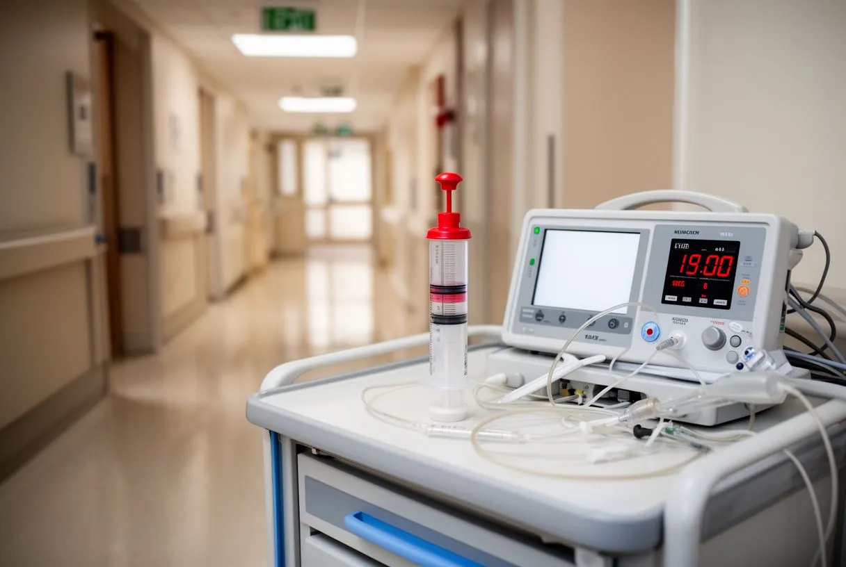
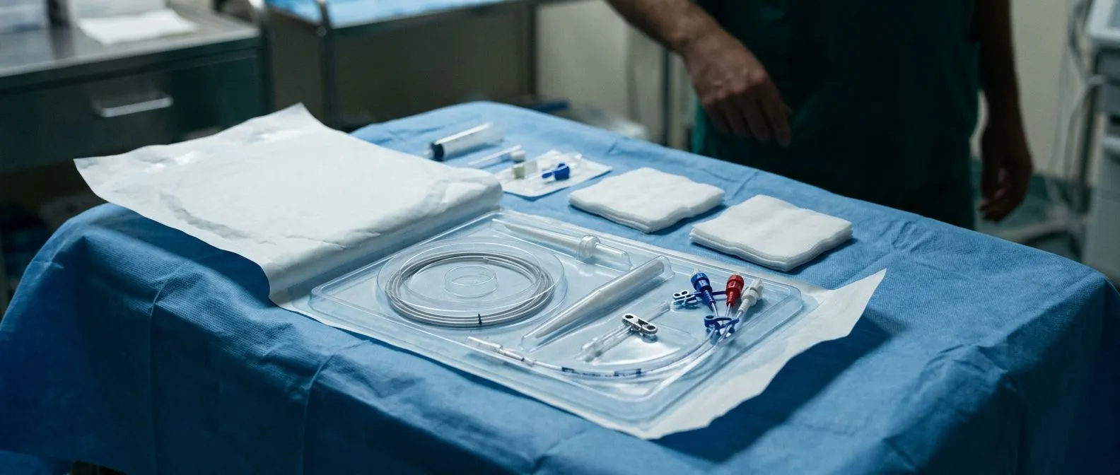
Central venous pressure monitoring is the measurement of the blood pressure in the thoracic vena cava, the large vein that returns blood from the body to the heart. To measure this, your cardiac team places a central venous catheter, usually into a large vein in your neck or upper chest. This catheter is advanced until its tip rests just outside the right atrium, which is the upper right chamber of the heart. By measuring the pressure at this exact location, we gain a clear picture of how much blood is returning to the heart and how effectively the heart is pushing that blood forward to the lungs.
During complex procedures, such as repairing an aneurysm in the ascending aorta, the heart is intentionally stopped and cooled. When the heart resumes beating, it is often temporarily stunned and requires time to regain its full strength. The right atrium receives all the systemic venous return, and if the heart is sluggish, blood will dam up behind it. Central venous pressure monitoring gives us real-time data on this backup. It tells us if the heart is keeping up with the volume of blood returning to it.
This monitoring is not just a standalone metric; it is the cornerstone of postoperative hemodynamic management. It allows us to see the immediate effects of medications, fluid boluses, and mechanical ventilation on the heart's filling pressures. By keeping a close eye on these pressures, your surgical team can make minute adjustments that protect the heart muscle and ensure organs receive the oxygen they need to heal.
Why Do We Measure Central Venous Pressure in the ICU?
In the intensive care unit, the body is transitioning from the extreme stress of surgery back to a stable baseline. Central venous pressure monitoring is used to guide fluid therapy and assess right heart function during this fragile period. When the heart is recovering from an operation, it is highly sensitive to volume changes. Too little fluid means the heart cannot generate enough pressure to perfuse the kidneys and brain. Too much fluid can overstretch the heart muscle, leading to failure and fluid backing up into the lungs or body tissues.
Assessing right heart function: The right side of the heart is responsible for pumping blood into the pulmonary circuit, which is a low-pressure, low-resistance system compared to the systemic circulation. If the right ventricle struggles, the central venous pressure rises because blood cannot be pushed forward. We use this data alongside an arterial line, which is a separate catheter placed in a wrist artery to monitor systemic blood pressure. The arterial cannula used during cardiopulmonary bypass is removed before you leave the operating room, but the central line stays for continuous surveillance.
We do not rely on numbers alone. Central venous pressure monitoring is always combined with clinical examination, including auscultation, or listening to your heart and lungs with a stethoscope. This dual approach ensures we have a complete understanding of your cardiovascular status, preventing us from treating a monitor rather than treating the patient.
How Does Central Venous Pressure Reflect Heart Function?
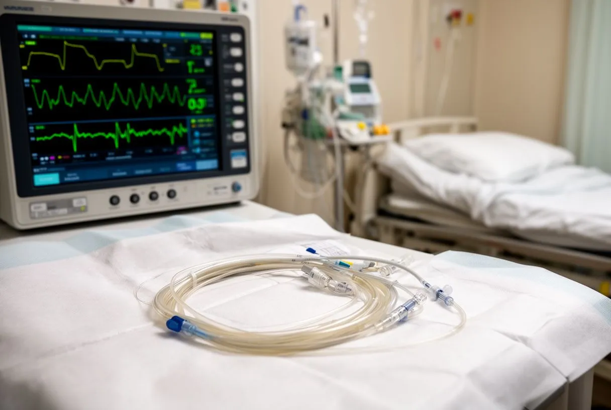
To understand how central venous pressure reflects heart function, it helps to understand the concept of preload. Preload is the degree of stretch of the cardiac muscle fibers at the end of diastole, right before the heart contracts. Central venous pressure is a direct surrogate for right ventricular preload. If the pressure is within a healthy range, the right heart is receiving an optimal volume of blood to pump efficiently. If the pressure is exceptionally low, it suggests the heart is underfilled, like a dry sponge unable to squeeze out any water.
Preload and afterload explained: While preload is the volume returning to the heart, afterload is the resistance the heart must overcome to eject that blood. Central venous pressure gives us a window into preload, but it also hints at right ventricular afterload. If the lungs are diseased or clamped down due to high ventilator pressures, the right heart has to work harder to push blood through them, which can elevate central venous pressure even if the blood volume is normal. This distinction is crucial for deciding whether to give more fluids or to use medications that open the blood vessels.
When the heart is not pumping effectively, fluid backs up into the venous system, causing the central venous pressure to climb. This backup is a primary indicator of right-sided heart dysfunction, which can occur after any cardiac surgery but is particularly common after operations involving the right coronary artery or procedures that require long bypass times. According to research published in American Heart Association Journals, careful hemodynamic management is critical to preventing long-term organ damage in these scenarios.
What Are Normal Central Venous Pressure Values?
In a healthy adult, normal central venous pressure values typically range between 2 and 8 mmHg. However, after cardiac surgery, the target range can shift depending on the specific operation and the patient's underlying heart condition. Your surgical team will establish a personalized target range based on your preoperative heart function, the type of valve or bypass surgery performed, and your body size. We do not treat a number; we treat the patient, using these targets as a guidepost rather than an absolute rule.
Low CVP readings: A reading below the target range usually indicates hypovolemia, meaning there is not enough blood volume in the circulation. This can happen due to bleeding, dehydration, or the effects of medications that dilate the blood vessels. To correct a low reading, we administer intravenous fluids. Whenever possible, surgical teams prefer to use autologous blood, which is your own blood salvaged during the surgery and given back to you, minimizing the risks associated with donor blood transfusions.
High CVP readings: A reading consistently above the target range suggests the heart is struggling to handle the returning blood volume. This can be a sign of right ventricular failure, fluid overload, or a condition called tamponade, where blood or fluid compresses the heart from the outside. High pressures prompt your team to investigate the cause, often using an echocardiogram to look at the heart's pumping action and rule out mechanical complications.
Fluid Management After Cardiac Surgery: Balancing the Scales
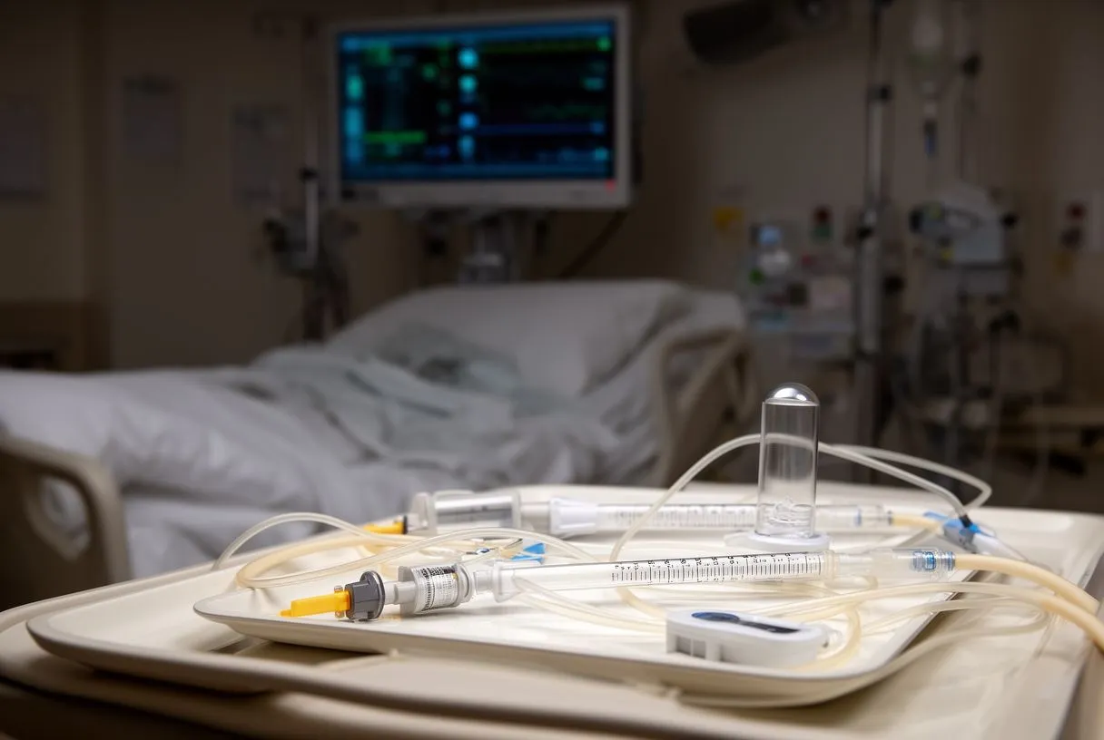
Fluid management after cardiac surgery is an intricate balancing act. The body naturally retains fluid after a major operation due to the stress response and the inflammatory cascade triggered by cardiopulmonary bypass. This retained fluid can seep into tissues, causing swelling, and can accumulate in the lungs, making breathing difficult. Central venous pressure monitoring is the compass that guides us through this fluid terrain, helping us give just enough fluid to keep the kidneys working without drowning the heart.
When central venous pressure readings are elevated, we often use diuretics, medications that help the kidneys excrete excess water and salt. The goal is to achieve a negative fluid balance, meaning more fluid leaves the body than goes in. This process must be gradual and closely watched. Dropping the pressure too quickly can cause low blood pressure and compromise blood flow to vital organs. Sometimes, an atypical infarction, which is a heart attack that presents with unusual symptoms or electrocardiogram patterns, can alter fluid management needs, making precise monitoring even more critical.
We also account for the effects of mechanical ventilation. Positive pressure ventilation pushes air into the lungs, which increases the pressure inside the chest cavity. This external pressure pushes against the heart and great vessels, artificially raising central venous pressure readings. Your intensive care team understands these dynamics and adjusts their interpretation based on ventilator settings, ensuring the numbers reflect true heart function rather than simply the pressure of the breathing machine.
Complications of CVP Monitoring and Catheter Care
While central venous pressure monitoring provides vital information, the catheter itself is a foreign object placed into a major vein, which carries certain risks. Complications can include infection, bleeding, blood clots, or even a punctured lung during insertion. To mitigate these risks, the placement is always done under sterile conditions, often using ultrasound guidance to visualize the vein and avoid neighboring structures. Once the line is in, meticulous care is required to keep the site clean and free from bacteria.
Preventing central line infections: Infection is one of the most serious risks associated with central lines. The intensive care team follows strict protocols, including full sterile barriers during insertion and daily antiseptic care at the insertion site. The need for the line should also be assessed every day. The moment the central venous pressure monitoring is no longer essential for your management, and you are stable enough to transition to less invasive monitoring, the catheter is removed to eliminate the infection risk.
Other medications also play a role in catheter care and overall postoperative health. Patients are often given Aspirin to prevent early clot formation in grafts or valves. If a patient has significant atheromatous plaque, which is a buildup of cholesterol and fats in the artery walls, the choice of insertion site is carefully considered to avoid dislodging any material. Understanding these risks helps families appreciate the rigorous standards maintained in the intensive care unit.
- Infection Control: Daily site care and prompt removal reduce bloodstream infections.
- Mechanical Risks: Ultrasound guidance minimizes the chance of lung puncture or arterial injury during insertion.
- Thrombosis: Blood thinners may be adjusted to prevent clots from forming around the catheter tip.
How Valve Disease Influences Right Atrial Pressure
Valve disease profoundly impacts the pressures inside the heart, and understanding this relationship is key to interpreting central venous pressure monitoring. The mitral valve, located between the left atrium and left ventricle, is a common site of disease. When the mitral valve leaks, a condition known as mitral regurgitation, blood flows backward into the left atrium when the left ventricle contracts. This backward flow increases pressures in the left atrium, which eventually transmits backward through the lungs to the right side of the heart, elevating central venous pressure.
Mitral regurgitation and right heart strain: As the right heart pushes against these elevated lung pressures, the right ventricle can become thickened and eventually fail. This dynamic is why central venous pressure monitoring is so crucial in patients recovering from mitral valve repair. Research on endoscopic approaches, such as the Endoscopic Mitral Repair for Degenerative Mitral Regurgitation: Effect of Disease Complexity on Short- and Mid-term Outcomes, shows that disease complexity directly influences postoperative hemodynamics. Complex repairs can temporarily alter right heart function, requiring careful fluid and pressure management.
The tricuspid valve, located between the right atrium and right ventricle, has an even more direct impact on central venous pressure. If the tricuspid valve leaks, blood flows backward into the right atrium and vena cava, immediately raising the central venous pressure reading. In cases of severe valve disease, a balloon-expandable valve might be used for transcatheter aortic valve replacement (TAVR), a procedure that changes aortic outflow but can still have downstream effects on right heart filling pressures. Genetic factors, as explored in studies on the Genetics and pathophysiology of mitral valve prolapse, also play a role in how these valves fail and how the right heart tolerates the stress.
Central Venous Pressure Interpretation: What the Numbers Mean
Interpreting central venous pressure is rarely about looking at a single static number. It is about understanding the trend. A central venous pressure that is steadily climbing over several hours is a warning sign, even if it has not yet crossed into the abnormal range. It suggests the heart is gradually falling behind in managing the blood volume. Conversely, a pressure that drops after a fluid challenge and then stabilizes suggests the heart is responsive to volume and is pumping effectively.
Dynamic fluid responsiveness: Rather than just looking at baseline numbers, we often perform a fluid challenge. We give a small, rapid bolus of fluid and watch the central venous pressure monitor. If the pressure rises significantly and stays high, the heart is on the flat part of the Starling curve, meaning more fluid will not help and could cause harm. If the pressure rises only slightly and then falls as the heart pumps the fluid forward, the patient is fluid responsive. This dynamic testing is far more informative than a single reading.
We also consider the impact of medications and arrhythmias. The ARISTOTLE trial highlighted the effectiveness of modern blood thinners in atrial fibrillation, a common arrhythmia after surgery. If a patient develops an irregular rhythm, the atrium does not contract effectively, which can alter venous return and change the central venous pressure waveform. The rhythm must be managed with medication or cardioversion before the numbers can be accurately interpreted as a reflection of volume status.
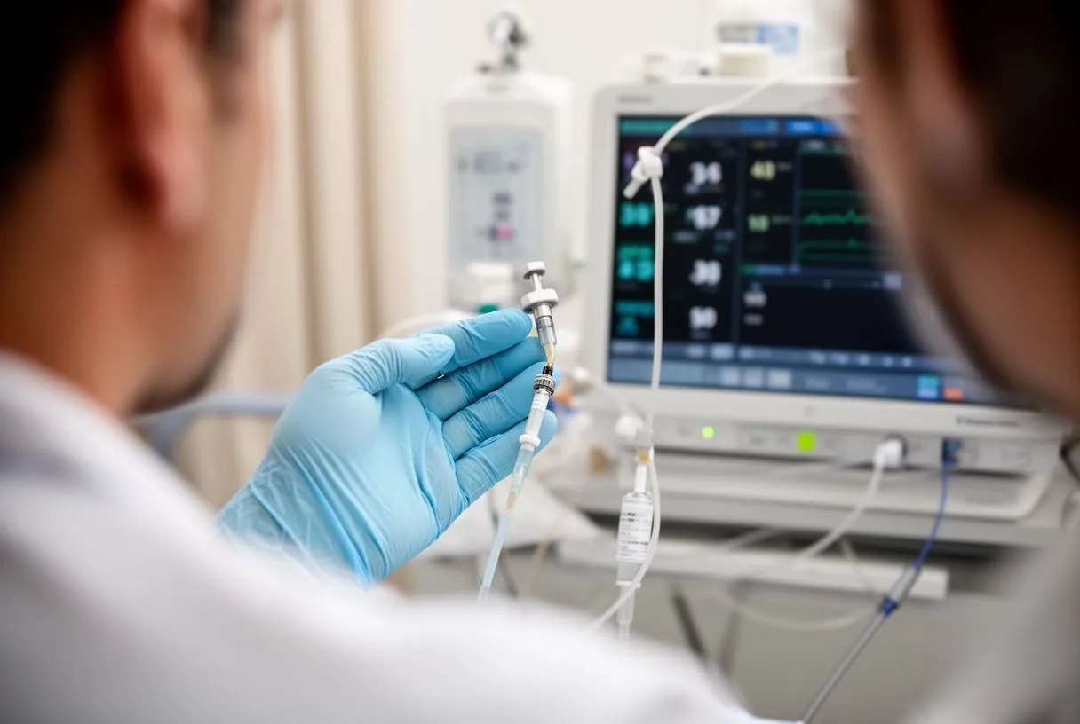
Post Cardiac Surgery Hemodynamics and Recovery Milestones
As you recover, the intensity of monitoring decreases. Central venous pressure monitoring is a feature of the intensive care unit, but as your hemodynamics stabilize, you will transition to a standard ward. This transition is a major recovery milestone. It means your heart has proven it can manage fluid and pressure without the continuous, invasive oversight of a central line. Your care shifts from minute-to-minute adjustments to oral medications and gentle physical activity.
Moving from ICU to ward: When you leave the ICU, the focus changes to rebuilding strength. You might start walking short distances. During this time, the team monitors your blood pressure with a standard cuff and watches for signs of fluid retention, such as weight gain or ankle swelling. If you had an arterial graft placed during bypass surgery, we ensure it is healing and functioning well. The management of cholesterol is also addressed, often starting with medications targeting apolipoprotein B, a protein that carries bad cholesterol in the blood.
Recovery is a multifaceted process. Some patients may have had a balloon angioplasty or a balloon aortic valvuloplasty prior to definitive surgery, and their hemodynamic baseline is different. Others might have received an autograft, using their own tissue for a valve replacement. Throughout this journey, we watch for arrhythmias like atrial flutter, a rapid beating of the atria, which can be managed with medication. In rare, severe cases, patients might need advanced support like Awake ECMO, where extracorporeal membrane oxygenation is used while the patient is awake, but for most, recovery follows a steady, progressive path.
- Extubation: Breathing tube removed, usually within 24 hours, marking the first step toward independence.
- Line Removal: Central and arterial lines removed as hemodynamics stabilize, reducing infection risk.
- Mobilization: Sitting up and walking with assistance to prevent blood clots and improve lung function.
When to Seek Help: Recognizing Abnormal CVP Readings
Once you go home, you will not have a central venous pressure monitor, but the concepts behind it remain relevant to your daily recovery. You become the monitor. The signs of high central venous pressure manifest as symptoms of fluid overload or heart failure. Recognizing these signs early allows you to alert your cardiac team before a minor issue becomes a crisis. You should watch for sudden weight gain over a few days, swelling in your legs or abdomen, or increasing shortness of breath, especially when lying flat.
Signs of fluid overload: If your body is retaining fluid, your heart might be struggling. You might notice your shoes fitting tighter or waking up in the middle of the night gasping for air. Other warning signs include a persistent cough that produces pink, frothy sputum, which indicates fluid in the lungs. You should also be aware of unusual arm pain, which, while sometimes musculoskeletal, can occasionally be a sign of a vascular issue related to your surgery or catheter site.
Remember that recovery is a partnership. At Cirugía de Corazón, Dr Alejandro Molina and our expert team use guideline thresholds, such as an aortic diameter greater than 5.5 cm or EuroSCORE II calculations, to drive decisions before, during, and after surgery. Even with the lowest risk for your situation, complications can arise. Always carry your discharge instructions and the automated external defibrillator (AED) locations in your community if you have severe heart disease. If you notice any concerning symptoms, do not hesitate to contact our team. Never self-diagnose; the safest step is to book a consultation with your qualified cardiac team to review your specific situation and ensure your recovery remains on track.