

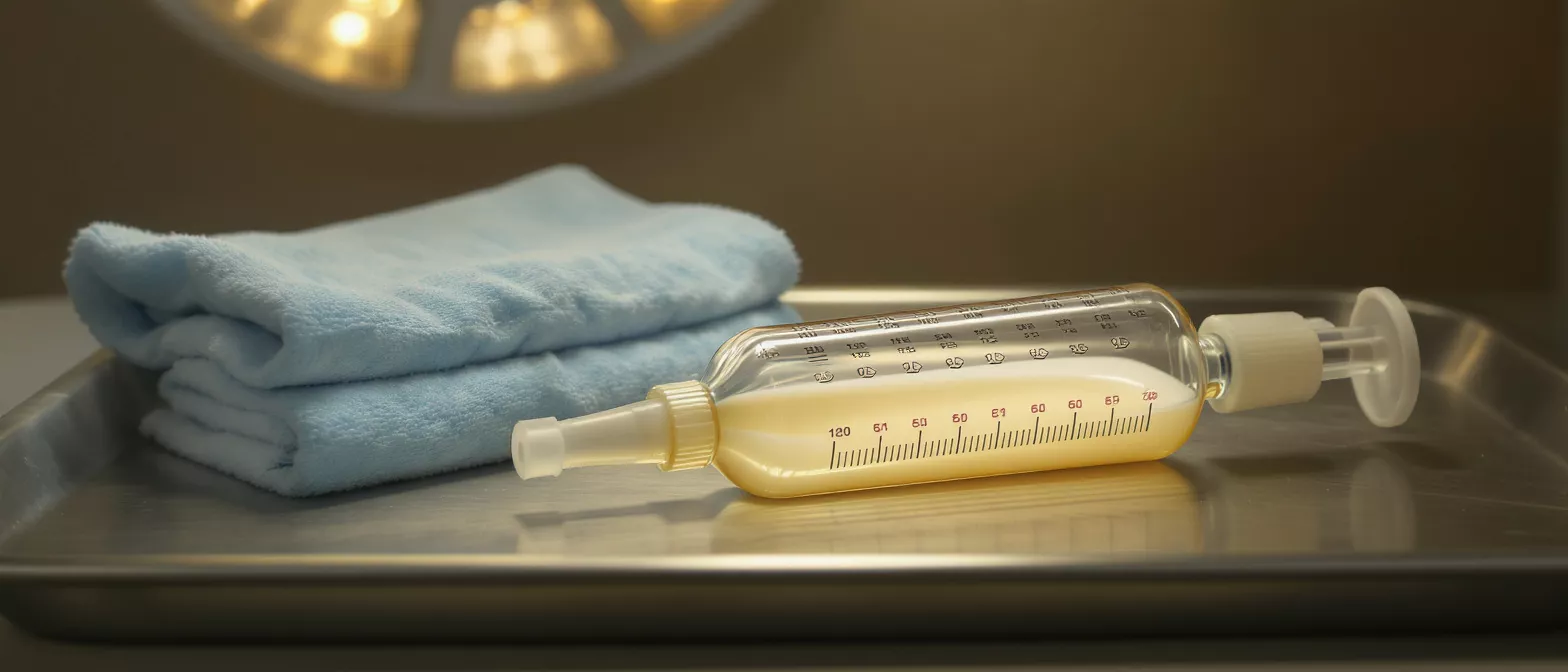
Chest Drain
Learn how a chest drain works after cardiac surgery, why it aids recovery, and what to expect in our full glossary guide from Cirugía de Corazón.
What is a chest drain?
A chest drain, also known as a chest tube or thoracic catheter, is a flexible plastic tube placed into the pleural space or mediastinum to remove unwanted air, blood, or fluid. During heart surgery, surgeons often need to open the chest cavity to access the heart and major blood vessels. Once the surgical repair is complete, the body naturally produces some fluid and bleeding in the operative area. The drain acts as an exit route for this material, preventing it from accumulating and putting pressure on the heart and lungs.
What does a chest drain look like? The setup consists of a clear plastic tube that exits the skin near the chest incision, connected to a collection system on the floor. This container is sealed with water or a one-way valve to create negative pressure. Patients in the intensive care unit will usually wake up with one or more of these drains in place, and they are a standard part of the immediate postoperative equipment.
Understanding the purpose of this device helps reduce anxiety for both patients and families. Seeing tubes draining from the chest can be alarming, but it is a routine and essential component of cardiac recovery. The drain is not a sign of a complication; it is a tool designed to keep the chest cavity clear and allow the heart and lungs to function normally as the body heals.
Why is a chest drain used after cardiac surgery?
The primary reason for using a chest drain after cardiac surgery is to prevent complications that could arise from fluid or air accumulation. When the chest is opened, the natural vacuum seal around the lungs is broken. The body also responds to surgical trauma by producing inflammatory fluid and bleeding. If blood or fluid collects in the pericardial or pleural space, it can compress the heart, restricting its ability to beat effectively, a condition known as cardiac tamponade.
Preventing lung collapse and heart compression is the main goal. By actively suctioning air and fluid out, the drain helps the lungs reinflate fully and keeps the heart from being squeezed by surrounding pressure. The drainage also allows the surgical team to monitor internal bleeding, providing crucial information about the patient's status in the first 24 to 48 hours.
- Lung Expansion: The drain removes air, allowing the lungs to reinflate and function properly.
- Heart Protection: It prevents fluid from compressing the heart, ensuring normal cardiac output.
- Bleeding Monitoring: The collection system gives surgeons real-time data on internal bleeding.
By maintaining a clear space around the heart and lungs, the drain significantly reduces the risk of postoperative emergencies and supports a smoother, safer recovery trajectory.
How does a chest drainage system work?
The mechanics of a chest drainage system rely on negative pressure, similar to a vacuum. The tube is inserted into the chest, and the other end connects to a sealed collection chamber on the floor. Because the chest cavity naturally has a lower pressure than the outside air, this setup helps pull air and fluid out of the body while preventing outside air from entering.
The water seal chamber and suction control are key components. The water seal acts as a one-way valve, allowing air and fluid to leave the chest but stopping air from going back inside. If there is an air leak from the lung, bubbles will pass through this water seal. The suction control chamber regulates the exact amount of vacuum pressure applied to the chest, ensuring the system removes material effectively without damaging internal tissues.
This closed system is entirely sterile and carefully monitored by the nursing staff. The transparent tubing allows nurses to visually inspect the color and consistency of the drainage, while the calibrated markings on the collection chamber help them accurately record the volume of fluid removed over time.
What to expect with a chest tube after heart surgery?
Waking up with a chest tube after heart surgery is a standard experience. Patients typically have one to three drains depending on the type of procedure performed. The tubes are secured to the skin with sutures to prevent accidental removal. While the thought of a tube in the chest can be frightening, most patients report that the drains are more of a nuisance than a source of sharp pain, though soreness around the insertion site is common.
Mobility and pain management are important aspects of early recovery. Patients are encouraged to sit up and walk with the drains, as movement helps expand the lungs and promotes drainage. The collection container is usually hung from the bed or carried on a wheeled stand during ambulation. Nurses administer pain medication to keep patients comfortable, making it easier to move and take deep breaths with the drains in place.
The drainage will initially be dark red, gradually changing to a lighter, yellowish fluid over the first day or two. The nursing team will regularly check the system, empty the collection chamber as needed, and ensure the tubing remains clear of clots. Observing these routine checks can provide peace of mind that the system is functioning exactly as intended.
Chest drain output and monitoring
Monitoring chest drain output is a critical nursing task after cardiac surgery. Nurses document the amount, color, and consistency of the fluid every few hours. The volume of drainage helps the medical team determine if internal bleeding has slowed to a safe level. High output or sudden changes in color can indicate that a patient needs further evaluation or intervention.
What is normal chest drain output? While every patient heals differently, drainage typically decreases over the first 24 hours. As the initial bleeding subsides, the fluid becomes thinner and lighter. If the drainage volume remains high or turns bright red again after slowing down, the surgical team will assess the patient for potential bleeding issues.
Another aspect of monitoring is checking for air leaks. If the lung has an air leak, bubbling will be visible in the water seal chamber. This observation helps the team track the healing of the lung tissue. As the leak resolves, the bubbling stops, signaling that the lung has sealed and the drain may soon be ready for removal.
How is a chest drain removed?
Chest drain removal happens when the medical team determines that the system has done its job. The criteria for removal include a significant drop in the volume of daily drainage, the absence of an air leak, and a clear chest X-ray showing that the lungs are fully inflated. This typically occurs within the first few days after surgery, often before the patient is transferred out of the intensive care unit.
The removal procedure itself is quick. The nurse or doctor will clean the site, clamp the tube temporarily, and ask the patient to take a deep breath and hold it. This breath-holding maneuver prevents air from entering the chest cavity during the split second the tube is pulled out. The tube is swiftly withdrawn, and a sterile dressing is immediately applied over the site to seal the wound.
While the removal process sounds stressful, it is over in seconds. Patients often feel a brief pulling sensation or a moment of mild discomfort, but sharp pain is uncommon. Once the drains are out, patients usually find moving and taking deep breaths much easier, marking a major milestone in their recovery journey.
Chest drain complications and safety?
Chest drains are highly effective and safe, but as with any medical device, complications can occur. The primary goal of the surgical and nursing team is to prevent issues such as infection, tube blockage, or accidental dislodgement. Strict sterile techniques are used during insertion, and the insertion site is regularly cleaned and dressed to minimize the risk of infection.
Preventing blockages and managing clots is a routine part of chest drain management. Nurses may gently squeeze the tubing to prevent blood clots from blocking the line and stopping the flow of drainage. If a clot does block the tube, the system may need to be flushed or the tube replaced, though this is rare. The nursing staff continuously assesses the tubing for kinks or blockages to ensure the system remains patent.
Accidental removal is a serious safety concern, which is why the drains are securely sutured to the skin. Patients and families are educated on how to move safely while the drains are in place. In the event a drain accidentally falls out, patients are instructed to call for a nurse immediately so they can apply pressure to the site and assess the situation to maintain chest safety.
Chest drain nursing care and management
Proper chest drain nursing care ensures the system functions correctly and safely. The intensive care team is specially trained to manage these systems, performing regular checks on the insertion site, the water seal, and the collection chamber. They also ensure the tubing is free of kinks and that the drainage is flowing in the right direction.
Preventing infection and maintaining patency are the two pillars of drain management. The insertion site is monitored for signs of redness, swelling, or discharge. The collection system is kept below the level of the patient's chest at all times to prevent fluid from flowing backward into the chest cavity, which is a critical safety measure for postoperative recovery.
- Site Inspection: Regular checks for redness or infection at the insertion point.
- Patency Maintenance: Mil






