

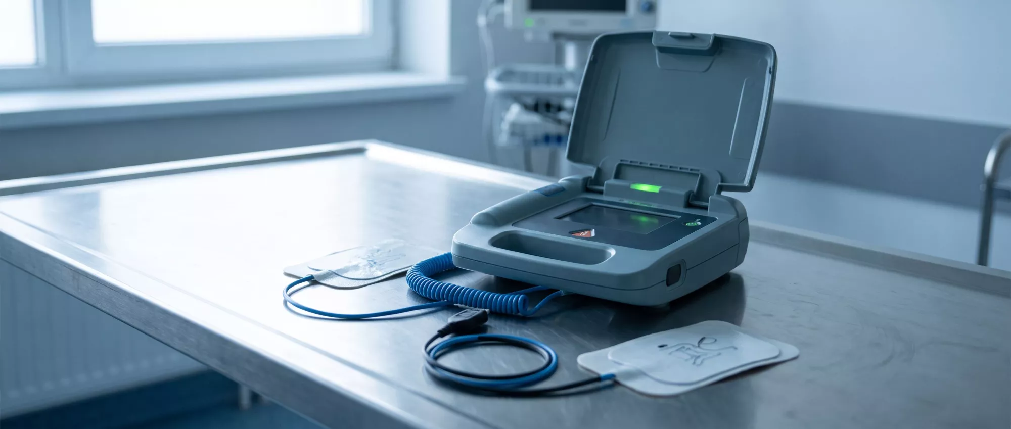
Defibrillation
Learn what defibrillation is, how it restores heart rhythm during cardiac arrest, its types, safety and recovery in our complete glossary guide.
What Is Defibrillation
Defibrillation is a critical medical intervention used to restore a normal heartbeat in life-threatening cardiac emergencies. When the heart's electrical system malfunctions, it can cause the lower chambers to quiver uselessly instead of pumping blood. This procedure delivers a controlled electric shock to the chest wall, effectively stopping the chaotic electrical activity and allowing the heart's natural pacemaker to resume a steady rhythm.
For patients and families navigating cardiac surgery, understanding this procedure is essential. If a loved one experiences a sudden cardiac arrest, immediate action is required.
It is entirely normal to feel frightened when hearing this term discussed in a medical context. The idea of electric shocks can sound jarring. However, defibrillation is a universally recognized, life-saving standard of care that has brought countless patients back from the brink of cardiovascular collapse.
How Defibrillation Work
The mechanism of action: The heart relies on organized electrical signals to contract and pump blood effectively. During an event like ventricular fibrillation, these signals become completely disorganized. The defibrillator sends a brief, high-energy pulse through the heart muscle, which depolarizes a critical mass of the cardiac tissue, essentially hitting a reset button on the electrical system.
This brief reset stops all electrical activity, including the chaotic signals. In the quiet moment that follows, the heart's natural pacemaker, the sinoatrial node, has the opportunity to fire first and re-establish a coordinated, effective heartbeat. The goal is never to start the heart, but rather to stop the dangerous arrhythmia so normal function can return.
The energy dosage is carefully calculated by medical professionals based on the patient's condition and the specific device used. Modern machines analyze the heart rhythm instantly, ensuring the shock is only delivered when absolutely necessary, maximizing both safety and efficacy.
Types of Defibrillation
Medical teams utilize different methods depending on the clinical environment and the patient's specific needs. The most common approach is external defibrillation, where pads are placed on the chest to deliver the energy. This is what occurs during emergency resuscitation efforts both inside and outside the hospital.
For surgical patients, internal defibrillation might be employed. During open-heart procedures, surgeons can apply paddles directly to the heart muscle. This requires significantly less energy and is sometimes needed if the heart struggles to restart naturally after being on bypass.
Additionally, some patients receive implantable cardioverter defibrillators, which are small devices placed under the skin to monitor heart rhythms continuously. These wearable devices can automatically deliver low-energy shocks to correct dangerous rhythms before the patient even notices symptoms.
Defibrillation vs Cardioversion
While both use electrical shocks, the distinction between defibrillation vs cardioversion is critical and based on urgency. Defibrillation is an emergency rescue maneuver used when the heart is in a pulseless, chaotic state like ventricular fibrillation. The shock is delivered immediately and without synchronization to the heart's natural cycle.
Cardioversion, by contrast, is a scheduled procedure used for unstable but still beating arrhythmias. The machine actually syncs the shock to the precise moment of the heart's electrical cycle to avoid causing further damage. This ensures the shock hits at the optimal time to restore normal rhythm without inducing a more severe emergency.
Understanding this difference helps families comprehend why a physician might wait or act immediately. If a patient is conscious and has a pulse, synchronized cardioversion is the safer path. If the patient is unresponsive, unsynchronized emergency defibrillation cardiac arrest protocols take over instantly.
When Is Defibrillation Used
Recognizing the critical window: The procedure is indicated exclusively for life-threatening ventricular arrhythmias, specifically ventricular fibrillation and pulseless ventricular tachycardia. In both conditions, the heart loses its ability to pump blood, leading to immediate loss of consciousness and the absence of a pulse.
Time is the most crucial factor in these scenarios. For every minute that passes without normal blood flow, the chances of survival decrease significantly. Medical teams undergo rigorous training to identify these rhythms on a monitor and apply the shock within seconds.
Outside the hospital, bystanders and first responders rely on automated external defibrillator AED defibrillation units. These machines are designed for public use and provide clear voice instructions, empowering even untrained individuals to act decisively during the most critical minutes before an ambulance arrives.
Defibrillation Pads and Device Placement
Proper placement of defibrillation pads is vital for the electrical current to pass effectively through the heart. The standard positioning is anteroposterior or anterolateral. In the most common anterolateral placement, one pad goes just below the right collarbone, and the other is placed on the left side of the chest, just below the armpit.
This configuration ensures the energy travels across the heart muscle evenly. If the pads are too close together or touch the skin directly, the current will not pass through the heart properly, and the shock will fail to reset the rhythm. Proper skin preparation is essential.
- Clear the chest: Wiping away sweat and drying the skin ensures optimal electrical contact.
- Avoid medication patches: Any transdermal patches must be removed to prevent burns and interference.
- Maintain distance: Pads must be placed at least one inch apart to prevent arcing between them.
Are Defibrillation Procedures Safe
The procedure is highly effective and absolutely necessary for survival, but it does carry some physical considerations. The shock itself can cause minor skin burns where the pads contact the chest. Fortunately, these burns are typically superficial and heal quickly, far outweighing the alternative of not treating the cardiac arrest.
There is also a small risk of the shock causing damage to the heart muscle, known as myocardial stunning, which can temporarily reduce pumping efficiency. Medical teams monitor for this closely in the intensive care unit following resuscitation.
Psychological safety is another crucial aspect. Patients who survive an arrest and subsequent defibrillation often experience anxiety or fear of recurrence. Cardiologists and specialized nursing teams provide extensive support to help patients process the trauma and regain confidence in their heart health.
Internal Defibrillation and Implantable Devices
Implantable cardioverter defibrillator function: For patients at high risk of sudden cardiac arrest due to severe structural heart disease or previous surgical repairs, an implantable defibrillator device is often recommended. This small unit, placed under the skin near the collarbone, continuously monitors the heart rhythm.
If it detects a lethal arrhythmia, it immediately delivers a shock directly inside the heart chamber, restoring normal rhythm before the patient loses consciousness. This internal approach requires significantly less energy than external pads and provides a constant safety net for vulnerable patients.
For surgical candidates, understanding how these devices integrate with surgical recovery is important. These garments offer gentle compression and stabilize the surgical area while the device leads heal into the heart muscle.
What to Expect During Emergency Defibrillation
Witnessing an emergency defibrillation can be alarming for family members. The procedure involves everyone stepping back from the bed, ensuring no one is touching the patient. When the shock is delivered, the patient's body may jerk slightly due to the sudden muscle contraction, which is a normal physical response.
Immediately after the shock, medical personnel resume chest compressions. This is because it often takes a few moments for the heart to establish a strong, effective rhythm. The team will continue high-quality CPR and check monitors until a stable pulse returns.
Family members are usually asked to wait outside during this active resuscitation. This protects the sterile environment and allows the clinical team to focus entirely on the patient. Once the situation stabilizes, the care team will provide a detailed update on the outcomes and the next steps for intensive care.
Recovery and Long-Term Heart Health
Surviving an event requiring defibrillation is a major medical milestone, but it is only the first step in a longer journey. The immediate recovery takes place in the intensive care unit, where specialists manage the underlying cause, such as blocked arteries or surgical complications, to prevent recurrence.
Long-term health requires a comprehensive approach, including medication adherence, lifestyle adjustments, and regular cardiac rehabilitation. Understanding why the heart went into a dangerous rhythm is the key to preventing future emergencies. Patients often undergo electrophysiology studies to map their heart's electrical pathways.
Finally, the emotional recovery cannot be overlooked. Surviving sudden cardiac arrest brings profound relief but also a need for psychological processing. Support groups, counseling, and open communication with the care team empower patients to reclaim their lives and face the future with confidence and resilience.






