

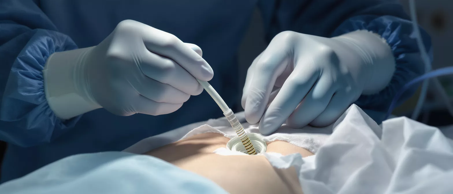
Femoral Cannulation
Learn how femoral cannulation works in cardiac surgery, including procedure steps, risks, and benefits.
What is femoral cannulation in cardiac surgery?
Femoral cannulation is a specialized medical technique used to connect a patient's circulatory system to a heart-lung machine or a life-support system. During complex cardiac procedures, the surgical team must temporarily take over the work of the heart and lungs. To achieve this safely, they insert large tubes called cannulas into the major blood vessels located in the groin. This process provides an alternative route for blood to exit the body, receive oxygen, and return without disrupting the surgical field in the chest.
Where the tubes are placed: The groin contains the femoral artery and the femoral vein, which are the primary highways for blood traveling to and from the lower body. Because these vessels are large and easily accessible, they serve as excellent entry points. The surgical team typically makes a small incision near the crease of the hip to locate these vessels.
Why this matters for your procedure: If you or a loved one is undergoing a minimally invasive heart surgery, the surgeon may choose this route to avoid opening the entire chest. By accessing the blood vessels through the leg, the medical team can still fully support the body while working on the heart through much smaller incisions.
How does the femoral cannulation procedure work?
The femoral cannulation procedure works by creating a secure, closed-loop circuit for blood circulation. Once the patient is under anesthesia, the surgeon exposes the femoral vessels. A specially designed tube is inserted into the femoral vein to drain deoxygenated blood away from the body. This blood is then pumped through an artificial lung, where carbon dioxide is removed and oxygen is added, before being returned to the body.
Returning oxygenated blood: The oxygen-rich blood is usually returned to the body through a second cannula placed in the femoral artery. This restores circulation to the lower body and vital organs, ensuring that tissue remains healthy while the surgical team repairs the heart. The entire circuit is carefully managed by a perfusionist, a specialist who monitors blood flow and pressure in real time.
Maintaining systemic circulation: This setup ensures that blood continues to flow to the brain, kidneys, and other critical organs. It is a highly coordinated effort that allows the heart to be safely stopped and emptied, giving the surgeon a still, bloodless environment to perform precise repairs. Utilizing surgical vascular grafts can sometimes be integrated into this process to ensure optimal vascular integrity.
Why is femoral artery cannulation used for ECMO?
Extracorporeal Membrane Oxygenation (ECMO) is a life-saving technique used when the heart or lungs are failing. Femoral artery cannulation is the most common approach for ECMO because it can be initiated quickly at the bedside in an intensive care unit. When a patient is in severe respiratory or cardiac distress, the rapid placement of these groin cannulas allows the ECMO machine to take over immediately.
Supporting lung and heart failure: The femoral route is highly effective for Veno-Arterial (VA) ECMO, which supports both the heart and lungs. Blood is drained from the femoral vein, oxygenated by the machine, and pumped back into the femoral artery. This bypasses the heart and lungs entirely, giving them time to rest and heal from severe illness or injury.
Why the femoral site is preferred: The anatomy of the groin offers relatively straightforward access to large vessels. This accessibility is critical during emergencies. In these high-stakes situations, maintaining vascular access is vital, and arterial blood pressure cuffs are often used to continuously monitor the patient's hemodynamic stability during the initiation of support.
Femoral vein cannulation and blood flow dynamics
Femoral vein cannulation focuses specifically on draining blood from the body back to the heart-lung machine. The femoral vein is a massive vessel that returns blood from the lower extremities. A long, flexible cannula is threaded through this vein, often advancing up into the abdomen and toward the heart, to ensure maximum blood drainage.
Draining deoxygenated blood: Efficient drainage is essential for a successful cardiac procedure. The perfusion team relies on this venous drainage to reduce the volume of blood in the heart, creating a quiet surgical field. The dynamics of this blood flow are driven by gravity siphon or vacuum assistance, depending on the specific equipment used by the surgical center.
Balancing the circulatory system: The amount of blood drained through the femoral vein must exactly match the amount returned through the artery. This balance prevents drastic shifts in the patient's blood pressure. To manage this delicate equilibrium, the surgical team frequently utilizes central venous catheter kits for precise pressure monitoring.
Types of femoral cannulation approaches
There are several distinct femoral cannulation approaches a surgeon might choose, tailored to the patient's anatomy and the surgical requirements. The strategy dictates how blood is removed and returned. Surgeons carefully evaluate the patient's vascular health prior to the operation to select the safest configuration.
- Veno-Arterial (VA) approach: Drains blood from the femoral vein and returns it to the femoral artery. This provides complete cardiopulmonary bypass, supporting both heart and lung function simultaneously.
- Veno-Venous (VV) approach: Drains and returns blood through the venous system. This is used primarily for lung support, such as in severe pneumonia, allowing the heart to continue beating naturally.
- Percutaneous approach: Involves inserting the tubes through the skin using needle punctures and guidewires, similar to a heart catheterization, avoiding large surgical incisions in the groin.
Each method carries specific hemodynamic implications. The surgical team uses preoperative imaging to ensure the selected vessels are free from severe blockages that could complicate the insertion process.
Step-by-step femoral cannulation steps
Understanding the femoral cannulation steps can help alleviate anxiety by demystifying the process. The procedure is methodical and precise. The surgical team follows a strict sequence to ensure the vessels are accessed safely without causing damage to the surrounding tissues.
Making the incision and identifying vessels: After cleaning the area, a small incision is made in the groin to expose the femoral artery and vein. If a percutaneous technique is used, a needle is guided directly into the vessel using ultrasound visualization. Once the vessel is accessed, a guidewire is passed through the needle.
Dilating and inserting the cannula: A series of progressively larger dilators are passed over the guidewire to gently stretch the tissue and vessel opening. Finally, the actual cannula is advanced into place. The guidewire is removed, and the tubes are secured to the skin to prevent accidental dislodgement during the operation.
Femoral cannulation risks and complications
While femoral cannulation is a routine practice in modern cardiac surgery, it involves major blood vessels and carries inherent risks. Being aware of potential femoral cannulation complications helps patients make informed decisions and recognize the importance of post-operative care. Surgeons employ meticulous techniques to minimize these risks.
Vascular injury and bleeding: The femoral vessels are large, and inserting tubes requires making an opening in them. There is a risk of bleeding or injury to the vessel wall. If a vessel is fragile or calcified, the risk increases. Surgeons use specialized imaging and careful suturing techniques to repair the vessels once the cannulas are removed.
Reduced blood flow to the leg: Because the cannula occupies space inside the femoral artery, it can restrict blood flow to the lower leg. To prevent this, the surgical team often places a smaller secondary tube directing blood down the leg. Compartment syndrome, a painful condition caused by inadequate blood supply, is a rare but serious risk.
Femoral cannulation catheter placement and monitoring
Accurate femoral cannulation catheter placement is critical for patient safety. The cannulas must be positioned securely within the vessels without causing trauma to the vessel walls. Incorrect placement can lead to inadequate bypass support or damage to internal structures. The anesthesiology and perfusion teams monitor the patient closely.
Using ultrasound and fluoroscopy: Modern cardiac surgery relies heavily on real-time imaging. Ultrasound allows the surgeon to visualize the vessel before insertion, ensuring they avoid plaque buildup. Fluoroscopy, a type of continuous X-ray, helps confirm that the tubes are threaded to the correct depth.
Monitoring for proper flow and pressure: Once the cannulas are in place, the perfusionist monitors the flow rates and pressures within the circuit. Any sudden change in pressure can indicate a kink in the tube, a blockage, or a malposition. Immediate adjustments ensure the patient remains hemodynamically stable throughout the heart surgery.
Recovering after peripheral cannulation femoral sites
Recovery after peripheral cannulation femoral procedures requires specific care for the groin incision. Once the heart-lung machine is no longer needed, the surgeon carefully removes the cannulas. The puncture sites in the blood vessels are repaired, usually with fine sutures, to prevent bleeding. The skin is then closed, and a sterile dressing is applied.
Leg mobility and discomfort: Patients may experience soreness or bruising in the groin area for several days. It is common to feel some stiffness when walking. The medical team will encourage gradual mobilization, but heavy lifting or strenuous activity must be avoided to allow the blood vessels to heal properly.
Watching for signs of complications: Patients are educated on what to watch for after discharge. Any signs of severe pain, swelling, or a cold, pale foot could indicate a problem with blood flow and require immediate medical attention. Proper wound care ensures the incision heals cleanly without infection.
Femoral cannulation benefits for high-risk patients
Despite the associated risks, femoral cannulation offers profound benefits, particularly for high-risk patients who might not tolerate traditional, open-chest approaches. By enabling minimally invasive procedures, this technique allows surgeons to perform complex repairs with significantly less trauma to the body.
Enabling minimally invasive surgery: Accessing the blood vessels through the groin means the surgeon does not need to fully open the sternum. This results in smaller scars, less post-operative pain, and a generally faster recovery time. For elderly or frail patients, avoiding a large sternal incision dramatically reduces the risk of infection.
Emergency support capability: In critical care settings, the ability to rapidly initiate femoral cannulation saves lives. When a patient suffers a massive pulmonary embolism or goes into cardiac arrest, this technique allows doctors to instantly restore circulation.
Ultimately, femoral cannulation is a cornerstone of modern cardiac care, providing the vital support needed to perform delicate surgeries and rescue failing hearts. By understanding how this technique works, patients and families can approach their treatment with greater confidence and clarity.






