

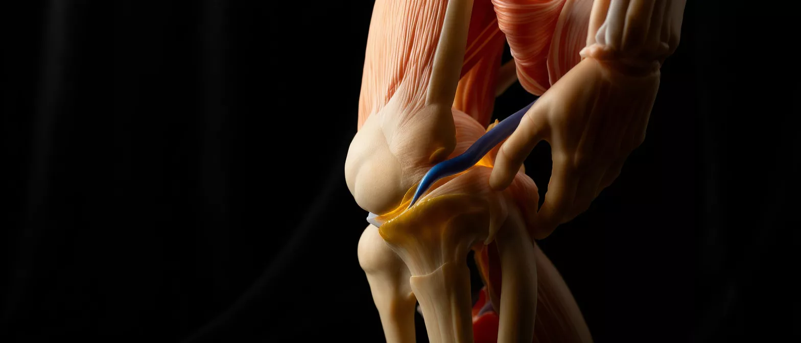
Femoral Vein
Learn femoral vein anatomy, function, thrombosis signs, and its role in cardiac surgery cannulation.
What is the femoral vein?
The femoral vein is a major blood vessel located deep within the upper thigh, playing a crucial role in returning deoxygenated blood from the legs back toward the heart. For patients preparing for cardiac surgery, understanding this vessel is vital, as it often serves as a primary access point for life-saving interventions and monitoring. It is one of the largest deep veins in the human body.
Femoral vein definition and medical terminology: In medical terms, the femoral vein is the continuation of the popliteal vein as it passes through the adductor hiatus, a gap in the thigh muscles. It travels upward through the groin, eventually becoming the external iliac vein once it crosses beneath the inguinal ligament. This anatomical transition point is significant for surgeons and cardiologists when planning vascular access.
Recognizing the importance of this vessel helps alleviate anxiety for patients facing heart procedures. Because of its large diameter and accessible location in the groin, medical teams rely on this pathway safely and effectively to support heart function during complex operations.
Femoral vein anatomy and location
Understanding femoral vein anatomy and location is essential for grasping how cardiac specialists approach various surgical procedures. The vein sits alongside the femoral artery and nerve within a structure known as the femoral sheath, deep within the upper thigh near the groin. Its central placement makes it a reliable landmark for both diagnostic imaging and emergency interventions.
Femoral vein placement in the femoral triangle: The femoral triangle is a key anatomical region in the upper inner thigh. Within this space, medical professionals utilize the mnemonic "NAVEL" (Nerve, Artery, Vein, Empty space, Lymphatics) to remember the exact placement of these critical structures. The vein rests medially to the artery, an important distinction during central venous access kits to ensure correct and safe puncture.
- Proximity to the heart: Allows rapid deployment of support catheters during cardiac surgery.
- Large diameter: Accommodates large-bore cannulas necessary for cardiopulmonary bypass.
- Superficial depth at the groin: Makes it accessible for percutaneous needle entry without deep surgical cuts.
By mapping this anatomy precisely, surgeons can navigate the vascular system safely. A thorough understanding of this regional anatomy minimizes complications and ensures precise cannulation when time is critical during cardiovascular events.
How does femoral vein function in venous return?
The primary femoral vein function is to act as a high-capacity highway for venous return, carrying oxygen-depleted blood from the lower limbs back to the heart. Without this vital pathway, circulation in the legs would quickly become compromised, leading to severe swelling and tissue damage. The vein relies on surrounding muscle contractions and one-way valves to push blood upward against gravity.
During cardiac surgery, this natural venous return is often temporarily rerouted. Surgeons place a cannula into this vessel to drain blood into a heart-lung machine, taking over the workload of the heart. By draining venous blood directly, the surgical team gains a bloodless, motionless field to operate on the heart with precision.
This process ensures that the patient's circulatory needs are met while the heart is intentionally stopped or opened. After the procedure is complete, the venous circuit is restored, allowing the heart to resume its natural pumping function without added stress.
Femoral vein vs femoral artery differences
Patients often ask about femoral vein vs femoral artery differences, as both are major vessels in the groin but serve opposite purposes. The artery carries oxygen-rich blood away from the heart to the legs, while the vein carries oxygen-poor blood back up to the heart and lungs for replenishment. This fundamental difference in direction is crucial for understanding cardiac access.
Pressure and flow characteristics: Because the artery is driven by the heart's pumping force, it has high pressure and a pulsing rhythm. The vein, relying on muscle movement and valves, operates under lower pressure with a steady, non-pulsatile flow. Recognizing these differences is vital during femoral vein cannulation, as accidentally accessing the artery instead of the vein can lead to significant bleeding complications.
Cardiac teams use ultrasound guidance and distinct anatomical landmarks to distinguish between the two before making an incision. This careful verification helps ensure that venous drainage tubes are placed correctly, protecting the patient's circulatory integrity.
What is femoral vein thrombosis?
Femoral vein thrombosis is a serious medical condition where a blood clot forms within this deep vessel, obstructing venous return from the leg. Also known as deep vein thrombosis (DVT), this condition requires immediate medical attention to prevent potentially life-threatening complications. Patients recovering from major heart surgery are at an increased risk for blood clots due to immobility.
Deep vein thrombosis femoral implications: When a clot blocks the femoral vein, blood backs up into the lower leg, causing sudden, severe swelling, pain, and redness. The greatest danger is that a fragment of this clot can break loose, travel through the heart, and lodge in the lungs, causing a pulmonary embolism. This risk necessitates vigilant monitoring and preventive care.
To mitigate this risk during cardiac recovery, doctors often prescribe blood thinners and encourage early mobilization. Understanding the signs of a clot empowers patients and families to seek immediate help if symptoms arise, ensuring rapid intervention and safety.
Signs of a femoral vein blood clot
Recognizing the signs of a femoral vein blood clot early is essential for patients recovering from cardiac surgery. Symptoms typically develop rapidly in one leg, causing noticeable asymmetry between the left and right limbs. Being aware of these warning signs allows for swift medical evaluation and treatment.
Femoral vein medical term symptoms: Clinically, patients may experience localized pain or cramping in the thigh or calf that worsens when standing or walking. The affected leg often displays visible swelling, a feeling of heavy warmth, and a pale or bluish discoloration. If a patient notices sudden leg swelling after surgery, they must alert their healthcare team immediately.
Early detection is the strongest defense against severe complications. Healthcare providers utilize rapid diagnostic tools to confirm the presence of a clot and begin life-saving blood-thinning therapy right away.
How femoral vein ultrasound diagnoses clots
To accurately diagnose a clot, doctors rely on a femoral vein ultrasound, a non-invasive imaging test that uses sound waves to visualize blood flow. This procedure is completely painless and can be performed right at the patient's bedside. It is the gold standard for identifying vascular obstructions quickly.
The diagnostic process: A technician applies gel to the skin over the groin and thigh, gliding a handheld transducer to capture real-time images of the vessel. The ultrasound machine shows the vessel's size, structure, and the movement of blood. If the vein cannot be compressed by the probe, it strongly indicates the presence of a solid blood clot inside.
For cardiac surgery patients, this vascular ultrasound provides immediate clarity, ruling out or confirming thrombosis without exposing the patient to radiation. It guides clinical teams to make fast, accurate decisions regarding necessary anti-embolism compression stockings or therapeutic anticoagulation.
Why femoral vein cannulation is used in cardiac surgery
Femoral vein cannulation is a critical technique used during cardiac surgery to establish extracorporeal circulation, commonly known as a heart-lung bypass. By inserting a cannula into this large vein, surgeons can drain deoxygenated blood out of the body, pass it through a machine to add oxygen, and return it to the body via an artery. This process allows the heart to be safely stopped during delicate repairs.
This method is particularly valuable in emergency situations or minimally invasive heart surgeries where opening the chest directly is not ideal. The vein's large diameter accommodates the high flow rates necessary to perfuse the entire body with oxygenated blood safely. It offers a reliable entry point for the extracorporeal circulation tubing packs required for bypass.
By utilizing this access route, surgical teams can maintain a patient's circulatory stability while operating on a still, bloodless heart. This technique represents a cornerstone of modern cardiac interventions, enabling complex procedures that save lives.
Femoral vein access complications and risks
While femoral vein access is generally safe, it carries potential complications that cardiac teams actively work to prevent. Because the groin area contains vital structures packed closely together, precision is required to avoid adjacent vessels and nerves. Understanding these risks helps patients engage in informed preoperative discussions with their surgeons.
Femoral vein complications to monitor: Potential issues include bleeding or hematoma formation at the puncture site, accidental puncture of the femoral artery, or localized infection. Additionally, the placement of a catheter can irritate the vein lining, increasing the risk of deep vein thrombosis. The medical team uses sterile techniques and real-time imaging to minimize these risks.
- Site care: Keeping the insertion site clean and dry to prevent infection.
- Bleeding watch: Monitoring for expanding bruising or firm lumps at the groin.
- Circulation checks: Assessing leg color, temperature, and pulse strength post-procedure.
Vigilant post-operative monitoring is key to catching and addressing these issues early. With proper care, the vast majority of patients experience no long-term access complications.
Recovery and care after femoral vein procedures
Recovery after femoral vein cannulation or catheterization focuses on protecting the access site and restoring normal circulation. Once the catheters are removed, firm pressure is applied to the groin to stop bleeding. Patients will be instructed to lie flat for a specific period to ensure a stable clot forms at the insertion site.
Post-procedure instructions: Healthcare teams monitor the patient's vital signs and check the pulse in the feet to ensure proper blood flow is maintained. Patients are encouraged to keep the leg still initially and avoid heavy lifting or strenuous activity for several weeks. Staying hydrated and gently moving the legs helps prevent new blood clots from forming during the healing phase.
By following these guidelines, patients can expect a smooth, uncomplicated recovery. Understanding the importance of this major vein and following post-operative care instructions ensures the best possible outcomes after heart surgery, allowing patients to return to their daily lives with a healthier heart.






