

Haemodynamic Study
Learn how a haemodynamic study evaluates heart function, blood flow, and pressure, plus what to expect from the procedure, results, and recovery.
What is a haemodynamic study?
A haemodynamic study is a specialized diagnostic test that measures how well blood flows through your heart and the surrounding major blood vessels. When preparing for cardiac surgery, your medical team needs a highly detailed picture of your cardiovascular system. This study provides real-time data regarding blood pressure, oxygen levels, and the overall pumping efficiency of your heart muscle.
Rather than relying solely on external symptoms or static imaging, cardiologists use haemodynamic assessment to see exactly how your heart performs under its normal physiological workload. By evaluating the force and volume of blood circulation, your surgical team can pinpoint specific areas of concern. This direct look at your circulatory dynamics is crucial for planning a safe and effective surgical intervention.
Haemodynamic study meaning in cardiology: In the context of cardiology, the term "haemodynamics" refers to the physical principles governing blood flow and the forces involved in circulation. A haemodynamic study translates these principles into measurable clinical data. It helps your surgeon understand the precise internal environment of your heart, ensuring that any structural repairs will be matched by improved functional blood flow.
How does a haemodynamic study work?
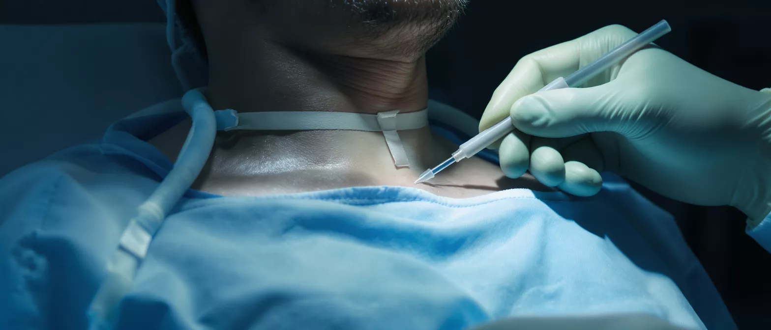
The haemodynamic study procedure relies on precise internal measurements taken via thin, flexible tubes called catheters. During the test, your cardiologist gently guides these catheters through a blood vessel in your wrist, neck, or groin, advancing them directly into the chambers of your heart and pulmonary arteries. This allows for direct, moment-to-moment observation of your cardiovascular system.
Cardiac catheterization haemodynamic study mechanics: Once the catheters are correctly positioned, they connect to external pressure transducers. These highly sensitive devices instantly translate the physical pressure of your bloodstream into electrical signals, displaying continuous waveforms on a monitor. This dynamic view reveals exactly how your heart valves are functioning and whether the muscle is pumping effectively with every beat.
The data gathered includes the exact pressure within the heart's right side, the left side, and the lungs. Additionally, small blood samples might be drawn directly from these chambers to compare oxygen saturation levels. By measuring blood flow volume against internal pressure, the medical team can map out a highly accurate functional diagram of your heart.
Why would a cardiologist order a haemodynamic study?
If you are facing heart surgery, your surgical team needs to understand the exact mechanical issues affecting your cardiovascular system. A cardiologist typically recommends a haemodynamic evaluation for heart disease when non-invasive tests like echocardiograms do not provide enough clear information. It offers the definitive data required to confirm a diagnosis and finalize a surgical plan.
Doctors frequently use this assessment to evaluate the severity of heart valve disease, pulmonary hypertension, or congestive heart failure. By measuring the specific pressure gradients across a narrowed or leaking valve, the surgical team can determine if a repair or replacement is necessary. It clarifies complex cases where the exact cause of a patient's breathing difficulties or chest pain might otherwise be ambiguous.
Haemodynamic monitoring for heart surgery preparation: Before any surgical intervention, establishing a baseline of your heart's current performance is essential. This monitoring reveals how much stress your heart can safely handle during an operation. It allows your surgeon to anticipate potential complications and select the most appropriate surgical approach for your specific anatomical needs, relying on a trusted haemodynamic monitoring system to ensure precision throughout the procedure.
How to prepare for a haemodynamic study procedure
Preparation for a haemodynamic study is similar to preparing for other minor cardiac catheterization procedures. Your medical team will give you specific instructions based on your overall health and current medication regimen. Generally, you will be asked to fast for a few hours before the test to prevent any discomfort or complications during the procedure.
Medication adjustments before your test: You must inform your doctor about all medications and supplements you are currently taking, especially blood thinners. Your cardiologist may temporarily adjust certain prescriptions a few days prior to the study to lower the risk of bleeding at the catheter insertion site. Always follow your medical team's exact instructions regarding dosage changes.
Because you may receive a mild sedative to help you relax during the test, arranging transportation home afterward is essential. Wear comfortable clothing and leave valuables at home. Upon arrival at the clinic or hospital, the nursing staff will review your medical history, check your vital signs, and place an intravenous line to easily administer any necessary medications.
What to expect during haemodynamic monitoring of the heart
The haemodynamic study procedure takes place in a specialized room called a cardiac catheterization laboratory. You will remain awake throughout the process, allowing you to follow simple breathing instructions from your medical team. The environment is highly controlled, featuring advanced imaging equipment that helps the cardiologist guide the catheters with extreme precision.
The insertion site is thoroughly cleaned and numbed with a local anesthetic, so you should only feel pressure, not sharp pain. As the catheter is gently advanced toward your heart, you might experience a brief sensation of warmth or a slight fluttering. The care team continuously monitors your electrocardiogram and oxygen levels, maintaining a state of careful cardiac monitoring equipment oversight to ensure your safety and comfort at every step.
Once the catheters are positioned, the actual measurements are taken. The process of recording pressures and drawing tiny blood samples from different heart chambers usually takes about 30 to 60 minutes. After the necessary data is collected, the catheters are gently removed, and pressure is applied to the insertion site to prevent bleeding.
Understanding your haemodynamic study results
After completing your haemodynamic study, your cardiologist will analyze the recorded waveforms, pressure readings, and oxygen saturations. These results are compared against established normal ranges to determine how well your heart is managing blood flow. Understanding the basic numbers can help you feel more confident when discussing your surgical options with your doctor.
Interpreting your haemodynamic study results: The results are typically presented as numerical values representing pressures in millimeters of mercury (mmHg) and oxygen percentages. Elevated pressures in specific chambers may indicate stiffening of the heart muscle or narrowed valves. Low oxygen saturation in one chamber might suggest a structural issue, such as a hole in the heart allowing oxygen-poor and oxygen-rich blood to mix.
- Right heart pressures: Elevated numbers here can point to pulmonary hypertension or right-sided heart failure.
- Cardiac output: This measures the total volume of blood your heart pumps per minute; lower values indicate weakened muscle function.
- Vascular resistance: High resistance indicates your heart is working harder than normal to push blood through your lungs or systemic arteries.
Your surgeon uses these exact measurements to calculate the severity of your condition and map out the surgical repair. If a valve replacement is deemed necessary, they may discuss the integration of a durable prosthetic heart valve tailored to your specific haemodynamic profile, ensuring optimal blood flow restoration post-surgery.
Are haemodynamic studies safe for heart patients?
Undergoing any internal medical procedure naturally causes some anxiety, especially for patients already dealing with cardiac concerns. However, a haemodynamic study is widely considered a routine and very safe procedure when performed by an experienced cardiology team in a controlled hospital environment. The diagnostic value it provides far outweighs the minimal risks involved.
Complications are rare, but as with any procedure involving a blood vessel, there is a small risk of bleeding, bruising, or minor infection at the insertion site. Some patients might experience temporary irregular heartbeats during the test, which usually resolve immediately once the catheter is repositioned or removed. Your medical team is fully trained to manage and immediately address any unexpected changes during the evaluation.
To ensure your complete safety, your vitals, heart rhythm, and oxygen levels are observed continuously from the moment you enter the laboratory until you are fully recovered. The clinical staff remains prepared for any scenario, providing a reassuring layer of professional oversight throughout your entire visit.
Right heart catheterization vs invasive haemodynamic evaluation
Patients often hear different medical terms when researching their upcoming tests, leading to understandable confusion. Right heart catheterization and invasive haemodynamic evaluation are frequently used interchangeably by medical professionals, but understanding the nuances can help clarify your specific surgical preparation plan.
A right heart catheterization is a specific type of invasive haemodynamic study that focuses entirely on the right side of your heart and the pulmonary arteries. It measures the pressures in the lungs and the right ventricle, which is critical for diagnosing pulmonary hypertension and evaluating fluid overload. It is the standard method used for most pre-operative heart surgery evaluations.
In contrast, a more comprehensive invasive haemodynamic evaluation might also involve crossing over to the left side of the heart to assess aortic or mitral valve gradients directly. Depending on your specific symptoms and preliminary imaging results, your cardiologist will determine which precise approach gives your surgical team the most accurate data for your upcoming operation.
Haemodynamic study recovery and what comes next
Recovery from a haemodynamic study is generally quick and straightforward. Because the procedure is minimally invasive and does not require general anesthesia, most patients are able to sit up and walk around within a few hours. Your medical team will observe you in a recovery area to ensure the insertion site stops bleeding and your vital signs remain perfectly stable.
Caring for the insertion site: You should keep the puncture site clean and dry for the first 24 hours following the test. Avoid heavy lifting, strenuous exercise, or submerging the area in water until your doctor gives you clearance. If you notice any sudden swelling, active bleeding, or increasing pain at the site, contact your healthcare provider immediately for guidance.
Once your surgeon reviews the complete study results, they will schedule a follow-up appointment to discuss the next steps. This conversation is where your surgical pathway becomes clear. The data gathered from this test empowers your medical team to finalize their surgical strategy, giving you a concrete roadmap for your journey toward better heart health.
Key takeaways about cardiac haemodynamic assessment
A haemodynamic study is a highly valuable tool that provides your surgical team with the exact physiological data needed to treat complex heart conditions safely. By directly measuring the pressures and oxygen levels inside your heart chambers, it removes the guesswork from surgical planning. It transforms static images into a dynamic understanding of your heart's true functional capacity.
Remember that experiencing anxiety before a diagnostic test is completely normal. This procedure is a standard, carefully monitored step designed to protect your safety and optimize your surgical outcome. Understanding how the test works, what to expect, and how the results are used can significantly reduce your stress and help you approach your treatment with confidence.
Ultimately, this study is a bridge between diagnosis and successful surgical intervention. By giving your surgeon a clear map of your internal cardiovascular pressures, the haemodynamic study directly contributes to a more precise, effective, and life-improving cardiac surgery experience.






