

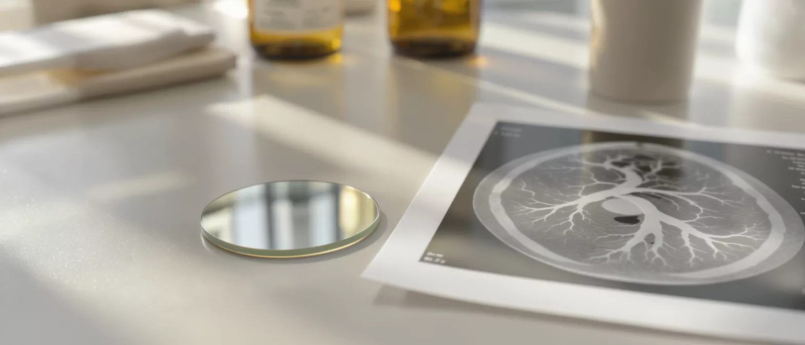
Imaging on Disc
Learn how imaging on disc works in cardiac care.
What Is Imaging on Disc in Cardiac Care?
When you undergo a heart scan or surgical procedure, your medical team generates detailed visual records of your cardiovascular system. Imaging on disc refers to the process of saving these digital heart scans onto a compact disc or DVD, allowing you to hold a portable, exact copy of your medical files. This practice ensures that the intricate details of your heart—whether captured through an MRI, CT scan, or echocardiogram—remain accessible without relying on a hospital's internal network.
For patients preparing for or recovering from heart surgery, having direct access to these records provides peace of mind. The disc contains the raw, unedited imaging data rather than a simple printed photograph or report summary. This means any specialist you visit can view the beating of your heart or the structure of your arteries in real-time motion, exactly as the original capturing machine recorded it.
Understanding this term helps demystify the discharge process. When a nurse hands you a disc before you leave the hospital, you are receiving a comprehensive digital copy of your cardiac health at that moment in time, which you can carry securely to any future medical appointment.
How Does a Heart Scan on Disc Work?
A heart scan on disc operates using specialized medical software embedded directly in the disc's interface. The viewing software: when you insert the disc into a computer's CD or DVD drive, an auto-run menu typically opens, allowing you to click a link that launches a proprietary viewer. This means you do not need to purchase or download complicated medical software to see your own scans.
The data stored on the disc includes far more than static images. The files contain dynamic loops, cross-sectional slices, and measurement markers that your cardiologist or surgeon used to evaluate your condition. Because the disc holds the original digital files, a specialist can adjust the contrast, zoom into specific arteries, and review the heart's mechanical function from multiple angles just as if they were standing in the imaging lab.
It is important to note that these discs are designed to be played on desktop or laptop computers rather than standard DVD players connected to televisions. The computer screen and processing power allow for the high-resolution viewing necessary to accurately examine the delicate structures of the cardiovascular system.
Why Is Medical Imaging on Disc Important for Heart Surgery Patients?
For a patient facing cardiac surgery, continuity of care is absolutely critical. Seamless specialist communication: carrying your medical imaging on disc allows a new surgeon, second-opinion specialist, or rehabilitation center to instantly view the baseline anatomy of your heart before beginning any new treatment. This immediate access eliminates the guesswork and waiting periods often associated with requesting physical films or waiting for hospital-to-hospital server transfers.
When a patient is anxious about an upcoming procedure, knowing they have a complete record of their heart's current state provides a tangible sense of control. The imaging provides a visual roadmap of any blockages, valve defects, or structural anomalies, helping the surgical team plan their approach with precision. It acts as a universal language between different departments, urgent care centers, and out-of-town clinics.
Furthermore, in the event of an emergency, having immediate access to your prior cardiac imaging disc can drastically reduce the time needed for a triage team to understand your medical history, especially if you are traveling far from your primary hospital network.
What Types of Cardiovascular Imaging Are Stored on a Disc?
Various diagnostic tests produce the files suitable for storage on a patient disc. Echocardiograms: these ultrasound recordings capture the live motion of the heart's walls and valves, often forming the bulk of the dynamic video files found on a disc. Cardiac MRI on disc provides highly detailed cross-sectional views of the heart muscle, helping identify tissue scarring or inflammation that might influence surgical planning.
Cardiac CT scan disc formats are frequently used to store angiograms, offering 3D rotational views of the coronary arteries to show exactly where plaque buildup has occurred. These different modalities are sometimes combined onto a single disc if a patient has undergone a comprehensive pre-surgical evaluation, giving the surgeon a complete picture of the heart's plumbing and electrical systems.
- Echocardiography: Live motion video of heart valves and pumping function.
- Cardiac MRI: High-resolution tissue detail to check muscle scarring or inflammation.
- CT Coronary Angiograms: 3D structural maps of blood vessels and arterial blockages.
Regardless of the test type, the disc preserves the data in a format that preserves the original resolution and measurement integrity, ensuring clinical accuracy is never lost in translation.
How to Use Your Cardiac Imaging Disc at a New Clinic
Presenting your imaging disc to a new medical provider is a straightforward process. The check-in handoff: when you arrive at a new clinic or specialist's office, simply provide the disc to the medical receptionist or nurse managing your intake. They will insert the disc into a clinical workstation to extract the relevant files directly into their patient chart system.
It is always a good idea to keep the disc in a protective sleeve and clearly label it with your name, date of birth, and the date the imaging was performed. While the disc itself contains digital metadata that identifies you, having a clear physical label ensures the medical staff routes the files to the correct chart instantly, preventing any administrative delays.
If you are attending a consultation before a major cardiac surgery, bringing your disc directly to the appointment ensures the surgeon can pull up your exact anatomy on their screen during the discussion, allowing them to point out specific areas of concern while explaining the procedure to you.
What Is the DICOM Format on an Imaging Disc?
The underlying technology that makes medical imaging portable is the DICOM standard, which stands for Digital Imaging and Communications in Medicine. Universal file compatibility: a DICOM imaging disc embeds your heart scans into a universally recognized file format that ensures any cardiac imaging machine or clinical software can read the data regardless of the manufacturer.
A DICOM file does more than just display a picture of your heart; it bundles the image pixels together with your personal metadata, such as your name, medical record number, and the specific machine settings used during the scan. This helps ensure that when a new doctor views the imaging, the picture is calibrated and scaled to your actual physical anatomy, lowering the risk of misdiagnoses related to distorted image sizes.
Patient education glossary sites often emphasize the importance of this standardization. By utilizing a DICOM imaging disc, your surgical team avoids the frustrating process of trying to convert proprietary, unreadable files, streamlining the pre-operative evaluation period significantly.
Are There Alternatives to a Heart Imaging CD?
While the compact disc remains a reliable standard in many hospitals, digital technology is rapidly shifting toward cloud-based patient portals. Secure cloud transfers: many modern cardiology imaging storage systems now allow hospitals to upload digital heart imaging directly to a secure online network, granting patients access via a login and password instead of a physical disc.
Despite these advancements, physical discs are still heavily utilized because they do not require an internet connection to operate. If a hospital's server is down, or if an older clinic lacks a secure portal to receive external digital transfers, a heart imaging CD serves as an unfailingly reliable backup method to transport crucial diagnostic data.
Some patients actually prefer the tangible nature of a disc because it feels more secure than an emailed link. When you cannot utilize a cardiac imaging viewer online, or if your internet access is limited, the disc ensures your medical history remains entirely in your hands.
What to Expect When Receiving an Imaging Disc After a Cardiac MRI
Receiving your records after a lengthy diagnostic test is often the final step before leaving the imaging center. Immediate physical handover: after a cardiac MRI, a technician or nurse will typically hand you a disc while you are still in the recovery area, confirming that all the captured data has been successfully written to the media. The disc will often be labeled with the date of the scan and the type of machine used.
You may notice that the disc looks identical to a standard blank CD you might purchase for home use, but the internal data structure is vastly different. You should not attempt to alter or add personal files to this disc, as doing so could corrupt the sensitive medical data and render the imaging unreadable for your future surgeons.
It is normal for the disc to contain folders with hundreds of individual image slices. These slices are stacked together by the viewing software to create the 3D models and dynamic beating heart loops that your doctor will review when planning your surgical intervention.
How to Store and Protect Your Medical Records on Disc
Properly caring for your digital cardiac records ensures they remain viable for future consultations. Environmental protection: medical imaging on disc should be stored in a cool, dry place away from direct sunlight, as extreme heat or excessive light can warp the plastic and permanently destroy the delicate data layers embedded within the media.
Always keep the disc inside a rigid plastic case or a soft protective sleeve to prevent scratches on the data side of the surface. Even minor surface damage can make portions of your heart scans unreadable, which might necessitate a repeat of the diagnostic test, causing unwanted delays in your surgical timeline and added stress.
It is also wise to keep a mental note of where the disc is located within your home, especially if you have a large volume of medical paperwork. When a sudden referral to a new specialist arises, knowing exactly where your digital records are stored saves valuable time.
Understanding the Role of Digital Heart Imaging in Your Recovery
The journey through heart surgery does not end when you leave the operating room; it continues through a carefully monitored recovery phase. Having a baseline heart scan on disc from before your surgery allows your rehabilitation team to compare your heart's current function against its pre-operative state, providing a clear visual metric of your healing progress.
These digital cardiac records serve as a bridge between your surgical history and your ongoing cardiovascular health management. By understanding exactly what is on your disc and how to share it with your care team, you actively participate in the safeguarding of your own health, ensuring that every future specialist operates with the full, unedited picture of your heart.
Ultimately, holding your imaging on disc is about maintaining continuity in your medical care. It is a simple piece of plastic that carries the detailed, life-saving blueprint of your cardiovascular system, ensuring your medical history follows you wherever your recovery journey leads.







Comments
This is incredibly reassuring. I was handed a disc after my MRI last week and had no idea what to do with it or if I should just wait for the online portal. Knowing it has the raw, unedited data makes me feel much better about bringing it to my surgical consult next week.
I always wondered why hospitals still use CDs in this day and age. The point about not needing an internet connection and avoiding server downtime makes total sense. I’ll be sure to keep my disc in a rigid case so it doesn’t get scratched before my next appointment.