Aortic Valve Disease: Stenosis, Leaks and Treatment
Aortic valve disease treatment in Guadalajara covers stenosis, leaks and minimally invasive options.
What Is Aortic Valve Disease and How Does It Affect the Heart?
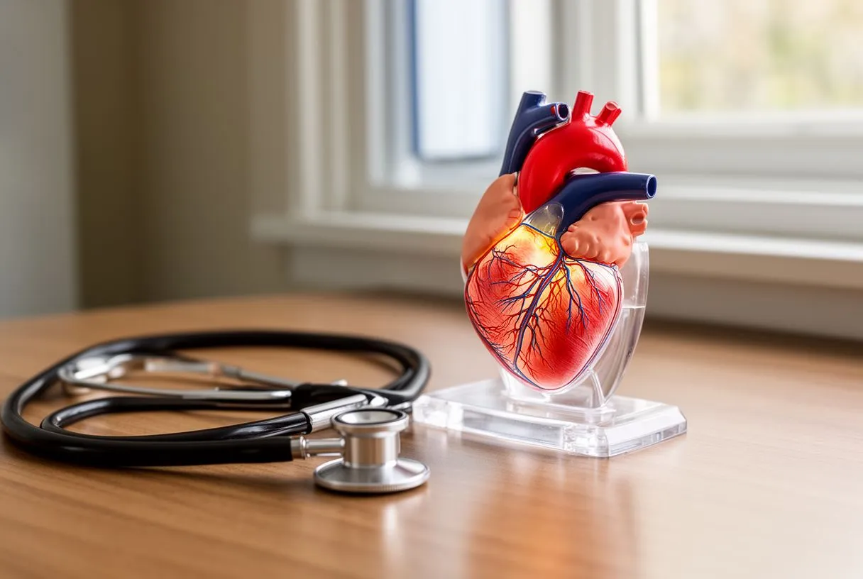
Receiving a diagnosis of aortic valve disease can feel overwhelming, but understanding the mechanics of your heart is the first step toward feeling confident in your care. The aortic valve is a crucial gateway in your heart. It sits between the left lower chamber, known as the left ventricle, and the ascending aorta, which is the main artery that carries oxygen-rich blood from the heart to the rest of your body. When this valve is healthy, its three leaflets open fully to let blood flow out and then snap shut tightly to prevent blood from leaking backward into the left atrium or ventricle. When disease strikes, this orderly flow is disrupted, forcing the heart muscle to work much harder than it should.
Understanding valve stenosis: Stenosis occurs when the valve leaflets become stiff or thickened. This narrowing restricts the opening, making it difficult for the heart to pump blood through the narrowed space. Over time, the heart muscle must generate immense pressure to push blood forward, which can lead to thickening of the heart walls and eventual weakening of the pump. Understanding valve regurgitation: Also known as insufficiency or a leak, this happens when the valve leaflets do not close completely. Blood slips backward into the left ventricle with each heartbeat, reducing the amount of oxygenated blood reaching your organs and increasing the volume of blood the heart must manage with each contraction.
Both conditions fall under the umbrella of aortic valve disease, and both can significantly impact your quality of life if left untreated. The specific experience varies from person to person, depending on the severity of the narrowing or leaking and how quickly the condition progresses. Recognizing the distinct types of valve dysfunction is vital because the treatment pathway, timing, and surgical approach differ considerably based on the exact anatomical problem. Here is a brief summary of the primary forms you might encounter:
- Aortic Stenosis: The valve opening is narrowed due to calcium buildup or congenital defects, restricting forward blood flow.
- Aortic Regurgitation: The valve leaks backward, causing the heart to handle excess blood volume and work harder to circulate oxygenated blood.
- Bicuspid Valve Disease: A congenital condition where the valve has only two leaflets instead of three, often leading to early stenosis or leaking.
What Causes Aortic Valve Stenosis and Leaks?
Age-related calcium deposits: The most frequent cause of aortic stenosis in older adults is calcific degeneration. Over decades, calcium deposits accumulate on the valve leaflets. While calcium is essential for strong bones, its presence on heart valves causes the delicate tissue to become rigid. Factors that accelerate this process include high cholesterol and the accumulation of atheromatous plaque in the arteries. Interestingly, researchers have found that high levels of Apolipoprotein B, a protein involved in lipid metabolism, can also play a role in the speed of valve calcification, linking systemic vascular health directly to valve deterioration.
Congenital heart defects: Some individuals are born with an aortic valve that has two leaflets instead of the standard three. This bicuspid aortic valve is a common congenital anomaly. Because the blood flows through a differently shaped opening, the leaflets are subjected to abnormal wear and tear. This often leads to early calcification and stenosis, sometimes requiring intervention in a patient's forties or fifties rather than their seventies or eighties. Rheumatic fever consequences: Though less common today due to antibiotics, a history of rheumatic fever can scar the valve leaflets, leading to a combination of both stenosis and regurgitation later in life. According to the American Heart Association, managing these underlying risk factors is a critical component of long-term cardiac care.
Endocarditis and connective tissue disorders: Valve leaks are frequently caused by structural issues that prevent the leaflets from closing properly. Endocarditis, an infection of the heart valves, can destroy valve tissue or create holes in the leaflets, resulting in severe regurgitation. Additionally, connective tissue disorders, such as Marfan syndrome, can cause the aorta itself to stretch and dilate. When the ascending aorta widens, it pulls the valve leaflets apart, preventing them from meeting in the center to seal the opening. This specific scenario often requires not just valve repair or replacement, but also the replacement of the enlarged aortic root to prevent future tearing or rupture.
What Are the Most Common Aortic Valve Disease Symptoms?
Many patients live with aortic valve disease for years without realizing it because early stages often produce no noticeable symptoms. The heart is incredibly resilient and can compensate for a gradual decline in valve function. Often, a doctor first detects the condition through auscultation, which is the simple act of listening to your heart with a stethoscope. An abnormal whooshing sound, known as a heart murmur, indicates turbulent blood flow and prompts further testing. For many, this incidental finding is the very first hint that a valve is not functioning perfectly, long before physical symptoms appear.
Classic exertion symptoms: As the disease progresses and the heart struggles to keep up with demand, symptoms become more apparent. The most common complaints are shortness of breath, especially during physical activity, and unusual fatigue. Patients often describe a heavy sensation in the chest or angina, which is chest pain caused by reduced blood flow to the heart muscle itself. Sometimes, this pain radiates to the neck or down the left arm, though in some populations, particularly women, arm pain without chest discomfort can be an atypical presentation of cardiac distress, sometimes overlapping with what physicians call an atypical infarction.
Syncope and advanced warning signs: In more advanced stages, patients may experience syncope, which is fainting or passing out, particularly during exertion. This happens because the narrowed valve cannot increase blood flow to the brain when the body demands more oxygen during activity. Fluid retention leading to swelling in the ankles and feet, known as edema, is another sign that the heart is beginning to fail. Preclinical research continually refines our understanding of these physiological changes; for instance, studies detailed in publications like Pig Models in Translational Surgery help surgeons comprehend the exact hemodynamic thresholds where symptoms transition from mild fatigue to dangerous syncope. If you experience fainting or severe chest pain, you should seek immediate emergency medical evaluation.
How Is Aortic Valve Disease Diagnosis Performed by Specialists?
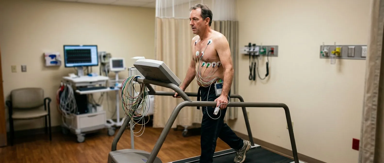
Diagnosing aortic valve disease is a meticulous process that combines physical examination with advanced imaging. Once a murmur is heard or symptoms are reported, the cornerstone of diagnosis is the echocardiogram. This non-invasive ultrasound allows the cardiologist to see the valve in real-time, measuring the speed of blood flow across the valve to determine the severity of stenosis. It also reveals the size of the heart chambers and the efficiency of the heart's pumping function, providing a comprehensive picture of how the valve disease is affecting the overall cardiac anatomy.
Risk scoring and rhythm analysis: Before any treatment decision is made, specialists assess your overall surgical risk using a calculation tool like EuroSCORE II. This system evaluates factors such as age, lung function, and previous surgeries to estimate the likelihood of complications, helping your team determine the safest intervention. During this phase, doctors also carefully monitor your heart rhythm. Aortic valve disease can stretch the heart chambers, leading to arrhythmias such as atrial flutter, a rapid beating of the upper chambers that requires management with medication or targeted procedures before valve surgery can safely proceed.
Additional imaging modalities: In some cases, a standard echocardiogram does not provide enough detail, especially if the patient's chest wall is thick or if the valve anatomy is complex. A transesophageal echocardiogram (TEE) might be ordered, where a specialized probe is passed down the esophagus to get a close-up, unobstructed view of the heart from behind. Coronary angiograms or CT scans are also frequently utilized to check for blocked arteries and to precisely measure the dimensions of the ascending aorta. This thorough anatomical mapping is essential; it ensures that if surgery is required, the surgeon knows exactly what they will encounter and can plan the repair or replacement with absolute precision.
What Are the Treatment Options for Aortic Valve Disease?
The approach to treating aortic valve disease depends heavily on the severity of the condition, your symptoms, and your overall health. For mild to moderate cases without symptoms, the primary strategy is watchful waiting. This involves regular echocardiograms, usually once a year, to monitor the progression of the disease. During this surveillance phase, doctors focus on optimizing cardiovascular health. Medications may be prescribed to manage blood pressure or control heart rate, easing the workload on the struggling heart muscle, though medication alone cannot fix a structurally damaged valve.
Bridging therapies: In some cases, patients need temporary relief before undergoing definitive surgery. Balloon Aortic Valvuloplasty is a procedure where a catheter with a small balloon is threaded into the heart and inflated to stretch the narrowed valve open. While the effects are often temporary, it can provide crucial symptomatic relief for patients who are too frail for immediate open-heart surgery. Other catheter-based techniques, similar to Balloon Angioplasty used for coronary arteries, might be employed to address other vascular issues concurrently. Medications such as Aspirin are routinely used to prevent blood clots, while blood thinners backed by clinical evidence from studies like ARISTOTLE are essential if atrial fibrillation is present, significantly reducing the risk of stroke.
Timing of surgical intervention: Surgery is usually recommended the moment symptoms develop, or when diagnostic tests show that the heart function is beginning to decline, even if the patient feels fine. Waiting too long once symptoms begin can lead to irreversible heart damage. The surgical decision is not a one-size-fits-all scenario. Your cardiac surgeon will weigh the benefits of restoring normal blood flow against the immediate risks of surgery. For patients with multiple health issues, a less invasive catheter-based approach might offer the lowest risk for your situation, while younger, healthier patients might benefit most from a highly durable surgical valve replacement that will last for decades.

Aortic Valve Repair vs Replacement: Which Is Right for You?
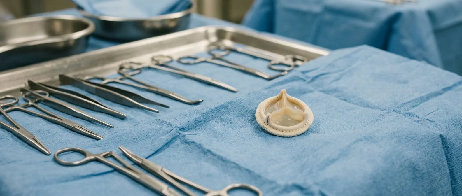
When surgery becomes necessary, one of the primary decisions is whether to repair your native valve or replace it entirely. Aortic valve repair is generally preferred when the primary issue is a leak caused by structural abnormalities that can be surgically reshaped. Surgeons can trim excess tissue, add supporting structures to help the leaflets close tightly, or use an autograft, which involves taking a healthy valve from another part of the heart to replace the damaged aortic valve. Repair offers the advantage of preserving your own tissue, which eliminates the need for long-term blood thinners and often provides excellent durability.
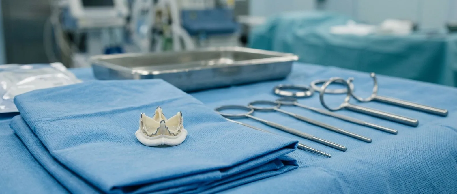
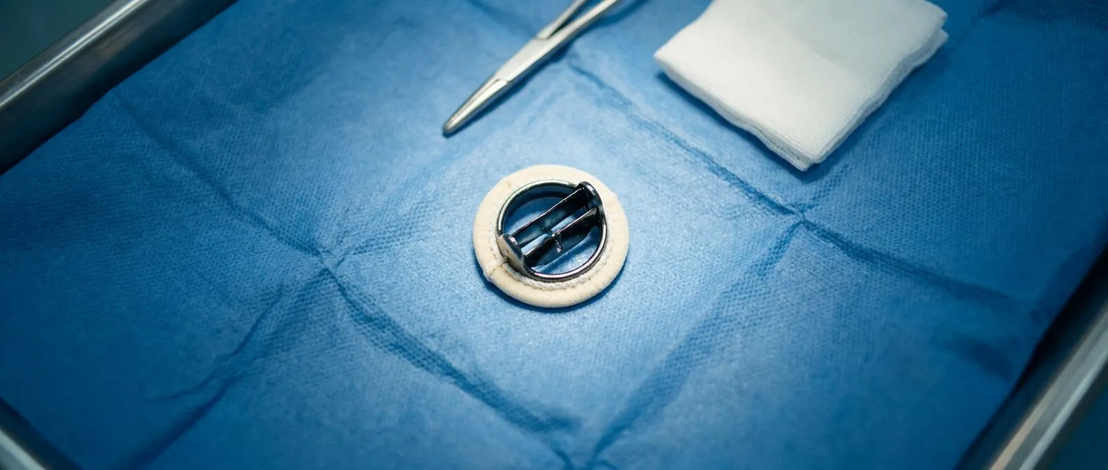
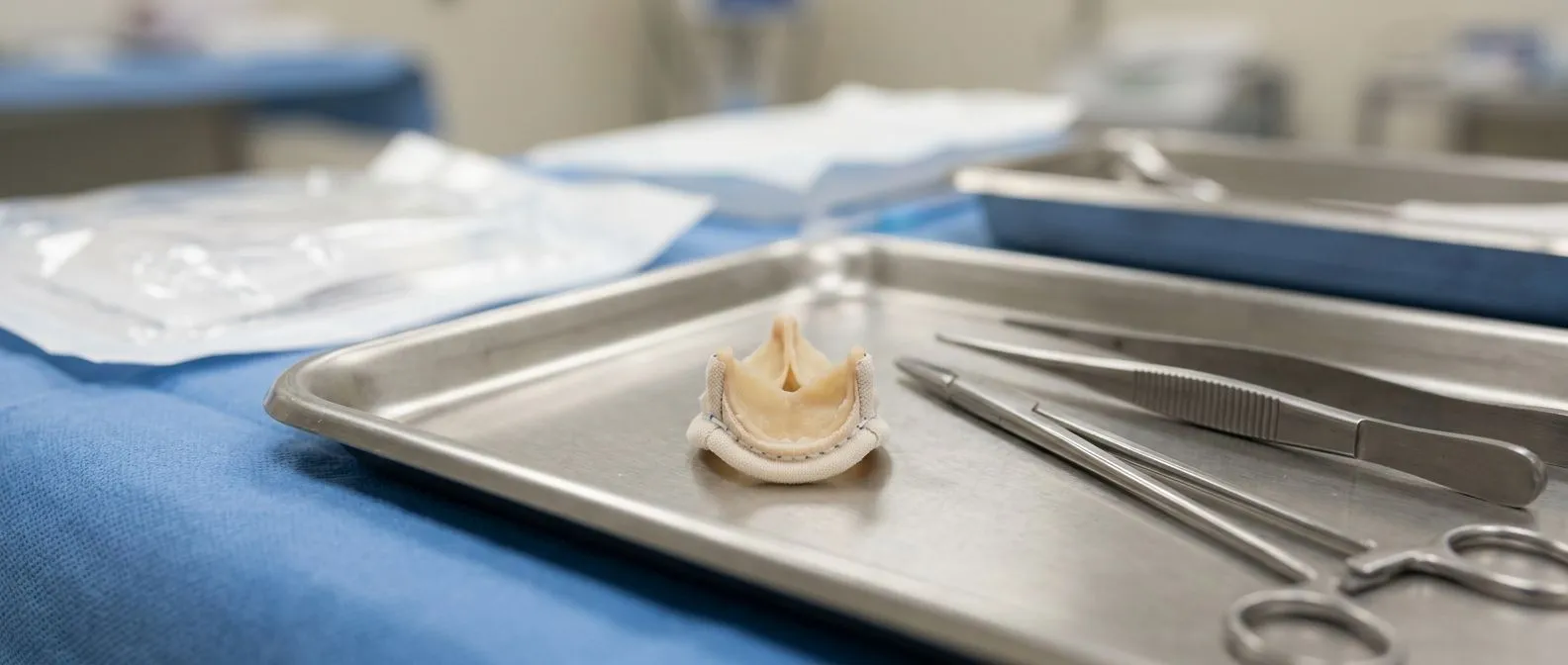
Replacement options: When the valve is heavily calcified, as seen in severe stenosis, repair is usually not feasible, and replacement is the standard of care. There are two main types of replacement valves: mechanical and biological. Mechanical valves are made from durable synthetic materials and can last a lifetime, but they require lifelong anticoagulation therapy to prevent clots. Biological valves are made from cow or pig tissue and do not require long-term blood thinners, but they have a limited lifespan and may need to be replaced after 10 to 15 years. Interestingly, the safety and precision of modern surgical environments cross disciplines; for instance, meticulous gas management in enclosed surgical spaces, as explored in research on Optimal carbon dioxide insufflation pressure during robot-assisted thyroidectomy in patients with various benign and malignant thyroid diseases, shares core principles with the careful atmospheric controls used in advanced cardiac operating rooms.
Choosing the right valve: The decision between a mechanical and biological valve depends on your age, lifestyle, and ability to safely take blood thinners. Younger patients often receive mechanical valves to avoid the risk of a second open-heart surgery later in life. Older patients, or those with a higher risk of bleeding, frequently receive biological valves. During surgery, advanced techniques are utilized to minimize blood loss, and the use of autologous blood—your own blood collected and recycled during the procedure—reduces the need for donor transfusions. Your surgical team will present these options clearly, guiding you toward the choice that provides the greatest long-term benefit with the least disruption to your daily life.
What Is Minimally Invasive Aortic Valve Surgery and TAVR?
Advancements in cardiac surgery have introduced minimally invasive techniques that avoid the traditional full sternotomy, where the entire breastbone is split open. Instead, surgeons can access the heart through a smaller incision in the upper chest or between the ribs. This approach causes less trauma to the body, leading to reduced postoperative pain, smaller scars, and a generally quicker return to normal activities. While not every patient is a candidate for this approach, those with suitable anatomy can benefit immensely from the less invasive surgical route for both valve repair and replacement.
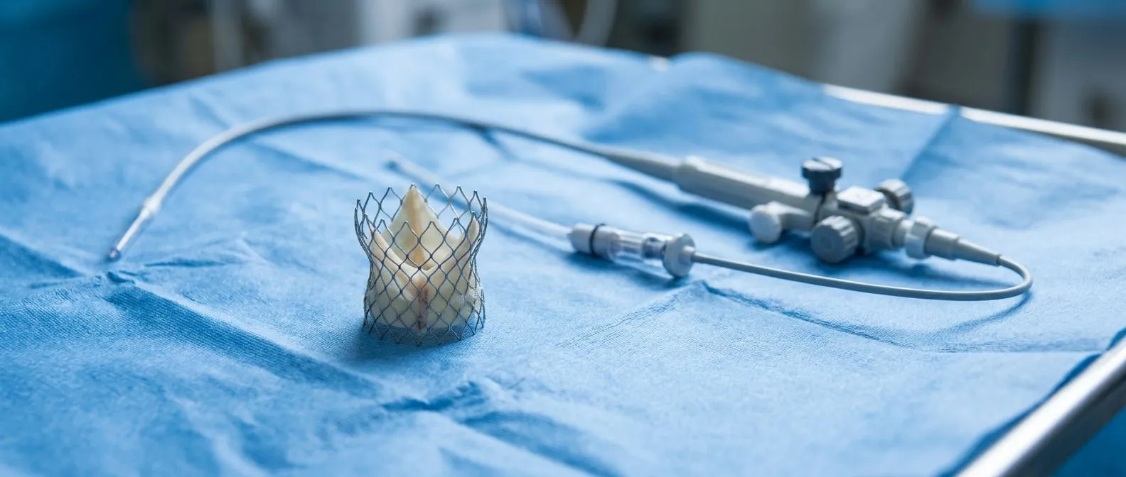
Transcatheter Aortic Valve Replacement (TAVR): For patients who are at high or intermediate risk for open-heart surgery, TAVR offers a remarkable alternative. During this procedure, a catheter is inserted through a blood vessel in the groin or chest, and a new valve is guided up to the heart. A Balloon-expandable Valve is often used; it is crimped onto the catheter, positioned directly inside the diseased aortic valve, and then expanded to push the old valve leaflets out of the way and establish a new, functional opening. TAVR does not require stopping the heart or using a heart-lung bypass machine, making it a vital option for frail patients who might not survive a traditional open procedure.
Recovery and outcomes: The recovery experience after minimally invasive procedures is typically much shorter than traditional surgery. Many TAVR patients are walking within a day of the procedure and go home within a few days. However, it is important to remember that TAVR valves are biological and may degrade over time, similar to surgically placed biological valves. Furthermore, some patients require a permanent pacemaker after TAVR if the new valve disrupts the heart's electrical pathways. The choice between minimally invasive surgery, TAVR, and traditional open surgery is highly personalized, based on a detailed evaluation of your heart's anatomy and your specific health profile.
What to Expect During Aortic Valve Surgery Recovery Time?
Recovery from aortic valve surgery is a gradual process that requires patience and careful adherence to your medical team's instructions. Immediately after surgery, you will be moved to the intensive care unit (ICU) for close monitoring. Here, your vital signs will be tracked constantly. An arterial line, a thin tube placed in an artery, will continuously monitor your blood pressure, while a temporary arterial cannula might be used if mechanical support was needed during the operation. This initial observation period usually lasts for one or two days, ensuring your heart is functioning stably before you are moved to a standard recovery room.
Cardiac rehabilitation: Once you are transferred out of the ICU, the focus shifts to regaining strength and mobility. Nurses and physical therapists will assist you with walking and gentle exercises to prevent blood clots and rebuild stamina. In rare instances where the heart needs extra support to recover its pumping strength, specialized centers might employ advanced extracorporeal membrane oxygenation techniques, such as Awake ECMO, which allows patients to remain conscious while their heart and lungs rest, though this is typically reserved for severe postoperative complications rather than routine recovery.
Home recovery timeline: Most patients spend about a week in the hospital before being discharged home. The complete recovery period generally spans six to twelve weeks. During the first few weeks at home, you will need to avoid heavy lifting and strenuous activities to allow your breastbone or chest incisions to heal properly. Walking is highly encouraged and forms the foundation of your daily routine. You will also have follow-up appointments to check your incisions, assess your valve function with an echocardiogram, and adjust any medications, ensuring your recovery stays on the safest and most effective track.
What Are the Aortic Valve Surgery Risks and Complications?
While aortic valve surgery is highly established and generally safe, it is a major medical procedure that carries inherent risks. The most common complications include bleeding, infection at the incision site, and adverse reactions to anesthesia. Because the heart is stopped and a heart-lung machine is used during traditional open surgery, there is a small risk of stroke or neurological issues related to altered blood flow. Your surgical team uses meticulous techniques and advanced monitoring to minimize these risks, carefully weighing the immediate dangers of surgery against the long-term certainty of progressive heart failure if the valve is left untreated.
Arrhythmias and electrical disruptions: A frequent complication after valve surgery is the development of atrial fibrillation or flutter. The manipulation of the heart tissue can irritate the electrical system, causing an irregular heartbeat. This is usually temporary and managed with medication or a brief electrical shock to restore normal rhythm. In the event of a sudden, life-threatening cardiac emergency in the hospital, staff are equipped with an Automated External Defibrillator to immediately reset the heart's rhythm. Furthermore, for patients with an enlarged ascending aorta, guideline thresholds—such as an aortic diameter greater than 5.5 cm—often dictate that the aorta is replaced concurrently with the valve to prevent future tearing.
Valve-related risks: Over the long term, replacement valves carry their own specific risks. Biological valves can deteriorate over time, potentially requiring a repeat procedure. Mechanical valves, while durable, increase the risk of blood clots, requiring lifelong anticoagulation therapy, which slightly increases the risk of bleeding. Endocarditis, an infection of the new artificial valve, is a rare but serious risk that requires patients to take prophylactic antibiotics before certain dental procedures. Knowing when to seek help is critical for long-term success. You should immediately contact your medical team if you experience any of the following:
- Fever or chills: Signs of a potential systemic infection or endocarditis around the new valve.
- Worsening shortness of breath: An indicator that the valve may be failing or fluid is accumulating in the lungs.
- Sudden chest pain or rapid heartbeat: Signs of a cardiac emergency or dangerous arrhythmia requiring immediate attention.
How to Choose the Best Heart Valve Surgeons in Guadalajara?
Selecting the right cardiac team is the most important decision you will make in your treatment journey. For international patients and families seeking care in Mexico, finding a team that combines deep surgical expertise with compassionate, clear communication is essential. Dr Alejandro Molina and the practice of Cirugía de Corazón offer specialized cardiac surgery, with consulting rooms in Guadalajara, Querétaro, and Hermosillo. The practice focuses on bridging complex cardiology with calm, clear explanations, ensuring that every patient understands their anatomy, their surgical options, and their recovery milestones without feeling rushed or overwhelmed by medical jargon.
Evaluating surgical expertise: When choosing a surgeon, it is important to look at their experience with specific procedures. A skilled surgeon will expertly navigate the complexities of the heart, whether performing a delicate valve repair, utilizing an arterial graft to bypass blocked vessels, or placing a replacement valve. The team at Cirugía de Corazón prioritizes patient education, taking the time to explain the trade-offs between repair and replacement, or between traditional and minimally invasive approaches. They address cost, travel logistics, and insurance concerns openly, providing a transparent pathway for international patients seeking high-quality cardiac care.
Your next steps: Aortic valve disease is a serious condition, but with modern diagnostics and surgical advancements, it is highly treatable. The goal of treatment is not just to fix a valve, but to restore your quality of life and return you to the activities you love. We encourage you to use this information as a foundation for your understanding, but not as a substitute for professional medical advice. Every heart is unique. If you or a loved one are experiencing symptoms or have been diagnosed with a valve condition, the safest and most effective next step is to discuss your specific situation with a qualified cardiac team. Book a consultation with Cirugía de Corazón today to receive a personalized evaluation and expert guidance on your path to heart health.