Calcific Aortic Stenosis: Progression and Treatment
Learn about calcific aortic stenosis treatment from early progression to TAVR and surgical valve replacement.
If you or someone in your family has been told the heart's aortic valve is narrowed by calcium deposits, you are not alone — and there is a clear, well-established path forward. Treatment for calcific aortic stenosis has changed enormously in the past decade, and many patients now have options that were not available to their parents' generation. This guide explains what the condition is, how it tends to progress, how it is diagnosed, and what the surgical and transcatheter choices actually involve, so you can walk into any consultation already understanding the conversation.
What Is Calcific Aortic Stenosis?
The aortic valve sits between the heart's main pumping chamber and the aorta, the large artery that carries blood to the whole body. A healthy valve has three thin, flexible leaflets that open wide with each heartbeat. In calcific aortic stenosis — the most common valve disease in older adults — calcium builds up on those leaflets over years, making them stiff and narrow. The heart then has to push blood through a much smaller opening.
Why the valve calcifies in the first place. This is not the same kind of calcium that forms kidney stones. It is an active, gradual process that resembles atherosclerosis — the hardening of arteries — and it is more common with age, high blood pressure, diabetes, smoking, and chronic kidney disease. The leaflets slowly stiffen until they no longer open fully, a condition doctors describe as narrowing or stenosis.
What the narrowing does to the heart. The left ventricle, the heart's strongest chamber, compensates by thickening its walls. That works for a while, but over time the muscle can tire, and symptoms appear. Understanding this natural history — and the anatomy behind it — is the first step in European Heart Journal-guided modern decision-making about this disease.
Is Aortic Calcification the Same as Aortic Stenosis?
This is one of the most common questions we hear, and the answer matters for how closely you need to be followed. Calcification and stenosis are related but not identical. Calcification means calcium is present on the valve; stenosis means the valve is narrowed enough to obstruct blood flow. Many people have mild valve calcification on an echocardiogram — an ultrasound of the heart — without any meaningful blockage.
Aortic sclerosis versus aortic stenosis. Doctors call a thickened, calcified valve that does not yet obstruct flow "aortic sclerosis." Studies suggest that a meaningful proportion of people with aortic sclerosis eventually develop stenosis, which is why a calcified valve is never ignored — it is monitored. If your report mentions calcification, ask how severe the narrowing is and when the next echocardiogram is due.
Calcium elsewhere in the aorta. Calcification can also affect the vessel wall itself, sometimes alongside conditions such as an Abdominal Aortic Aneurysm, where the aorta below the chest bulges and weakens. These are separate problems that an imaging study can pick up at the same time, so a complete evaluation of the whole aorta is worthwhile, not just the valve.
What Are the Symptoms of Calcific Aortic Stenosis?
In the early stages, most people feel completely normal, which is exactly why the condition is often found by accident. Symptoms tend to arrive gradually, and the classic trio is breathlessness on exertion, chest discomfort, and fainting or near-fainting during activity. Because they come on slowly, many patients adjust their lifestyle without realizing the valve is the cause.
Does aortic stenosis affect the brain? It can. When the valve is severely narrowed, the heart may not pump enough blood forward, and fainting during exertion — sometimes preceded by dizziness or blurred vision — is a warning sign. There is also a small risk that calcium debris or formed clots can travel upward to the brain, which is one reason symptoms should never be dismissed as "just age."
Symptoms that warrant urgent attention. Fainting with exercise, breathlessness at rest, new chest pain, or episodes of irregular heartbeat with palpitations deserve prompt evaluation. In our consulting rooms in Guadalajara, Querétaro and Hermosillo, we tell patients that any new exertional fainting in someone known to have a calcified valve should be reported the same day, not at the next routine visit.

How Does Calcific Aortic Stenosis Progress Over Time?
Progression is individual, but the pattern is well described. The narrowing is measured as the "valve area" — the opening the blood must pass through — and also by the pressure gradient, the extra pressure the heart needs to push blood across the valve. A valve area below about 1.0 cm², or a mean gradient above roughly 40 mmHg, defines severe stenosis, the threshold at which guidelines generally recommend valve replacement once symptoms appear.
How fast does it move? On average, severe stenosis can develop from moderate disease over a few years, and once symptoms begin, they typically worsen over the following months to years. Some valves calcify faster — for example in people on dialysis or with abnormal valve anatomy from birth. This is why serial echocardiograms are scheduled at intervals that depend on severity: yearly for moderate disease, more often when it is approaching severe.
Why timing matters. Waiting until the heart muscle has already weakened raises the risk of any procedure and can limit how well the heart recovers. The goal of modern care is to intervene at the right moment — after severe stenosis is confirmed and symptoms have appeared, but before the heart has paid a permanent price.
How Is Calcific Aortic Stenosis Diagnosed?
The first clue is often a heart murmur heard with a stethoscope — a rough sound as blood squeezes through the narrowed valve. The cornerstone test is the echocardiogram, which measures the valve area, the gradient, how well the heart muscle is squeezing, and whether the valve leaks backward as well as failing to open. It is painless, uses no radiation, and gives almost everything needed for a first decision.
Additional tests before treatment. Once severe stenosis is confirmed, most teams order a CT scan of the chest and aorta to look at the valve's calcium load and the size of the access vessels, plus a coronary angiogram or CT coronary study to check the heart's own arteries. Cardiac catheterization — thin tubes threaded from the wrist or groin into the heart — may be used when the echocardiogram and symptoms do not agree.
Interesting research directions. Researchers are even exploring opportunistic screening, such as measuring valve calcification during routine imaging; one example is a study of early detection of aortic stenosis in the community during flu vaccination (NCT03250806). The practical takeaway for patients is simpler: if you have a murmur or known calcification, keep your echocardiogram appointments.
What Can Be Done for a Calcified Aortic Valve?
Medication alone cannot open a stenotic valve. Drugs can control blood pressure, rhythm problems, and fluid retention, and they matter while you are being evaluated — but no medicine reverses the calcification. When severe stenosis causes symptoms, replacing the valve is the only definitive treatment. What has changed is how the replacement can be done.
The main options today are:
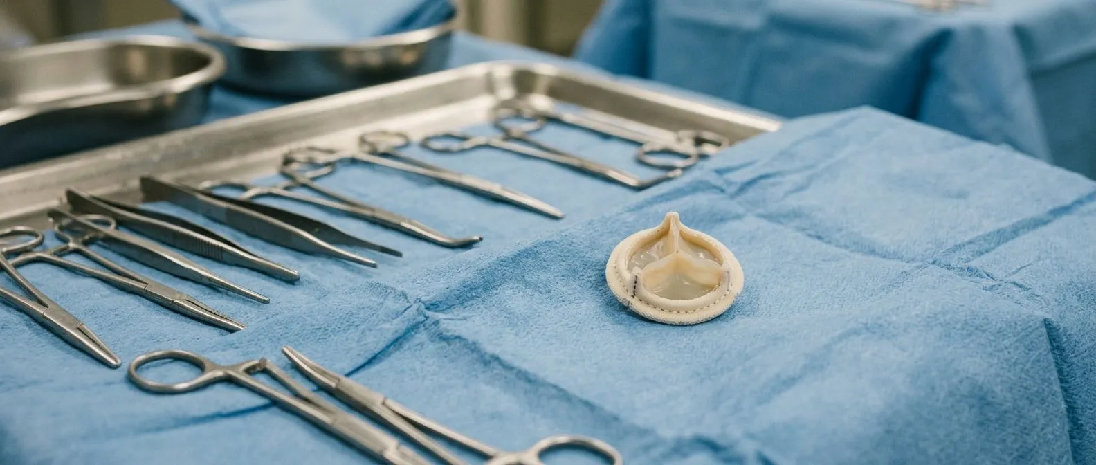
- Surgical Aortic Valve Replacement: the classic approach through the chest, replacing the valve with a tissue or mechanical prosthesis while the heart is stopped, sometimes supported by an Aortic Cross-clamp during the operation.
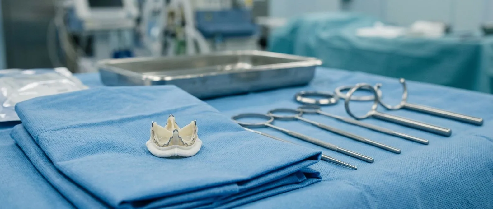
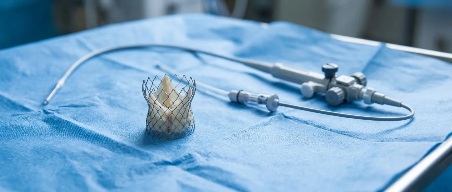
- TAVR (transcatheter aortic valve replacement): a new valve is delivered through a catheter, most often from an artery in the groin, and expanded inside the old valve — no chest incision and usually no need to open the heart.
- Balloon Aortic Valvuloplasty: inflating a balloon to widen the valve temporarily; useful in selected situations, but the valve re-narrows, so it is a bridge rather than a definitive fix.
Choosing between them is not a matter of preference alone. It depends on your age, anatomy, other conditions, the amount of calcium, the state of the aortic root — the ring and sinuses at the base of the aorta — and whether other valves or vessels need attention at the same operation. Every case at our team is reviewed by the multidisciplinary group against international practice guidelines before a recommendation is made, and the patient receives a clear proposal with the alternatives, the final decision always remaining theirs.
TAVR vs Surgical Aortic Valve Replacement: Which Is Right for You?
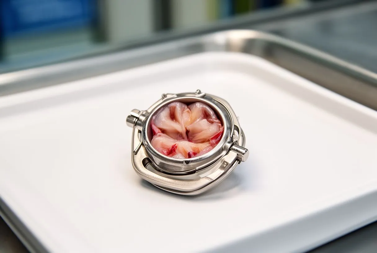
Both approaches replace the valve; they differ in how they get there. Surgery offers decades of proven durability and lets the surgeon address the coronary arteries, other valves, or an enlarged aorta in the same sitting — relevant if there is a Thoracic Aortic Aneurysm or a need for Aortic Root Replacement. TAVR avoids a chest incision and generally means a shorter hospital stay and faster return to daily life, which matters greatly for older or frailer patients.
How devices are compared. Modern TAVR valves come in balloon-expandable and self-expanding designs, and registries such as the EVAL registry comparing balloon-expandable and self-expanding transcatheter valves in severe aortic stenosis have shown both can deliver good outcomes when the right device is matched to the right anatomy. Valve selection also affects whether a permanent pacemaker may be needed afterwards, which is one of the details a good team will explain.
Who is best suited to each. In general terms, younger patients with a long life expectancy and no other cardiac problems often do well with surgery because of its long-term durability record, while older patients, those with heavily calcified anatomy, previous chest surgery, or significant frailty often benefit from TAVR. Our team performs conventional, minimally invasive, robotic and transcatheter procedures, and we recommend the less invasive route only where the evidence supports it for that specific patient — never as a one-size answer.
What Is Recovery Like After Aortic Valve Replacement?
After TAVR. Most patients walk the same day or the next morning, and many go home within two to four days. The groin puncture site needs a few days of care, and heavy lifting is restricted for roughly a week or two. Cardiac rehabilitation — supervised, gradually increasing exercise — is strongly recommended and helps rebuild strength and confidence. Breathlessness often improves within weeks.
After surgery. Surgical recovery is longer: typically a few days in hospital, several weeks of sternal precautions to let the breastbone heal, and about six to twelve weeks before returning to normal activity. Driving, work, and travel resume step by step. Both paths include lifelong follow-up with echocardiograms to confirm the new valve is working well.
What we watch for afterwards. The new valve should open well and not leak significantly; blood thinners or antiplatelet medication is prescribed according to the type of valve and any rhythm issues. In the consultation, the team explains what the studies show, in plain words, and answers every question before any decision, because patients do best informed, not anxious.

Are the Treatment Options Safe?
No heart procedure is risk-free, and honest discussion of risk is part of good care. Both surgery and TAVR carry risks of bleeding, stroke, kidney injury, rhythm problems requiring a pacemaker, and — rarely — damage to the aorta, including aortic dissection or rupture, which are among the emergencies cardiac teams train for. The overall risk depends heavily on age, other illnesses, frailty, and anatomy.
How risk is measured before deciding. Teams use scores such as the STS or EuroSCORE II, which combine age and health factors to estimate surgical risk, alongside frailty assessment and imaging of the access vessels. If complications occur, the intensive care setting is prepared for them — from an Intra-aortic Balloon Pump supporting the circulation to emergency conversion from a catheter procedure to surgery.
Risk in context. For a patient with severe, symptomatic calcific aortic stenosis, the risk of doing nothing is usually higher than the risk of treatment, because untreated severe symptomatic stenosis progresses relentlessly. That comparison — your individual procedural risk versus the natural history of the disease — is exactly the conversation a good team should walk you through with numbers, not slogans.
When Should You Seek Help for Calcific Aortic Stenosis?
Seek evaluation if you have a known murmur or calcified valve and develop breathlessness, chest discomfort, fainting, or swelling in the legs. If you have already been diagnosed with severe stenosis and symptoms have begun, do not delay — this is the window in which treatment offers the most benefit. And if any test result confuses you, a second opinion is always reasonable.
The value of a second opinion. Bringing your echocardiogram and CT images to another team often confirms the plan — or reveals an option you had not been told about. We encourage second opinions and treat seeking one as a normal part of deciding about heart surgery; the first contact is a free video consultation where we get to know the patient, talk through the condition, and say which studies are still needed so no unnecessary ones are ordered.
Taking the next step. Calcific aortic stenosis is common, its progression is predictable, and treatment for calcific aortic stenosis — whether surgical or transcatheter — is safer and more accessible than ever, including here in Mexico for international patients weighing cost, travel, and insurance. The single most useful thing you can do is discuss your own images and symptoms with a qualified cardiac team rather than self-diagnose. If you would like that conversation, you can book a consultation with our team and start with your questions, not with a scalpel.