

Inferior Vena Cava Filter
Learn how an inferior vena cava filter catches clots to prevent pulmonary embolism.
What Is an Inferior Vena Cava Filter?
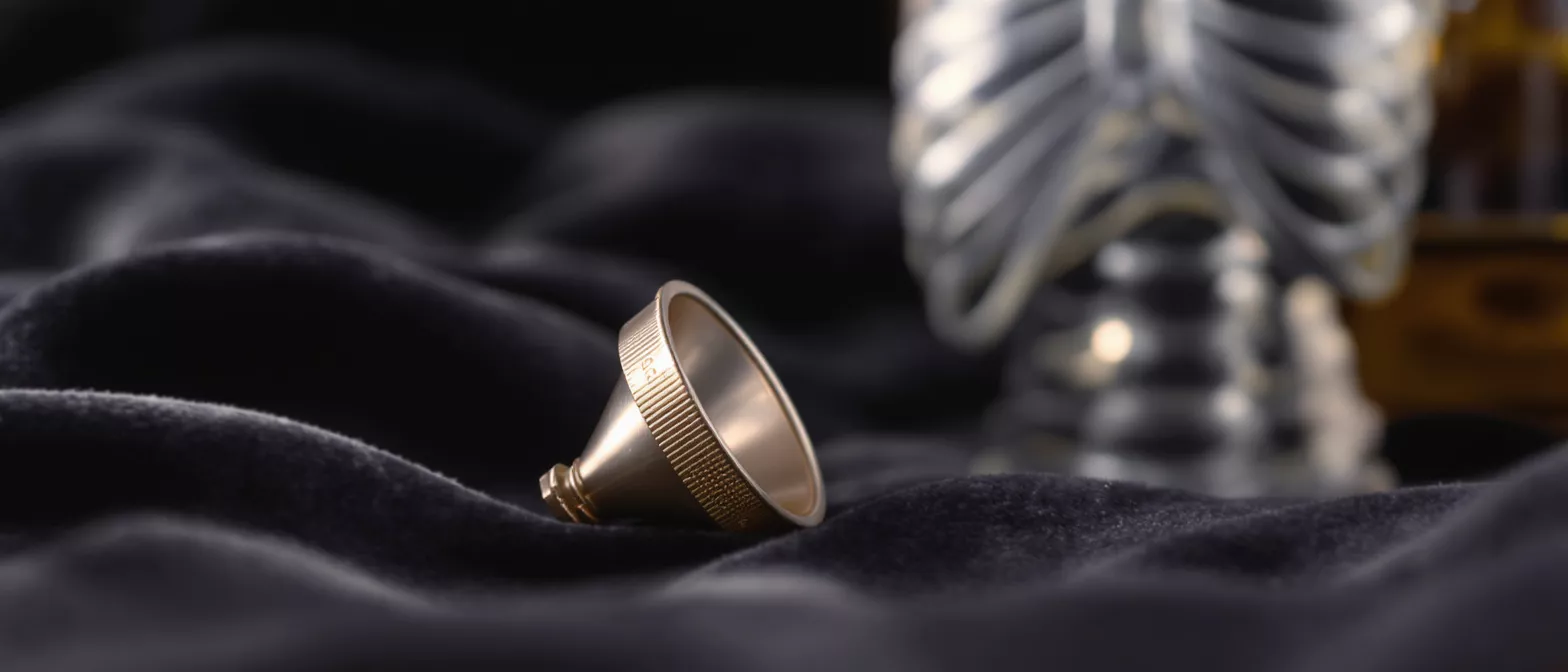
An inferior vena cava filter is a small, cone-shaped medical device implanted in the body's largest vein to catch dangerous blood clots before they can travel to the lungs. The inferior vena cava (IVC) is the major blood vessel responsible for returning deoxygenated blood from the lower half of your body back to your heart and lungs. When a blood clot forms in the legs or pelvis—a condition known as deep vein thrombosis (DVT)—it can break loose and travel upward. The filter acts as a physical trap, designed to let blood flow freely while snaring these migrating clots.
Who needs an IVC filter? This device is typically reserved for patients who cannot tolerate standard blood-thinning medications. If you are preparing for cardiac surgery or managing a complex cardiovascular condition, your surgeon may evaluate whether your risk of bleeding is too high for anticoagulants. In such cases, a mechanical filtering solution becomes a crucial safety net. For patients exploring options to manage these risks, utilizing a blood clot prevention device provides a mechanical safeguard when pharmacological approaches are unsafe or ineffective.
Understanding the role of this device helps alleviate much of the anxiety surrounding its placement. The filter is not a permanent cure for blood clots, but rather a protective measure designed to prevent a life-threatening pulmonary embolism. By mechanically intercepting clots, it shields your lungs from debris that could otherwise cause severe respiratory distress or cardiac strain.
How Does an IVC Filter Prevent Pulmonary Embolism?
A pulmonary embolism occurs when a blood clot travels through the bloodstream and lodges in the arteries of the lungs, a complication that demands immediate medical attention. An IVC filter pulmonary embolism prevention strategy works on a simple but highly effective mechanical principle. The device is strategically positioned in the inferior vena cava, located below the renal veins. As blood flows from the lower body toward the heart, any clot fragments are forced through the metallic struts of the filter.
How does the filter trap clots safely? The cone-shaped design of the filter allows small clots to become ensnared within its wires. Once trapped, the clot remains in the filter until the body's natural fibrinolytic system gradually breaks it down over time. This process keeps the debris safely confined, preventing it from migrating through the right side of the heart and into the pulmonary arteries. The blood continues to flow around the trapped clot without major obstruction.
By physically blocking the pathway, the filter significantly reduces the risk of a massive pulmonary embolism. This is especially vital for heart surgery patients whose cardiovascular systems are already under stress. Preventing a sudden blockage in the lungs helps maintain stable blood pressure and reduces the workload on a recovering heart, supporting a smoother postoperative journey.
When Is IVC Filter Placement Surgery Necessary?
The decision to proceed with IVC filter placement surgery is made after careful evaluation of a patient's specific risk factors. The primary indication is an acute venous thromboembolism where standard anticoagulant therapy is explicitly contraindicated. This means patients who have severe bleeding disorders, recent internal bleeding, or those who require urgent cardiac surgery cannot safely take blood thinners. In these scenarios, mechanical interception is the safest available intervention.
What are the clinical guidelines for placement? Inferior vena cava filter guidelines suggest the device should be deployed when a patient experiences recurrent clots despite optimal anticoagulation, or when severe trauma prevents the use of standard medications. It is also utilized as a temporary measure for high-risk surgical patients. Understanding these inferior vena cava filter indications helps patients and their families grasp why their surgical team chose this route over traditional drug therapies.
In the context of cardiac care, a surgeon might place a filter preemptively if a patient has a history of DVT and is about to undergo a procedure that requires stopping blood thinners. This ensures the patient remains protected during the vulnerable perioperative window. It acts as a bridge, maintaining safety until the patient can safely resume anticoagulant therapy during recovery.
Types of Blood Clot Filter Devices
When discussing vena cava filter placement, it is important to distinguish between the two primary types of devices available. The choice depends on the patient's expected duration of risk and overall medical profile. Your surgical team will select the most appropriate option based on your long-term cardiovascular needs.
- Retrievable IVC filters are designed for temporary use. They feature specialized hooks or loops that allow a doctor to safely remove the device once the risk of pulmonary embolism has passed and the patient can safely resume blood thinners.
- Permanent IVC filters are built from durable materials meant to last a lifetime. These are typically chosen for patients who have chronic conditions that will permanently prevent them from taking anticoagulant medications.
Can a retrievable filter become permanent? Yes, if a retrievable IVC filter is left in place for an extended period, it can eventually become incorporated into the vessel wall, making retrieval difficult. Because of this, modern practice favors retrievable filters, as they offer flexibility and can be left in temporarily or permanently based on the patient's response to treatment. This adaptability makes them highly valuable for dynamic cardiac surgery recoveries.
What to Expect During Vena Cava Filter Placement
Patients often feel relieved to learn that vena cava filter placement is a minimally invasive procedure, not an open surgery. It is typically performed in a hospital's interventional radiology suite. The patient is given a local anesthetic and a mild sedative to ensure comfort throughout the process. Using ultrasound and fluoroscopy—real-time X-ray imaging—the doctor guides a slender catheter through a small incision, usually in the neck or groin.
How long does IVC placement take? The actual placement of the filter takes only a few minutes, though the entire process from preparation to final imaging usually takes about an hour. The doctor carefully advances the catheter through the venous system into the inferior vena cava. Once the catheter is perfectly positioned below the renal veins, the filter is carefully deployed and expands to anchor itself against the vessel walls.
Because there are no large surgical incisions, the trauma to the body is minimal. The incision is so small that it only requires a few minutes of pressure and a small bandage. This quick, image-guided approach makes it a highly suitable intervention for fragile patients who are preparing for, or recovering from, major cardiac surgery. Patients looking into these supportive interventions often explore a retrievable vena cava filter for temporary clot management.
IVC Filter Placement Recovery Time
One of the main advantages of this procedure is the remarkably short recovery period. Most patients can sit up and walk within a few hours. The IVC filter placement recovery time is generally just a day, with many patients returning to normal activities within 24 hours. You will be observed briefly to ensure there is no bleeding at the insertion site and that your vital signs remain stable.
What should you avoid after placement? Patients are advised to avoid heavy lifting and strenuous exercise for the first few days to allow the puncture site to heal properly. Mild discomfort or slight bruising at the neck or groin incision is entirely normal. Your care team will provide specific discharge instructions regarding when it is safe to resume more rigorous physical activities or physical therapy.
It is vital to attend all follow-up appointments so your surgical team can monitor the filter's position and function. These visits also help your doctor determine the optimal timing for retrieval, if a temporary device was used. Consistent communication with your cardiologist ensures the filter is doing its job effectively and safely as you recover.
Inferior Vena Cava Filter Risks and Complications
While generally safe and life-saving, it is crucial to understand the potential inferior vena cava filter risks. Like any medical device placed inside the body, there are associated vena cava filter side effects. Immediate risks include minor bleeding at the insertion site, infection, or an allergic reaction to the contrast dye used during imaging. These acute complications are rare and usually managed quickly by the medical team.
What are serious IVC filter complications? Over time, more complex issues can arise. One rare but noted complication is IVC filter migration, where the device shifts from its original position. Another risk is filter fracture, where a metallic strut breaks off. Additionally, in some cases, the filter can cause a narrowing of the vena cava or trap so much clot that it blocks blood flow entirely, leading to swelling in the legs.
- Filter migration: The device moves from its placement, though usually only a short distance.
- Filter fracture: Metal components break, which rarely causes symptoms but requires monitoring.
- Vessel thrombosis: The filter traps too many clots, causing a blockage in the vena cava.
These risks underscore the importance of diligent medical follow-up. By keeping a close watch on the device, doctors can intervene early if any structural changes occur. The benefits of preventing a fatal pulmonary embolism usually heavily outweigh these potential complications, particularly in high-risk surgical patients.
Vena Cava Filter Retrieval and Removal Procedure
For temporary devices, vena cava filter retrieval is a planned part of the treatment cycle. The IVC filter removal procedure is very similar to the initial placement, utilizing image guidance and a minimally invasive approach. A doctor threads a catheter through a vein in the neck or groin, locates the filter, and uses a specialized snare to collapse the device back into the catheter for safe extraction.
When should an IVC filter be removed? The standard recommendation is to remove a retrievable filter once the risk of pulmonary embolism has resolved, or once the patient can safely tolerate blood thinners again. This is typically a few weeks to a few months after placement. Delaying retrieval increases the risk that the device will become embedded in the vein wall, making the removal procedure more technically challenging for the surgeon.
The removal process is usually quick and carries a low risk of complications. Patients are often discharged the same day. For patients recovering from cardiac surgery, removing the filter is a positive milestone, indicating that their cardiovascular health has stabilized enough to manage clot risks naturally or with medications. Some patients navigating complex cardiovascular care consider a permanent IVC filter option when long-term anticoagulation remains unsafe.
Inferior Vena Cava Filter Alternatives
Before placing a mechanical filter, doctors exhaust all inferior vena cava filter alternatives. The standard first-line treatment for blood clots is anticoagulation therapy, commonly known as blood thinners. These medications effectively stop existing clots from growing and prevent new ones from forming. For patients who can tolerate them, medications are highly effective and completely non-invasive.
What are the alternatives to filter placement? Beyond standard blood thinners, other alternatives include compression stockings and intermittent pneumatic compression devices, which physically squeeze the legs to promote blood flow and prevent clot formation, especially during or after surgery. For patients with anatomical or medical reasons that preclude a filter, advanced catheter-based clot-busting drugs can sometimes be delivered directly to the clot site.
When these alternative therapies fail, are medically contraindicated, or are not tolerated by the patient, the mechanical filter becomes the necessary choice. The decision is always highly personalized, balancing the risks of bleeding against the risks of clotting, ensuring the safest path for the patient's specific heart surgery recovery.
Living With an IVC Filter: Long-Term Outlook
For patients who retain a permanent filter, life can continue largely as normal. The device is designed to be silent and unnoticeable, quietly performing its job without interfering with daily activities. You will not set off metal detectors at airport security, and the filter will not limit your physical mobility once fully healed from the placement procedure.
What is the long-term outlook with an IVC filter? The key to living comfortably with an inferior vena cava filter is consistent medical follow-up. Your cardiologist will periodically order imaging, such as a CT scan, to ensure the filter remains in place and intact. Long-term complications are rare, but early detection is the best way to manage any structural changes to the device before they cause symptoms.
Ultimately, an IVC filter is a powerful tool in a cardiac surgeon's arsenal to keep high-risk patients safe. By intercepting dangerous clots, it provides a crucial layer of protection when the body is most vulnerable. Understanding how it works, the risks involved, and the recovery process empowers patients and families to approach heart surgery with greater confidence and peace of mind.






