

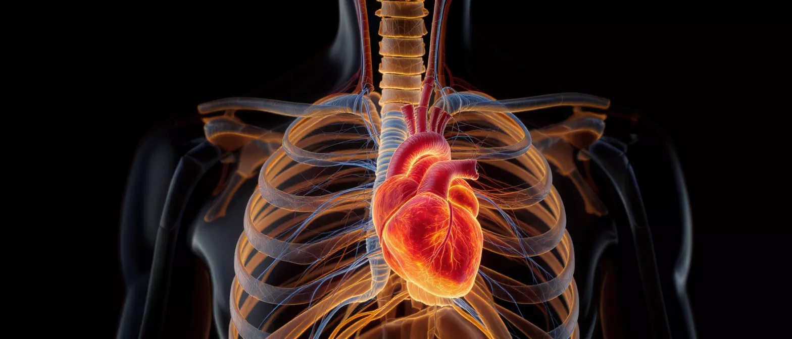
Inferior Vena Cava
Learn about the inferior vena cava, its anatomy, blood flow function, and related conditions like thrombosis and syndrome.
What Is the Inferior Vena Cava?
Understanding the inferior vena cava begins with recognizing it as the largest vein in the human body, playing a foundational role in your cardiovascular system. When you face a cardiac procedure, knowing the basic structures helps alleviate the fear of the unknown. This vessel is responsible for carrying deoxygenated blood from the lower half of your body back to the heart.
Where the journey begins: Blood from your legs, pelvis, and abdomen converges into this massive vein, which acts as a primary highway returning blood to the right atrium. From there, the blood moves to the lungs to receive fresh oxygen. This continuous cycle is strictly monitored by your cardiology team before any surgical intervention.
It is entirely normal to feel anxious about heart anatomy, but understanding this vessel reveals how remarkably well-designed your body is. The inferior vena cava function is purely about return transport—it does not actively pump blood itself, relying instead on muscle movements and pressure changes in the chest to keep blood moving upward against gravity.
Inferior Vena Cava Anatomy and Body Location
To visualize the inferior vena cava anatomy, imagine a large, vertical tube running just to the right side of your spine. It forms deep in the lower abdomen from the joining of the left and right common iliac veins, which drain the legs. As it ascends, it collects blood from various major abdominal organs.
Key anatomical landmarks include:
- Formation: Forms at the level of the fifth lumbar vertebra (L5) from the merging of the iliac veins.
- Heart drainage: Empties directly into the right atrium of the heart, delivering deoxygenated blood to be pumped to the lungs.
- Organ tributaries: Receives blood from the renal veins (kidneys), hepatic veins (liver), and adrenal veins along its path upward.
Because of its central location, the vein sits close to several major organs. Cardiac surgeons must carefully navigate this intricate anatomy during procedures to ensure safe and precise access to the heart without disrupting surrounding structures.
How Inferior Vena Cava Blood Flow Works
Understanding inferior vena cava blood flow is highly reassuring, as it explains how your body naturally manages circulation. Unlike arteries, veins operate under low pressure and rely on a combination of valves and surrounding muscle contractions to push blood upward. When you walk or breathe, you are actively helping this blood move.
The respiratory pump mechanism: Every time you inhale, your diaphragm drops, creating negative pressure in the chest. This pressure drop acts like a gentle vacuum, drawing blood upward through the inferior vena cava vein toward the heart. This is why deep breathing exercises are often recommended during postoperative recovery.
If you are resting in a hospital bed before or after surgery, your care team may encourage you to move your legs periodically. This movement prevents blood from pooling in the lower extremities and keeps the flow steady, protecting your veins from complications.
Inferior Vena Cava Function in the Heart
The inferior vena cava heart connection is essential for maintaining balanced circulation. The right atrium receives blood from both the superior vena cava (upper body) and the inferior vena cava (lower body). Once the blood enters this chamber, it flows through the tricuspid valve into the right ventricle, ready to be sent to the lungs for oxygenation.
Maintaining volume balance: The body constantly regulates how much blood returns to the heart to ensure it does not overload the cardiac chambers. If there is ever a disruption in the drainage system, the heart struggles to pump efficiently, which can lead to fluid buildup or swelling.
During heart surgery, cardiologists pay close attention to how blood drains into the right atrium. Bypass machines are used to temporarily take over the role of the heart and lungs, ensuring that blood from the inferior vena cava continues to be circulated safely throughout the body while the surgeon operates.
What Is Inferior Vena Cava Syndrome?
Inferior vena cava syndrome occurs when blood flow through this major vein is partially or completely blocked. Patients may experience significant swelling in the legs, lower back pain, or a feeling of fullness in the abdomen. Recognizing these symptoms early is vital for effective treatment.
How it affects the body: When drainage is obstructed, blood seeks alternative pathways through smaller, collateral veins, which can become engorged. While the body is remarkably adaptable, this increased pressure can cause discomfort and visible swelling in the lower extremities. Diagnostic imaging is usually required to confirm the exact cause and location of the blockage.
If you are experiencing these symptoms before a planned surgery, it is crucial to inform your medical team immediately. They can evaluate the vein and determine if any additional interventions are needed prior to your cardiac procedure to ensure a smooth recovery.
Inferior Vena Cava Obstruction Causes
An inferior vena cava obstruction can result from several different factors, ranging from physical compression to internal blockages. Sometimes, an enlarged organ or a mass in the abdomen can press against the vein from the outside. In other cases, a blood clot within the vein itself narrows the passageway.
Common reasons for an obstruction include:
- Blood clots: Deep vein thrombosis (DVT) can extend into the larger vessel, creating a direct internal blockage.
- External pressure: Tumors, cysts, or an enlarged liver can physically compress the vein walls and restrict flow.
- Post-surgical changes: Scar tissue from previous abdominal or vascular surgeries can occasionally narrow the vein over time.
Identifying the root cause of the obstruction allows your surgical team to tailor their approach. They may use advanced imaging or ultrasounds to view the exact nature of the blockage before deciding on the safest surgical path.
Inferior Vena Cava Thrombosis Explained
Inferior vena cava thrombosis is a specific condition where a blood clot forms within this major vein. This is a serious medical condition because clots can break loose and travel to the lungs, causing a pulmonary embolism. Your cardiology team takes immediate action if they suspect thrombosis.
Risk factors for thrombosis: Prolonged immobility, inherited blood clotting disorders, and certain medications can increase the risk of developing clots. Patients preparing for cardiac surgery are often evaluated for these risks well in advance. If a clot is found, doctors may prescribe blood thinners to stabilize the clot before the operation begins.
If you have a history of clotting, be sure to discuss this thoroughly with your surgeon. Understanding your history helps them create a safe, comprehensive plan to manage your blood flow before, during, and after your heart procedure.
How Inferior Vena Cava Imaging Guides Surgery
Inferior vena cava imaging is a critical tool used by surgeons to plan your procedure. By using ultrasound, CT scans, or MRIs, your medical team can clearly see the size, shape, and path of the vein. This clear visualization is essential for navigating around complex vascular structures safely.
Accurate preoperative mapping: Detailed imaging ensures that surgeons know exactly where the vein enters the right atrium. If there are any anatomical variations or blockages, the surgical team will adjust their approach accordingly. This level of preparation significantly reduces surgical risks and improves overall outcomes.
Imaging also helps doctors determine the exact placement of catheters or bypass cannulas. Knowing the precise dimensions of the vessel allows them to choose the correct equipment, ensuring that your blood is efficiently circulated while your heart is being repaired.
What Is an Inferior Vena Cava Filter?
An inferior vena cava filter is a small, cone-shaped device placed inside the vein to catch blood clots before they reach the lungs. It is often recommended for patients who cannot safely take blood thinners. The filter allows normal blood to flow through while trapping clots, preventing severe complications.
Temporary versus permanent placement: Some filters are designed to stay in permanently, while others can be removed once the risk of clotting decreases. Your medical team will decide the best option based on your specific health needs. Understanding the purpose of this device helps reduce anxiety if one is recommended before your surgery.
Knowing that a physical safeguard is in place provides immense peace of mind. It serves as a protective barrier, catching debris before it can enter the heart and lungs, thereby adding an extra layer of safety to your cardiovascular care.
Inferior Vena Cava Definition and Cardiology Care
At its core, the inferior vena cava definition highlights a vessel of immense importance, acting as the body's primary drainage pipe for deoxygenated blood from the lower half of the body. Recognizing its role helps you understand why your cardiology team monitors it so closely during heart surgeries. Maintaining its patency is crucial for your overall cardiovascular health.
Your surgical care team: Cardiologists, anesthesiologists, and surgeons work together to protect this vessel throughout your treatment. From preoperative imaging to intraoperative monitoring, every step is taken to ensure that blood flow remains uninterrupted. This coordinated effort is what makes modern cardiac surgery so successful and safe for patients.
Remember that understanding your anatomy is an empowering step in your healthcare journey. By learning how your body functions, you become an active participant in your recovery, working alongside your medical team to ensure the best possible outcome after your heart procedure.






