

Arterial Line in Heart Surgery: Monitoring Blood Pressure
An arterial line in heart surgery gives beat-by-beat blood pressure data your team needs.
If you or a family member is preparing for heart surgery, the word "arterial line" will almost certainly come up. An arterial line in heart surgery is a thin, flexible tube placed into an artery — usually at the wrist — so the care team can see your blood pressure beat by beat, second by second. It sounds intimidating, but it is one of the most routine and most valuable pieces of monitoring in modern cardiac care. This guide explains, in plain language, why it is used, how it goes in, what it feels like, and what to expect when it comes out.
What Is an Arterial Line in Heart Surgery?
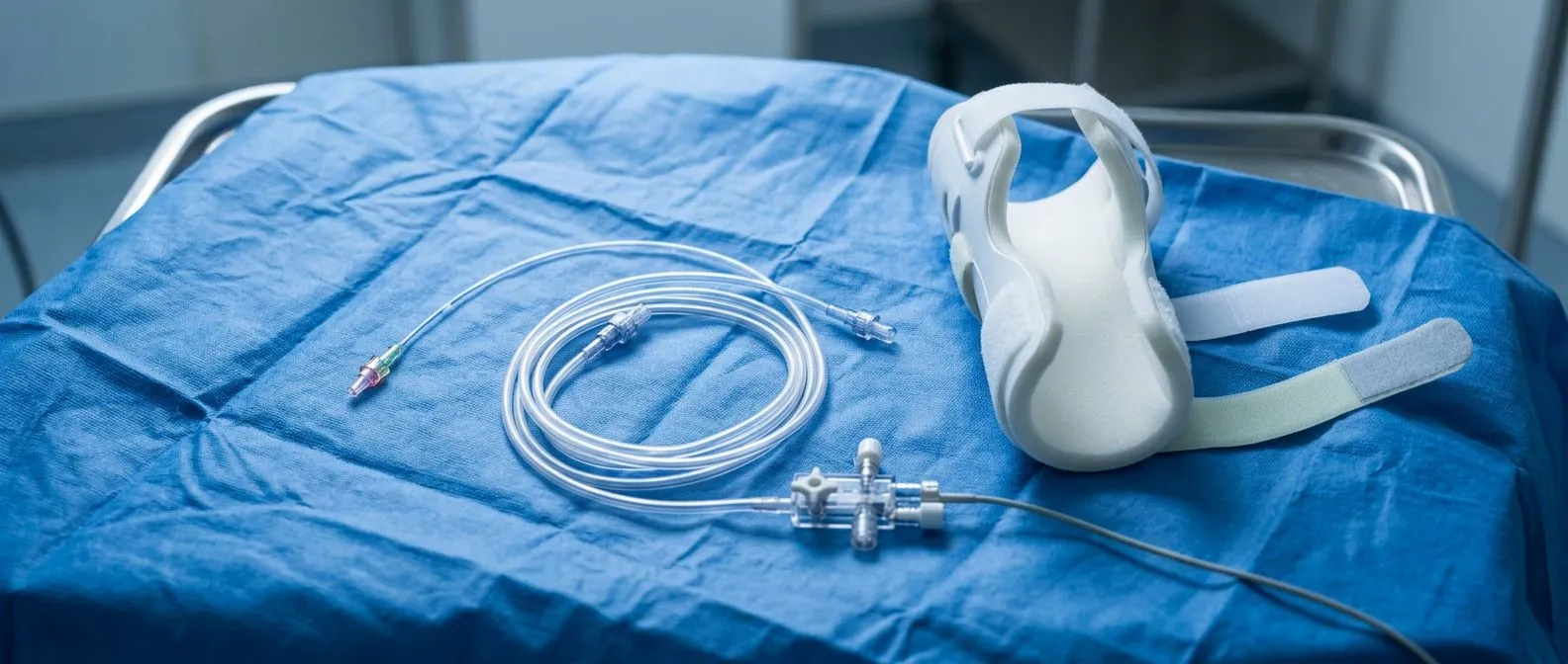
A definition in everyday terms. An arterial line — sometimes called an arterial catheter or "A-line" — is a slender plastic tube, about as wide as the lead in a pencil, inserted into an artery. Unlike a regular intravenous drip, which sits in a vein and carries fluids toward the heart, an arterial line measures pressure inside the arteries themselves. It connects through sterile tubing to a device called a transducer, which converts the pressure of each heartbeat into a live waveform on the operating room monitor.
The most common site is the radial artery, the artery you can feel on the thumb side of your wrist. If that artery is not suitable, the team may use the brachial artery at the elbow or the femoral artery in the groin. The choice depends on your anatomy, the type of surgery planned, and whether other lines or catheters are already needed.
It helps to think of the difference between a thermometer you dip in and out of water and one that reads continuously. A standard blood pressure cuff takes a snapshot every few minutes. Invasive blood pressure monitoring through an arterial line gives the anesthesiologist a continuous picture — every single heartbeat, in real time, throughout the operation.
Why Do Heart Surgery Patients Need an Arterial Line?
Precision matters in the moments that count. During cardiac surgery, blood pressure can change quickly and dramatically. The anesthetic drugs, the heart-lung machine, blood loss, and the surgical handling of the heart itself all shift circulation from one moment to the next. A cuff that updates every few minutes would be like driving at night with the headlights flicking on only occasionally. The care team needs to see changes as they happen so they can respond within seconds.
The arterial line also allows frequent blood sampling without repeated needle sticks. During open heart surgery, the team draws blood to check oxygen levels, acidity, clotting, and blood counts — often many times. With an arterial catheter in place, each sample comes from the line itself, sparing you dozens of needle punctures.
Finally, the waveform itself carries information. The shape of the pressure wave tells an experienced anesthesiologist about the heart's rhythm, the strength of each contraction, and even how well certain valves are working. International guidance on managing arterial hypertension emphasizes accurate measurement as the foundation of treatment decisions, and nowhere is that accuracy more important than in the operating room 2013 ESH/ESC Guidelines for the management of arterial hypertension. In our practice, every surgical plan is reviewed by the multidisciplinary team against international guidelines before we recommend it, and arterial monitoring is part of the standard we hold ourselves to.
Arterial Line vs Blood Pressure Cuff: What's the Difference?
Two tools, two jobs. You know the blood pressure cuff well — the soft wrap on the arm that squeezes, releases, and gives a number. It is non-invasive, painless, and perfectly adequate for most situations. But it has real limitations in cardiac surgery, and understanding them helps explain why the arterial line exists at all.
- Frequency: a cuff takes a reading every few minutes; an arterial line shows every heartbeat, continuously.
- Accuracy: cuff readings can drift when blood pressure is very low, very high, or when the patient has an irregular rhythm; arterial measurement stays reliable across those extremes.
- Detail: the cuff gives two numbers; the arterial waveform gives a full picture of how the heart and circulation are behaving.
- Blood sampling: the arterial line doubles as a sampling port, avoiding repeated needle sticks during long operations.
One practical note: blood pressure can differ between the two arms, and the arterial line is usually placed on the side that gives the truest reading. If your team measures your pressure on both arms before surgery, this comparison is one reason why. At our consulting rooms in Guadalajara, Querétaro and Hermosillo, we take time in the first consultation to explain what each monitoring device does, in plain words, before you ever enter the operating room.
How Is an Arterial Line Placed Before Surgery?
Setting up the line. The insertion usually happens in the operating room or the preparation area, after you have received sedation so you are calm and drowsy. The wrist is cleaned with antiseptic, the area is numbed with local anesthetic, and a fine needle guides the thin catheter into the radial artery. Many teams use an ultrasound probe to see the artery and guide the needle — this has become the standard approach because it improves first-pass success.
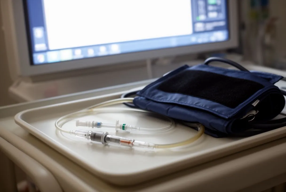
Once the catheter is in, it is secured with a clear dressing and connected to a sterile tubing set filled with salt water. The tubing runs to the transducer, which sits at a fixed height level with the heart. The monitor then displays the pressure as both a number and a moving wave. Before the reading is trusted, the team "zeros" the transducer against atmospheric pressure and checks that the waveform looks correct.
For you, the sequence feels like this: an injection of numbing medicine, a brief feeling of pressure at the wrist, and then it is done. Most patients remember little of it because of the sedation. The whole setup typically takes a few minutes and happens while other members of the team prepare the rest of the equipment.
What Does the Arterial Waveform Show During Surgery?
Reading the wave. Each heartbeat produces a characteristic pressure wave: a sharp rise as the heart pumps blood out, a peak, and then a decline with a small secondary bump. That bump — the dicrotic notch — reflects the aortic valve closing and blood rebounding briefly against the closed valve. Its presence and shape tell the anesthesiologist a great deal about vascular tone and cardiac output.
The team watches the waveform for much more than blood pressure. Changes in the wave's shape can signal low blood volume, a leaky heart valve, an irregular rhythm, or the effects of medications. In cardiac surgery, the waveform also helps confirm that the heart-lung machine is delivering adequate flow and that blood pressure is being managed within safe limits at every stage.
You do not need to understand the wave yourself — but knowing it exists explains why your team seems glued to those monitors. The 2018 European hypertension guidelines stress that out-of-office and continuous measurements often reveal what occasional readings miss, and the same principle applies dramatically inside the operating room, where conditions change by the minute 2018 ESC/ESH Guidelines for the management of arterial hypertension.

How Painful Is an Arterial Line?
Honest answer: brief discomfort, then nothing. The insertion itself is not painful once the local anesthetic takes effect. Patients most often describe a sting from the numbing injection and a dull pressure as the catheter enters. After that, the line sits quietly in the artery and you generally stop noticing it — including while you sleep in intensive care afterward.
The wrist may feel slightly tender for a day or two after the line is removed, similar to the ache after a blood draw. Bruising at the site is common and usually settles within a week. If you feel sharp or increasing pain at the site while the line is in place, tell your nurse immediately — that is easy to check and easy to fix in most cases.
Many patients are more bothered by the tubing and dressings than by any pain. The line is secured firmly so it does not pull, but it does limit wrist movement on that hand. Let your nurse know if the dressing feels tight or the hand feels cold or numb; those sensations are worth checking right away.
How Long Can an Arterial Line Stay In?
A short-lived tool by design. Arterial lines are not meant for long-term use. In cardiac surgery patients, they typically stay in from the operation through the first day or two in the intensive care unit, and are removed as soon as continuous monitoring is no longer needed — often the morning after surgery, once you are stable and breathing on your own.
The reason for the short duration is infection and injury risk. An artery is a high-pressure, high-importance vessel, and leaving a foreign tube in it for days increases the chance of local complications. Clinical references describe arterial catheters as short-term monitoring devices precisely because the benefit is greatest in the unstable early period, then falls away quickly as the patient recovers. Harvard Health guidance on hospital monitoring and blood pressure measurement echoes the broader principle: more measurement is only valuable while decisions are actually being made from it.
If your recovery is straightforward, expect the line out within a day or so. If your early course is more complicated — for example, if medications are still being finely adjusted — the team may keep it in a little longer, and that is a deliberate choice, not a sign of trouble.

Arterial Line Complications During Heart Surgery
The risks, stated plainly. Serious complications from arterial lines are uncommon, and the benefits during cardiac surgery far outweigh them in almost every case. But you deserve a candid account of what can go wrong, because pretending otherwise helps no one.
- Bruising and local bleeding: the most common issue; almost always minor and self-limiting.
- Infection at the insertion site: minimized by sterile technique and early removal.
- Reduced blood flow to the hand: rare, because the hand has two arterial supplies — the radial and the ulnar artery — which usually cover for each other.
- Blood clot in the artery: uncommon with modern short catheters and careful monitoring; checked for by watching the hand's color and warmth.
Before the line is placed, many teams perform a quick bedside check of circulation to the hand — pressing on the wrist to confirm both arteries are doing their job. This takes seconds and adds a layer of safety. The catheter is also flushed with a slow, continuous trickle of sterile salt water to keep it open, which further protects against clotting. I place arterial lines myself in nearly every cardiac case I anesthetize or supervise, and I always check the hand's circulation before and after insertion — it is a habit that costs nothing and prevents problems.
What Happens When the Arterial Line Comes Out?
Removal is simple. Taking the line out is far easier than putting it in. A nurse or doctor removes the dressing, withdraws the catheter in a few seconds, and presses firmly on the site for several minutes — arteries bleed more briskly than veins, so sustained pressure matters. A small bandage goes on, and that is essentially it.
You may be asked to keep the arm still for a short while afterward. Watch for a small bruise over the next day or two; that is normal. What is not normal — and what you should report — is a growing, painful, or pulsing lump at the site, numbness or color change in the hand, or bleeding that soaks the bandage. Those findings are rare, but reporting them early makes any problem easy to manage.
Once the line is out, your blood pressure returns to cuff readings, checked regularly by your nurses. For most patients this transition happens quietly overnight, and by the next morning the arterial line is a memory. Some patients notice a tiny puncture scar at the wrist; it fades over weeks.
What to Ask Your Cardiac Team About Monitoring
Questions that are always reasonable. Good heart surgery care invites questions, not silence. Before your operation, consider asking your team the following — any team worth its name will welcome them.
- Where will my arterial line be placed, and will ultrasound be used to guide it?
- How long do you expect my line to stay in after surgery?
- What should I tell the nurse if my hand feels numb, cold, or painful?
- How will you check my circulation before the line goes in?
Your decision, your team. Monitoring technology is only as good as the people interpreting it. When you are choosing where to have heart surgery, ask how the team decides between conventional, minimally invasive, robotic, and transcatheter approaches — and whether monitoring plans are tailored to your specific anatomy. Our team performs conventional, minimally invasive, robotic and transcatheter procedures, and we recommend the less invasive route only where the evidence supports it for that specific patient — and we actively encourage second opinions as a normal part of deciding about heart surgery. If you would like to talk through your own situation, our first contact is a free video consultation: we get to know you, discuss your condition, and tell you which studies you still need so that no unnecessary ones are ordered. Bring your questions — especially the ones in this article — and book a consultation rather than trying to self-diagnose. Your heart deserves a team that explains, listens, and decides with you.






